Reading...
![]()
Play button
![]()
Play button
![]()
Use LEFT and RIGHT arrow keys to navigate between flashcards;
Use UP and DOWN arrow keys to flip the card;
H to show hint;
A reads text to speech;
70 Cards in this Set
- Front
- Back
|
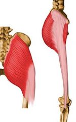
Muscle: glueteus maximus m.
Insertion: gluteal tuberosity of the posterior surface of the femur; iliotibial tract of fascia latae Origin: posterior gluteal line of the ilium Action: Extends thigh at the hip; assists in laterally rotating the thigh |
|
|
Insertion: gluteal tuberosity of the posterior surface of the femur;
iliotibial tract of fascia latae Origin: posterior gluteal line of the ilium Action: Extends thigh at the hip; assists in laterally rotating the thigh |

gluteus maximus m.
|
|
|
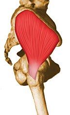
Muscle: gluteus medius m.
Insertion: lateral and superior surfaces of the greater trochanter Origin: dorsal ilium inferior to iliac crest Action: major abductor of thigh; anterior fibers help to rotate hip medially; posterior fibers help to rotate hip laterally |
|
|
Insertion: lateral and superior surfaces of the greater trochanter
Origin: dorsal ilium inferior to iliac crest Action: major abductor of thigh; anterior fibers help to rotate hip medially; posterior fibers help to rotate hip laterally |

gluteus medius m.
|
|
|
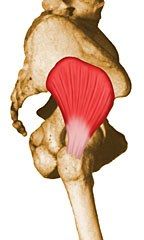
Muscle: gluteus minimus m.
Insertion: Anterior surface of greater trochanter Origin: Dorsal ilium Action: abducts and medially rotates the hip joint |
|
|
Insertion: Anterior surface of greater trochanter
Origin: Dorsal ilium Action: abducts and medially rotates the hip joint |

gluteus minimus m.
|
|
|
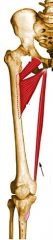
Muscle: piriformis m.
Insertion: superior border of the greater trochanter Origin: anterior sacrum Action: rotates hip joint laterally |
|
|
Insertion: superior border of the greater trochanter
Origin: anterior sacrum Action: rotates hip joint laterally |

piriformis m.
|
|
|
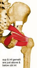
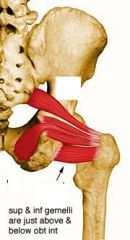
Muscle: quadratus femoris m.
Insertion: Quadrate tubercle and adjacent bone of intertrochanteric crest of proximal posterior femur Origin: Lateral margin of obturator ring Action: Rotates the hip laterally; also helps adduct the hip |
|
|
Insertion: Quadrate tubercle and adjacent bone of intertrochanteric crest of proximal posterior femur
Origin: Lateral margin of obturator ring Action: Rotates the hip laterally; also helps adduct the hip |

quadratus femoris m.
|
|
|
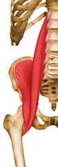
Muscle: iliopsoas m.
Insertion: Lesser trochanter Origin: iliac fossa Action: flex the torso and thigh with respect to each other |
|
|
Insertion: Lesser trochanter
Origin: iliac fossa Action: flex the torso and thigh with respect to each other |

iliopsoas m.
|
|
|
Muscle: pectineus m.
Insertion: pectineal line of the femur Origin: pectineal surface of the pubis Action: Adducts the thigh |
|
|
Insertion: pectineal line of the femur
Origin: pectineal surface of the pubis Action: Adducts the thigh |

pectineus m.
|
|

|
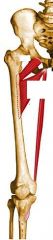
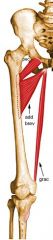
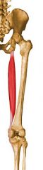
Muscle: adductor longus m.
Insertion: Middle third of linea aspera Origin: Anterior surface of body of pubis, just lateral to pubic symphysis Action: adducts thigh; lateral rotation of hip joint |
|
|
Insertion: Middle third of linea aspera
Origin: Anterior surface of body of pubis, just lateral to pubic symphysis Action: adducts thigh; lateral rotation of hip joint |
adductor longus m.
|
|

|
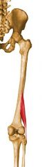
Muscle: adductor brevis m.
Insertion: superior linea aspera Origin: Anterior surface of inferior pubic ramus Action: adducts thigh, laterally rotates thigh |
|
|
Insertion: superior linea aspera
Origin: Anterior surface of inferior pubic ramus Action: adducts thigh, laterally rotates thigh |
adductor brevis m.
|
|
|
Insertion: superior linea aspera
Origin: Anterior surface of inferior pubic ramus Action: adducts thigh, laterally rotates thigh |

adductor brevis m.
|
|

|
Muscle: adductor magnus m.
Insertion: linea aspera and adductor tubercle Origin: Inferior pubic ramus Action: adduction of thigh |
|
|
Insertion: linea aspera and adductor tubercle
Origin: Inferior pubic ramus Action: adduction of thigh |
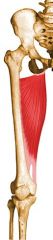
adductor magnus m.
|
|
|
Muscle: tensor fascia latae
Insertion: iliotibial band Origin: Anterior superior iliac spine Action: puts tension on the iliotibial band and stabilizes the hip joint and knee joint |
|
|
Insertion: iliotibial band
Origin: Anterior superior iliac spine Action: puts tension on the iliotibial band and stabilizes the hip joint and knee joint |

tensor fascia latae m.
|
|

|
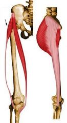
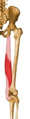
Muscle: sartorius m.
Insertion: Superior aspect of the medial surface of the tibial shaft Origin: anterior superior iliac spine Action: Flexes and laterally rotates the hip joint and flexes the knee |
|
|
Insertion: Superior aspect of the medial surface of the tibial shaft
Origin: anterior superior iliac spine Action: Flexes and laterally rotates the hip joint and flexes the knee |
sartorius m.
|
|

|
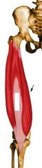
Muscle: rectus femoris m.
Insertion: patellar ligament Origin: anterior inferior iliac spine Action: Extends the knee |
|
|
Insertion: patellar ligament
Origin: anterior inferior iliac spine Action: Extends the knee |
rectus femoris m.
|
|

|
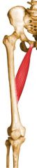
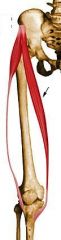
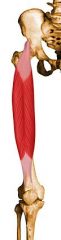
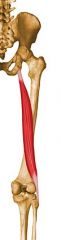
Muscle: gracilis m.
Insertion: Medial surface of tibial shaft Origin: Inferior margin of pubic symphysis Action: Flexes the knee, adducts the thigh, and helps to medially rotate the tibia on the femur |
|
|
Insertion: Medial surface of tibial shaft
Origin: Inferior margin of pubic symphysis Action: Flexes the knee, adducts the thigh, and helps to medially rotate the tibia on the femur |
gracilis m.
|
|
|
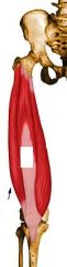
Muscle: vastus lateralis
Insertion: Lateral base and border of patella Origin: anterior and inferior borders of greater trochanter, superior portion linea aspera, and gluteal tuberosity of femur Action: Extends the knee |
|
|
Insertion: Lateral base and border of patella
Origin: anterior and inferior borders of greater trochanter, superior portion linea aspera, and gluteal tuberosity of femur Action: Extends the knee |

vastus lateralis m.
|
|
|
Muscle: vastus intermedius m.
Insertion: Lateral border of patella; also forms the deep portion of the quadriceps tendon Action: Extends the knee Origin: Superior 2/3 of anterior and lateral surfaces of femur; |
|
|
Insertion: Lateral border of patella; also forms the deep portion of the quadriceps tendon
Action: Extends the knee |

vastus intermedius m.
|
|
|
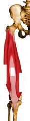
Muscle: vastus medialis m.
Insertion: base of patella Origin: Inferior portion of intertrochanteric line and wraps around to posterior femur all the way down femur Action: Extends the knee |
|
|
Insertion: base of patella
Origin: Inferior portion of intertrochanteric line and wraps around to posterior femur all the way down femur Action: Extends the knee |

vastus medialis m.
|
|

|
Muscle: semitendinosus m.
Insertion: anterior superior medial tibial shaft Origin: From common tendon with long head of biceps femoris to ischial tuberosity Action: Extends the thigh and flexes the knee, and also rotates the tibia medially |
|
|
Insertion: anterior superior medial tibial shaft
Origin: From common tendon with long head of biceps femoris to ischial tuberosity Action: Extends the thigh and flexes the knee, and also rotates the tibia medially |
semitendinosus m.
|
|
|
Muscle: biceps femoris long head m.
Insertion: fibular head Origin: Common tendon with semitendinosus to the ischial tuberosity Action: Flexes the knee, and also rotates the tibia laterally; long head also extends the hip joint (same as short head) |
|
|
Insertion: fibular head
Origin: Common tendon with semitendinosus to the ischial tuberosity Action: Flexes the knee, and also rotates the tibia laterally; long head also extends the hip joint (same as short head) |

biceps femoris m. long head
|
|

|
Muscle: biceps femoris m. short head
Insertion: fibular head Origin: linea aspera Action: Flexes the knee, and also rotates the tibia laterally; long head also extends the hip joint (same as long head) |
|
|
Insertion: fibular head
Origin: linea aspera Action: Flexes the knee, and also rotates the tibia laterally; long head also extends the hip joint |
biceps femoris m. short head
|
|
|
Muscle: semimembranosus
Insertion: Posterior surface of the medial tibial condyle Origin: Ischial tuberosity Action: Extends the thigh, flexes the knee, and also rotates the tibia medially |
|
|
Insertion: Posterior surface of the medial tibial condyle
Origin: Ischial tuberosity Action: Extends the thigh, flexes the knee, and also rotates the tibia medially |

semimembranosus m.
|
|

|
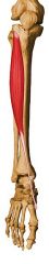
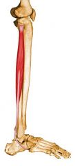
Muscle: tibialis anterior m.
Insertion: Medial and plantar surfaces of 1st cuneiform and on base of first metatarsal Origin: Lateral condyle of tibia, proximal 1/2 - 2/3 or lateral surface of tibial shaft, interosseous membrane Action: Dorsiflexor of ankle and invertor of foot |
|
|
Insertion: Medial and plantar surfaces of 1st cuneiform and on base of first metatarsal
Origin: Lateral condyle of tibia, proximal 1/2 - 2/3 or lateral surface of tibial shaft, interosseous membrane Action: Dorsiflexor of ankle and invertor of foot |
|
|
|
Muscle: tibialis anterior m.
Insertion: Medial and plantar surfaces of 1st cuneiform and on base of first metatarsal Origin: Lateral condyle of tibia, proximal 1/2 - 2/3 or lateral surface of tibial shaft, interosseous membrane Action: Dorsiflexor of ankle and invertor of foot |
|
|
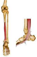
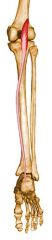
Muscle: tibialis posterior
Insertion: tuberosity of the navicular bone and sometimes medial cuneiform; deeper slip divides again into slips inserting on plantar surfaces of metatarsals 2 - 4 and second cuneiform Origin: Posterior aspect of interosseous membrane, superior 2/3 of medial posterior surface of fibula, superior aspect of posterior surface of tibia Action: Principal invertor of foot; also adducts foot, plantar flexes ankle |
|
|
Insertion: tuberosity of the navicular bone and sometimes medial cuneiform; deeper slip divides again into slips inserting on plantar surfaces of metatarsals 2 - 4 and second cuneiform
Origin: Posterior aspect of interosseous membrane, superior 2/3 of medial posterior surface of fibula, superior aspect of posterior surface of tibia Action: Principal invertor of foot; also adducts foot, plantar flexes ankle |

tibialis posterior m.
|
|

|
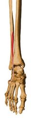
Muscle: fibularis (peroneus) brevis m.
Insertion: Lateral surface of styloid process of 5th metatarsal base Origin: Inferior 2/3 of lateral fibular surface Action: Everts foot and plantar flexes ankle |
|
|
Insertion: Lateral surface of styloid process of 5th metatarsal base
Origin: Inferior 2/3 of lateral fibular surface Action: Everts foot and plantar flexes ankle |
fibularis (peroneus) brevis m.
|
|
|
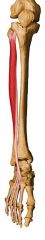
Muscle: fibularis (peroneus) longus m.
Insertion: Plantar posterolateral aspect of medial cuneiform and lateral side of 1st metatarsal base Origin: Head of fibula, upper 1/2 - 2/3 of lateral fibular shaft surface Action: Everts foot and plantar flexes ankle |
|
|
Insertion: Plantar posterolateral aspect of medial cuneiform and lateral side of 1st metatarsal base
Origin: Head of fibula, upper 1/2 - 2/3 of lateral fibular shaft surface Action: Everts foot and plantar flexes ankle |

fibularis (peroneus) longus m.
|
|

|
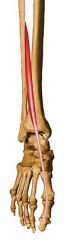
Muscle: fibularis (peroneus) tertius m.
Insertion: Dorsal surface of the base of the fifth metatarsal Origin: medial fibular shaft surface Action: dorsiflex, evert and abduct the foot |
|
|
Insertion: Dorsal surface of the base of the fifth metatarsal
Origin: medial fibular shaft surface Action: dorsiflex, evert and abduct the foot |
fibularis (peroneus) tertius m.
|
|

|
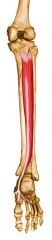
Muscle: extensor digitorum longus m.
Insertion: Splits into 4 tendon slips after inferior extensor retinaculum, each of which insert on dorsum of middle and distal phalanges as part of extensor expansion complex Origin: Lateral condyle of fibula, upper 2/3 - 3/4 of medial fibular shaft surface, upper part of interosseous membrane Action: Extend toes 2 - 5 and dorsiflexes ankle |
|
|
Insertion: Splits into 4 tendon slips after inferior extensor retinaculum, each of which insert on dorsum of middle and distal phalanges as part of extensor expansion complex
Origin: Lateral condyle of fibula, upper 2/3 - 3/4 of medial fibular shaft surface, upper part of interosseous membrane Action: Extend toes 2 - 5 and dorsiflexes ankle |
extensor digitorum longus m.
|
|

|
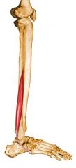
Muscle: extensor hallucis longus m.
Insertion: distal phalanx Origin: Anterior surface of the fibula and the adjacent interosseous membrane Action: Extends great toe and dorsiflexes ankle |
|
|
Insertion: distal phalanx
Origin: Anterior surface of the fibula and the adjacent interosseous membrane Action: Extends great toe and dorsiflexes ankle |
extensor hallucis longus m.
|
|

|
Muscle: flexor digitorum longus m.
Insertion: plantar surface of bases of 2nd - 5th distal phalanges Origin: Posterior surface of tibia Action: Flexes toes 2 - 5; also helps in plantar flexion of ankle |
|
|
Insertion: plantar surface of bases of 2nd - 5th distal phalanges
Origin: Posterior surface of tibia Action: Flexes toes 2 - 5; also helps in plantar flexion of ankle |
flexor digitorum longus m.
|
|

|
Muscle: flexor hallucis longus m.
Insertion: Plantar surface of base of distal phalanx of great toe Origin: Inferior 2/3 of posterior surface of fibula, lower part of interosseous membrane Action: Flexes great toe, helps to supinate ankle |
|
|
Insertion: Plantar surface of base of distal phalanx of great toe
Origin: Inferior 2/3 of posterior surface of fibula, lower part of interosseous membrane Action: Flexes great toe, helps to supinate ankle |
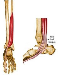
flexor hallucis longus m.
|
|
|
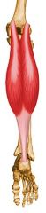
Muscle: gastrocnemius m.
Insertion: The two heads unite into a broad aponeurosis which eventually unites with the deep tendon of the soleus to form the Achilles tendon, inserting on the middle 1/3 of the posterior calcaneal surface Origin: Medial head from posterior nonarticular surface of medial femoral condyle; Lateral head from lateral surface of femoral lateral condyle Action: plantar flexor of ankle |
|
|
Insertion: The two heads unite into a broad aponeurosis which eventually unites with the deep tendon of the soleus to form the Achilles tendon, inserting on the middle 1/3 of the posterior calcaneal surface
Origin: Medial head from posterior nonarticular surface of medial femoral condyle; Lateral head from lateral surface of femoral lateral condyle Action: plantar flexor of ankle |

gastrocnemius m.
|
|

|
Muscle: soleus m.
Insertion: Eventually unites with the gastrocnemius aponeurosis to form the Achilles tendon, inserting on the middle 1/3 of the posterior calcaneal surface Origin: Posterior aspect of fibular head, upper 1/4 - 1/3 of posterior surface of fibula, middle 1/3 of medial border of tibial shaft, and from posterior surface of a tendinous arch spanning the two sites of bone origin Action: plantar flexor of ankle |
|
|
Insertion: Eventually unites with the gastrocnemius aponeurosis to form the Achilles tendon, inserting on the middle 1/3 of the posterior calcaneal surface
Origin: Posterior aspect of fibular head, upper 1/4 - 1/3 of posterior surface of fibula, middle 1/3 of medial border of tibial shaft, and from posterior surface of a tendinous arch spanning the two sites of bone origin Action: plantar flexor of ankle |
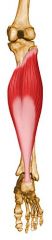
soleus m.
|
|
|
Muscle: plantaris
Insertion: Middle 1/3 of the posterior calcaneal surface, just medial to Achilles tendon Origin: Inferior aspect of lateral supracondylar line of distal femur Action: Plantar flexor of ankle; also flexes knee |
|
|
Insertion: Middle 1/3 of the posterior calcaneal surface, just medial to Achilles tendon
Origin: Inferior aspect of lateral supracondylar line of distal femur Action: Plantar flexor of ankle; also flexes knee |

plantaris m.
|
|

|
Muscle: popliteus m.
Insertion: Posterior surface of tibia in a fan-like fashion, just superior to the popliteal line Origin: Anterior part of the popliteal groove on lateral surface of lateral femoral condyle Action: Rotates knee medially |
|
|
Insertion: Posterior surface of tibia in a fan-like fashion, just superior to the popliteal line
Origin: Anterior part of the popliteal groove on lateral surface of lateral femoral condyle Action: Rotates knee medially |
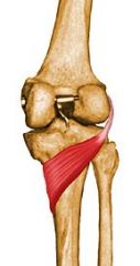
popliteus m.
|