Reading...
![]()
Play button
![]()
Play button
![]()
Use LEFT and RIGHT arrow keys to navigate between flashcards;
Use UP and DOWN arrow keys to flip the card;
H to show hint;
A reads text to speech;
86 Cards in this Set
- Front
- Back
|
Muscle composition and function
|
Made of muscle cells and connective tissue. (conn. contains blood and supplies nerves to the muscle and serves as a "harness to transmit forces of musclular contraction)
Contractiility and conductivity |
|
|
Muscle Tissue
|
Smooth (unstriated;involuntary; visceral
Skeletal (striated voluntary) Cardiac (striated involuntary) |
|
|
Embrylogically Muscle comes from
|
Mesoder
|
|
|
Sarcoplasm
|
cytoplasm
|
|
|
sarcolemma
|
plasma membrane
|
|
|
sarcoplasmic reticulum
|
S-ER
|
|
|
sacromere
|
linear unit
|
|
|
myofilaments
|
myofibrils
|
|
|
myfibers
|
muscle cells
|
|
|
Sketal Muscle General Features
|
Striated (voulntary)
T-tubule system well developed sarcoplasmic reticulum multinucleated nuclei located at cell periphery (just inside sarcolema) |
|
|
Skeletal muscle Fibers (myofiber) Shape
|
Cylindrical (variations in length 1mm to 4cm) Bulky muscles have larger cells; smaller are smaller.
Several hundred nuclei just inide sarcolemma, flattened oval and elongated in appearanc |
|
|
Myofibrils are?
|
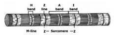
Tiny cylindrical rods within sarcoplasm of muscle cell, responsible for appearance of cross striations of muscle (due to registry of alternating ligh and dark portions of myofibrils adjacent to one another)
Appear as small dots when viewed cross sectionally Bands, zones, lines and functional units of myofibril |
|
|
A-band of myofibril
|
Broad, dark staining band, anisotropic band
|
|
|
I band of myofiril
|
Narrower light staining band, isotropic bond
|
|
|
z disk (z-line) myofibrins
|
Dense staining;narrow
Bisects I band, distance between 2 z disks=sarcomere |
|
|
Sarcomere
|
Distance between 2 z disks, functional unit of myofibril. Many, many sacro. line up end to end to form myofibril.
LENGTH varies as a function of contration |
|
|
H band
|
Pale staining; narrow
bisects A band In middle of sacromere |
|
|
M line
|
thin, bisects H band
|
|
|
Myofibrils are comprised of?
|
Finer myofilaments
1. Actin filaments (thin) extend from z disks to edge of H band 2. Myosin Fialaments (thick!!) extend from one end of A band to other end of A band, forms thin cross bridges that extend from each mysoin filament towards actin filaments. This creates M line (at center of H band) |
|
|
M line
|
At center of H band, contains myomesin, c protein and other proteins that interconnect thick myosin filaments to maintain specifi lattice arrangement
|
|
|
Z- disk
|
region where attachment of ends of actin filaments in adjacent sacromeres, appears filamentous and somewhat dense, contains a-actinin
|
|
|
Sarcoplasm
|
cytoplasm of muscle fibers
A. between myofibrils, beneath sarcoloma, and around nuclie has Golgi and Ribosomes, mitochondria are near nuclie beneath sarcolemma between myofibrils |
|
|
Sarcoplasmic Reticulum
|
Network of cisterns or membrane tubules running between and around myofibriles, tends to form 'collars' at A-I junctions. Binds and releases Ca+
|
|
|
Collars at A-I junctions
|
1. INterconnect by longitudinal components
2. Two collars at A-I junction 3. Collars seperated from each other by T-tubules |
|
|
sarcolemma
|
Cell membrane muscle fibers
Numerous invaginations along cell surface. Creates T-tubules (transverse tubules) |
|
|
T-tubules
|
Lumen of T-tubule is continuous with extracellular space.
Wind between myofibrils Typically seen at A-I junction between the 2 collars of the Sarco. reticulm |
|
|
Triads
|
T-tubule +2 lateral cisternaie of s. Ret.
Membranes of T-tubules appear to be closely coupled to Sar. Ret. memranes (this facilitates transmission of electical impulses from sarcolmma to interior deps of cell) 2 triads/sacromere |
|
|
Myfibrls are made of
|
Actin,-- G actin (Globular actin)which makes F-actin (fibrous actin) 2 helically wound strands of polymerized G-actin
Myosin (4 Light chains and 2 heavy) Regulatory Molecules |
|
|
Light meromysin
|
Long molecular subunits rigid, packed together longitudinally to form backbone of myosin filament
|
|
|
Heavy meromysin
|
Long felxible moleculare subunits
2 components 1. rod like portion parallel to backbone of filament 2. Globular head (extends laterally as a cross bridge from thick myosin fil. to actin fil. no cross bridges at M-line, ATPase activity has actin binding sites. **Polarized, globular heads directed away from midpoint of myosin filament. |
|
|
Regulatory molecules of myofibrils
|
Tropomyosin
troponin complex Ca++ |
|
|
Tropomyosin
|
fibrous; arranged head to tail in linear series.
Helically wound along grooves of F-actin helix |
|
|
Troponin Complex
|
(3 Subunits)
Tnt: binds entire troponin molecular to tropomyosin TnC: great affinity for Ca++ TnL: binds to actin,preventing actin-myosin interactions |
|
|
TnT
|
Binds entire troponin molecular to tropomyosin
|
|
|
TnC
|
great affinity for Ca
|
|
|
TnL
|
binds actin preventing Actin-myosin interactions
|
|
|
Ca++
|
binds to TnC, binding to TnC induces a con. shift in tropomyosin exposing previously blocked active sites on actin filament
|
|
|
a-actinin
|
component of Z-disk
Holds actin filaments in register by binding them in parallel array |
|
|
Titin
|
large, linear elastic protein
helps position myosin filaments precisely with the sacromere extends from each ahlf of myosin filament to z-disk |
|
|
nebulin
|
long non elactic protein
2 molecules of nebulin wrap around each thin filament help anchor thin filament to z-disk |
|
|
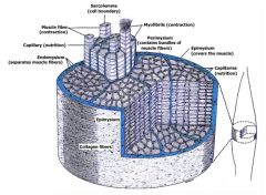
Types of gross muscle organization
|
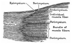
Endomysium, perimysium, epimysium
|
|
|
What is the mechanism of action of NPNs?
|
Urea + urease ---> NH4+. this is normal.
if there are not enough H+, Urea + urease --> NH3 (ammonia). that is bad! you will see rumen alkalosis and systemic acidosis. |
|
|
Vascular Supply to muscle??
|
Larger arteries follow perimysium, arterioles penetrate fascicles, capillaries // individul fibers
Lymphatics in connective tissue septa |
|
|
Nerve muscle association
|
Motor End Plates (form myo-neural junctions)
Sensory Structures |
|
|
Motor end plate components
|
Myelin sheath of axon is lost as nerve fiber approaches surface of muscle cell
Axon branches close to surface of cell Subneural Apparatus synaptic vesicles Motor units |
|
|
Axon branches characterisics
|
Branches occupy recesses in surface of muscle fiber
RECESSES ARE Synaptic troughs or primary synaptic clefts |
|
|
Subneural apparatus
|
secondary synaptic clefts caused by infolding of sarcolemma
acetylcholinesterase located at surface of sarcolemma |
|
|
synaptic vesicles
|
in axon terminals, contain acetylcholine
|
|
|
Motor Unit
|
Motor neuron and muscle fibers innervated by it Ratio of muscle to fiber reflects control
1:1 very fine control 1:1600 very coarse (gross control) |
|
|
Mysathenia Gravis
|
Disease characterized by weakness and easy fatigue of muscles
Autoimmune response to Acetyl Choline receptor Treatment::: administer ACHe inhibtors, cause both diagnostic and therapeutic values |
|
|
Sensory Structures
|
Muscle Spindles
Golgi Tendon Organ |
|
|
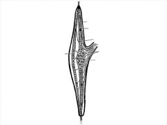
Muscle Spindle
|
Small speciallized muscle fibers in c.t. capsule (SMALL fibers= inrafusal fibers
Two zones.... Central region of Intrafusal fibers--Nuclei chain fibers -----------nuclear bag fibers Polar regions are long and tapered |
|
|
Muscle Spinde Capsules
|
Internal capsule-immediately surrounds intrafusal fibers; encloses inner axial space
External capsule surrounds internal capsule and creates periaxial space between inner and outer capsules |
|
|
Innervation of Muscle Spindle
|
Sensory-large fibers form annulospiral or primary endings around nuclear bag and nuclear chain regions...smaller diameter afferts form flower spray or secondaryendings on intrafusal fibers on polar segments
Motor-efferent fibers form motor end plates on either side of mid region of inrafusal fibers. |
|
|
Muscle SPindle
|
|
|
|
Muscle Spindle Functions
|
Act like minature strain gauges
|
|
|
Golgi Tendon Organs
|
Spindle shaped body comprised of collagen and enclosed by thin capsule
Afferent (sensory) nerve fibers penetrate between collagen fibers Sensitive to stretching forces on tendon |
|
|
Embryological origins of skeletal muscle
|
loose mesenchyme>myoblasts
myoblasts fuse>mutinucleated myotubes Myofilaments appear--irregularily arrange at first, gradually align into myofibrils |
|
|
Hypertrophy of muscle
|
due to size increse of fibers not number
|
|
|
Cardiac Muscle General Features
|
Striated (involuntary)
T-tubule system sarcoplasmic ret. less well develped than Skeletal muscle Single Nucleus/Cell Nucleus at center of cell Intercalated disks are a diagnostic feature of this tissue |
|
|
KEY features of cardiac muscles
|
Cells are elongated and brancing, messy placement, one nucleus/cell at center of cell an oval and fairly large...
Have myofibrils that branch and sometimes blend with adjacent myofibrils (less distinct) Sacromeres A, I bands and Z disks Sarcoplasm typically seen at each pole of nucleus Interclated disks-dark cross brands, frequently appear step like occur where a z disk should, mark spots of cell to cell attachment |
|
|
Myofilaments of Cardiac Muscle and Mitochondria Content
|
Actin and myosin present
Many mitochondria, very regularily spread out between myofibrils |
|
|
T-tubule system and Sarcoplasmic Reticulm of cardia muscle
|
T-tubules-- larger than those in skeletal muscle, at Z line instead of A-I juncti.
Sarco. Ret. Less well developed than skeletal muscle, simple plexiform arrangement of tubular elements no terminal cisternae |
|
|
Intercalated disk of cardiac muscle cells
|
Represent a complex of several types of cell2cell junctions,
Gap junction, Desmosomes fascia adherens (serve as anchoring points for actin filaments) |
|
|
Cardiac muscle characteristics
golgi? Glcycogen? Fat? Atrial Granule? |
Has golgi apparataus,
Glycogen is very abundant Lots of fat droplets Atrial granules-are unique to atrial cardiac muscle cells, contain atrial natrueric peptide which fuctions to lower BP |
|
|
Atrial natriuretic Peptide
|
Functions to lower BP
Decreases renal tubule capacity to reabsorve (conserve) Na and H2O |
|
|
Cardiac muscle and connective tissue?
Vascular Supply? Nerve Supply? Conductive system? |
CM is surrounded by CT that form a fine net of reticualr and collagenous fibers, collagenous and elastic fibers occur between bundles of cells
EXCELLENT VS, come from coronary artiers and lymphatic drainage Nerves branch from sypathetic and parasympathetic nerves to terminate on muscle cells Conductive: composed of modified muscle fibers, (Purkinje Fibers) |
|
|
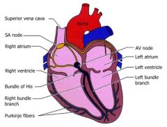
Purkinje Fibers?
|
Specialized muscle cells
1. myfibrils are reduced in #, 2. contain more sacroplasm 3.nuclie are rounded 4.large diameter 5.lack T-tubules 6. more glycogen |
|
|
Heart Diagram with Purkinje Fibers
|
|
|
|
Cardiac muscle comes from where??
|
Mesoderm>myoblasts, no fusion of myoblasts
|
|
|
SMooth muscles General features
|
No striations or T-Tubules.
Found in walls of hollow viscera, (GI tract, portions of urinary and reproductive tracts, walls of BV, larger ducts of cmp. glands) Acts involuntary, is regualted by autonomic NS, hormones, or local physiological conditions. Acts for contractility, conductivity, and production of EC products(collagen, elastin, glycoaminglycans etc.) |
|
|
Smooth Muscle Appearance
|
Fusiform shape, ends are tapered, oval nucleus in center of cell takes on a corkscrew appearance durin contraction, form sheets or clusters
Cytoplasm is eosinophilic CT contibutions, every cells is surrounded by thin external lamina that seperates the PM of ajacent SM cells Reticular Fibers are prevalent in external lamina, help harness force of contration. |
|
|
Smooth Muscles Electron micros..
|
Mitochondria and other organells cluster at the poles of nuclei,
lots of interweaving filaents , thin filaments (tropomysin present, troponin absent) thick filaments (myosin type 2) difficult to see. NB- the actin and myosin filaments are not arranged in the percise manner noted is SKM or CAM, they tend to form bundles that run obliquely to cytoplasm. IF's are numerous |
|
|
SM Cells have a special characteristic of??
|
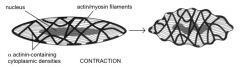
Dense bodies, located along inner aspects of sarcolemma, and scattered in sarcolemma.
Similar to z-disks of SKM and CAM contain alpha-actinin serve as anchor sites for actin-myosin filament bundles as well as IF's |
|
|
SM cells also have Caveolae for?
|
Pincytotic activities, may function as the equivalent of T-tubule in SKM and CAM, may work in concert with Sarcoplasmic RETiculum to modulate Ca availability
|
|
|
Caveolae in SM Cells?
|
Used to regulate Ca+ availability with The SR. Stores the Ca+
|
|
|
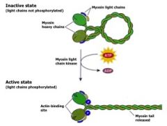
Contractile method of SM Cellsq
|
1. Ca+ +calmodulin form complex
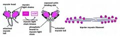
2. Ca+-calmodulin complex activates myosin light chain kinase 3.Mysone LC kinase phosphorylates one of the 2 LC's associated with myosin molecule head 4.Phosphor. of LC exposes 'actin-binding' site on myosin head. a. also allows myosin head to straighten out and form small bipolar filaments. b.phosph. is slow process (takes time for SM contraction) c. ATP hydrolysis occurs more slowly also, so SM requires less muscle to contract |
|
|
Innervation of SM cells caused by?
|
Sympathetic Innervation
Parasympathetic Innervations Variations of number of cells in a group that are inervated. |
|
|
Regeneration of SM cells
|
Reatains mitotic capability, can also be formed from pre-existing pericytes
|
|
|
Sympathetic innervations of SM cells?
|
Caused by synaptic vesicles contain norepinephrine
|
|
|
Parasympathetic innervation of SM cells?
|
synaptic vesicles contain acetylcholine
|
|
|
Variation in # of cells in group that are innervated?
|
Iris SM: every muscle fiber is innervated
SM in GI/uterus: only small % of SM cells are innervated. (impulse transmission from muscle cell to muscle cell accomplished via gap junctions between adj. muscl. cells |
|
|
CONTRACTILE MECHANISM OF SM======Myosin activation,
|
|
|
|
CONTRACTILE MECHANISM OF SM======Myosin activation,
|
|
|
|
Myosin activation second view
|
|
|
|
SM contraction
|
|