![]()
![]()
![]()
Use LEFT and RIGHT arrow keys to navigate between flashcards;
Use UP and DOWN arrow keys to flip the card;
H to show hint;
A reads text to speech;
125 Cards in this Set
- Front
- Back
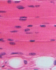
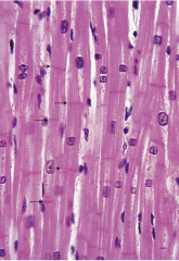
Describe. Type Reason Staining |
Skeletal muscle Multinucleated (nuclei at outet erdges, peripheries) pINKS TAINED MYOFILAMENT PRoteins with mainly Eosin |
|
|
Skeletal muscleMultinucleated (nuclei at outet erdges, peripheries)pINKS TAINED MYOFILAMENT PRoteins with mainly Eosin |
|
Describe. |
Developing skeletal muscle tissue Reason: Multnuclei (at peripheries), and myotubes but lack myofilaments |
|
|
Development skeletal myofibers |
Myoblasts --fuse together to form multinucleated --> myotubes |
|
|
Functional unit of striated muscle. |
Sarcomere (myosin, actin units) |
|
|
Function muscle |
Generate contraction |
|
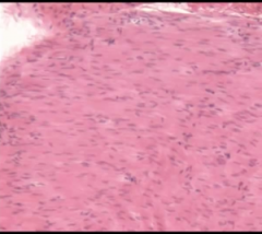
Describe. |
Smooth muscle |
|
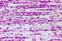
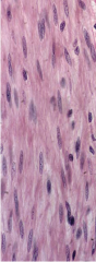
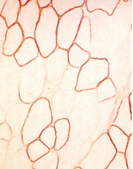
Describe. What staining |
PAS staining smooth muscle |
|
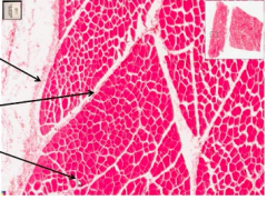
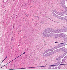
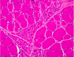
Describe. Label arrows: top, middle, bottom |
Top: epimysium middle: perimysium Bottom: endomysium |
|
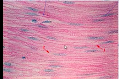
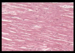
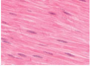
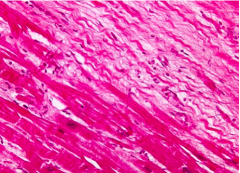
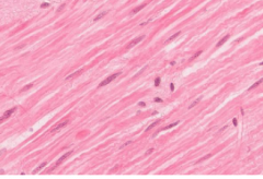
Describe, reason |
Skeletal. Nuclei on peripehries |
|
|
What gets wider or thinner during muscle contraction? |
A band |
|
|
What changes during skeletal muscle contractions Does not? |
I band H line Does not: Everything but I H |
|
|
What found in skeletal muscle. Distintuighins |
Mitochondria: darker Glycogen: lighter |
|
|
Properties cardiac muscle |
Intercalated discs Central nuclei (along with smooth) Branching alSO Has: gap junctions |
|
|
Different between T tubule SR in skeletal and cardiac |
Triad: 2 t tubules 1 terminal cisternae in skeletal in AI junctions Diad: 1 t tubule 1 terminal cisternae in skeletal in z lines |
|
|
Structure cardiac muscle |
fascia adherens (anchor thin filaments, make transverse component Macula adherens (desmosomes); connect cells Gap junctions (allow cells to communicate) |
|
Describe. dark, light areas |
Dark: regular cardiac Light: conducting zone > containing purkinje fibers |
|
|
What muscles undergo proliferation, divide |
Smooth muscle Not skeletal, cardiac |
|
Describe. Characteristics |
Skeletal muscle Properties: > Arranged longitudinally striaght (non branched) > Nuclei are periphery > striation (light I and dark A bands) > multinucleated > muscle arranged around epineurisum |
|
Describe. Characteristics. |
Smooth muscle Properties: > 'cigar shaped' nuclei in center > No striations Diff from C/T > CT Blue with trichrome, not smooth muscle |
|
|
Describe. Characteristics |
Cardiac muscles > Striations (dark A band and light i band) > Intercalated discs (lines) > Nuclei in center > branching, no stacking |
|
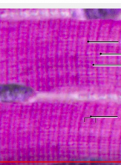
Describe. from top to bottom. 1, 2, 3, 4 |
A band I band or light band Z line Sarcomere, extends from 1 z line to the next z line |
|
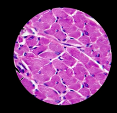
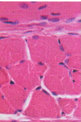
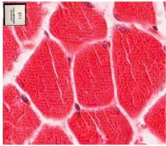
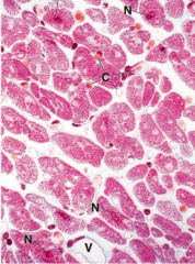
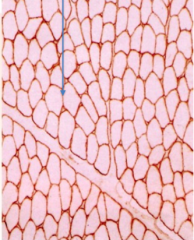
Describe. |
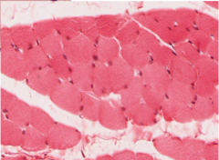
Cross section of skeletal muscle > Nuclei on peripheries |
|
|
Characteristics of muscle |
> pink (Eosinophilic) fibers (acidophilic b/c stains with - eosino dye) |
|
Describe. |
Cardiac muscle properties: > branching > intercalated discs > striated (+ skeletal) > central nuclei (uni nucleated + smooth) |
|
Describe. |
Smooth muscle > non striations , no branching > central nuclei = 'cigar shaped' nuclei > 'spindle shaped' smooth muscle fibers, |
|
Describe. |
Cross section skeletal muscle > peripherally nuclei nucleated > striations |
|
Describe. |
Skeletal > peripherally located nuclei |
|
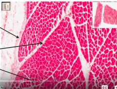
Label arrows. |
Epimysium Perimysium Endomysium |
|
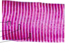
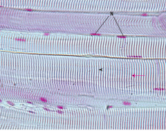
Describe. |
From left to right: Sarcomere (between z lines, middle white line to next middle white line) I band (actin only) A band (myosin only) |
|
|
Parts troponin |
T troponin: attached to troponin C troponin: binds Ca2+ I troponin: inhibits actin-myosin interaction |
|
|
Which is attached to tropomyosin? a. C troponin b. I troponin c. T troponin d. M troponin |
c. T troponin |
|
|
What binds to calcium? a. C troponin b. I troponin c. T troponin d. M troponin |
a. C. troponin |
|
|
What inhibits actin and myosin interaction? a. C troponin b. I troponin c. T troponin d. M troponin |
b. I troponin |
|
|
Components of ca2+ regulation in muscles? |
> sarcolemma = plasma membrane of muscle > T tubules = invagination of Sarcolemma > Sarcoplasmic reticulum > Triad = T tubule and 2 ends of sER |
|
|
Function of intercalated discs?
|
Allows coordination between gap junctions of cardiac cells |
|
Describe. |
Cardiac muscle > branching > uni nuclear > intercalated discs > sarcomere arrangement |
|
Describe. |
Smooth muscle. |
|

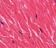
Describe. |
Smooth muscle |
|
|
Which of the following is true regarding cardio hypertrophy? a. hypertrophy is always maladaptive b. hypertension is an example of adaptive physiological hypertrophy c. a sarcomere mutation leads to compensation d. exercise leads to decompensation |
c. a sarcomere mutation leads to compensation explan: sarcomere mutation > pathological hypertrophy > decompensation elim: a. incorrect, adaptive hypertrophy is an example of a good adaptation b. incorrect, HTN is an example of pathological hypertrophy c. correct d. exercise leads to adaptive hypertrophy |
|
|
What are the steps in interatrial and interventricualr septa formation? |
> Sepation of atria > Septation of ventricle > Growth of endocardial cushion > Growth of bulbar ridges that divide bulbus growth (aorta and PA regions) |
|
|
Which of the following is correct? a. transition from tubular to 4 chambered heart occurs between 12 and 16 weeks b. Growth of endocardial cushion results in an outflow of blood in the aortic and pulmonary artery regions c. Growth of the bulbus cordis allows outflow of blood in the aorta and pulmonary tract regions d. Growth of the bulbus region separating certain regions allows outflow of blood in the aorta and pulmonary artery regions |
d. Growth of the bulbus region separating certain regions allows outflow of blood in the aorta and pulmonary artery regions Elim: a. incorrect, transition from tubular to 4 chambered heart occurs 4-8 weeks b. incorrect, this is the 3rd step, but does not form the Aorta/PA regions. Growth of bulbar ridges separating bulbus cordis is what forms the A/PA regions. c. incorrect, the growth of the bulbar ridges separating the bulbus cordis forms the outflow tract into future aorta and Pul. tract d. correct |
|
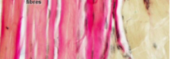
Describe. |
epimysium of skeletal muscle |
|
Describe. |
fascicle of skeletal muscle |
|
Describe. |
nerve |
|
Describe. |
skeletal muscle |
|
|
How are skeletal muscle cells formed? |
myoblasts |
|
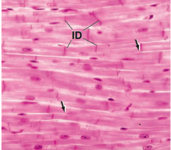
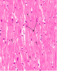
1. Describe. a. cardiac muscle b. smooth muscle c. skeletal muscle d. bone e. cartilage f. dense irregular c/t g. dense regular c/t h. blood 2. What do the arrows point to? a. gap junctions b. connexons c. intercalated discs d. endoplasmic reticulum e. golgi apparatus |
1. a. cardiac muscle 2. c. intercalated discs Reason: arrow points to intercalated discs |
|
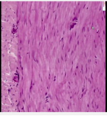
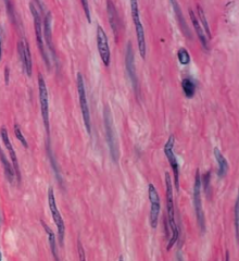
Describe. |
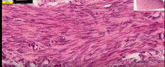
Longitudinal section of smooth muscle |
|
Describe. |
Cross section of smooth muscle |
|
Describe. |
Smooth muscle |
|
Describe. |
Cross section smooth muscle |
|
Describe. |
long section cardiac musce |
|
Describe. |
cross section cardiac. |
|
|
Which of the following is true for mammalian skeletal muscle? a. t tubules located at z disc b. t tubules absent c. troponin absent d. it possesses triads e. it possesses caveolae. |
d. it posseses triads |
|
|
Muscles
-Structure -Function -Properties |
Muscle: structure: > actin, myosin (arranged differently in each) (most ordered: skeletal in sarcomeres, cardiac in less arranged sarcomeres, poor to no arrangement smooth as peripehral-cytoplasmic densities) function: > contraction Skeletal muscle: Structure: > triads: narrow central T tubule flanked by terminal cisternae of the SR located at the A-I junction Function: Cardiac muscle: Structure: T tubules > Larger t tubules than in skeletal muscle > Comprise dyads: each with 1 t tubule and 1 profle of SR (found in viscinity of Z discs) Function: Smooth Muscle: Structure: Function: > T tubule + SR = Caveolae (but no T tubules) + some SER (limited SER) Properties > spindle (fusiform shaped) > 1 - 2 central nuclei > regeneration capability (limited Sk. muscle, no cardiac (fibrosis/scarring only)) > no sarcomere or myofibril, but cytoplasmic and peripheral densities arrangement = no Z lines, A bands, I bands, H bands |
|
|
Where are triads in skeletal muscle located? a. I band b. A band c. near Z line d. At IH junction e. At AI junction |
e. AI junction |
|
Describe. |
Skeletal muscle |
|
|
Diferent skeletal muscle bands |
A bands: Mainly myosin (darkbands), but some actin I band: Only actin (light bands) H band: Only myosin (dark bands) M line: middle of M line (adjacent thick filaments) Z disc > desmin (anchors Z disks to each other) |
|
|
Dyads -Structure -Function |
Cardiac muscle Structure: 1 t tubule and 1 SR (dyad) going into Z line |
|
|
Triads -Structure -Function |
Skeletal muscle Structure: (T tubule going to AI junction) 1 triad: 1 t tubule and 2 SR cisternae |
|
|
T-tubule + SR orientation of the muscle types |
Skeletal muscle: > Triad (1 t tubule and 2 cistaernae) going into AI junction Cardiac muscle: > Dyad (1 t tubule and 1 cistaernae) going into Z line Smooth muscle: > Caveolae (no T tubule) + SER |
|
|
Which of the following is incorrect regarding muscles?
a. there is regenerational capability in smooth muscle b. there are peripherally located nuclei in smooth muscles c. there are dyads at Z discs in smooth muscle d. there are caveolae (but no T tubules) and some sER in smooth muscle e. There are central nuclei in cardiac muscle |
c. there are dyads at Z discs in smooth muscle This refers to cardiac Elim: abde all correct |
|
Describe. |
Cardiac muscle tissue |
|
Describe. |
Smooth muscle |
|
|
Which muscle tissue has extensive regenerational capability? a. cardiac b. skeletal c. smooth |
c. smooth explanation a: incorrect, no regeneration at all, only fibrosis/scarring b: incorrect, limited generation c: correct , extensive regeneration |
|
|
Characteristics of muscle -function -structure -staining |
Function: > Aggregates of specialized cells with primary role of contraction Method: > Shortening of muscle cells (fibers) produce movement Structure: > Muscle fibers typically arranged in parallel. = Smooth muscle arranged in layers of different orientation Actin (6-8 nm) Myosin (15 nm diameter) Staining: eosinophilic with H&E b/c presence of proteins |
|
|
How are muscle fibers generally arranged? |
Parallel But smooth muscle cells typically arranged in layers of different orientation. |
|
|
How are smooth muscle fibers arranged |
Arranged in layers of different orientation. |
|
|
Striated muscle not striated |
Skeletal muscle: > voluntary movement Visceral striated: > associated with gut (tongue, esophagus, etc) Cardiac muscle: > Heart involuntary contraction Non striated: Smooth |
|
|
Types striated * not striated |
skeletal visceral striated, associated with GUT (tongue, GI) cardiac x: smooth |
|
|
Smooth muscle char. -structure -location -function |
Structure: non striated Location: viscera Function: Lining hollow organs (involuntary contraction) |
|
Describe. |
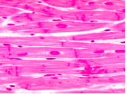
Skeletal muscle |
|
Describe. |
Cardiac muscle |
|
Describe. |
Smooth muscle Non striated, non parallel (different orientations) |
|
|
Division, Repair, and renewal of muscle tissues |
Skeletal: > cells do not divide > Fibroblasts may produce scar tissue > Satellite cells on edges of cells = Can differentiate into myogenic precursors = Show limited capacity for skeletal muscle fibers to repair Cardiac: > cells do not divide > little capacity for repair beyond childhood = In case of cell death, cardiac muscle replaced with scar tissue |
|
|
1. Which is nonstriated? 2. Which contains cells capable of cell division? 3. What has limited regeneration? 4. Which has limited repair beyond childhood? 5. What has satellite cells along edges of muscle fiber capable of differentiating into myogenic precursors? 6. Innervation is necessary for muscle integrity of what type of muscle? a. cardiac muscleb. skeletal muscle c. smooth muscle |
1. c. smooth 2. c. smooth 3. b. skeletal muscle 4. a. cardiac 5. b. skeletal muscle 6. b. skeletal muscle |
|
Describe. |
Dense regular c/t |
|
Describe. |
Dense irregular c/t |
|
Describe. |
Smooth muscle |
|
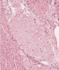
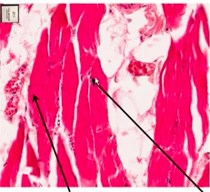
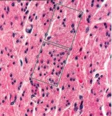
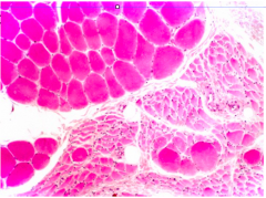
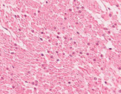
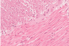
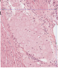
Describe middle and regions surrounding middle. |
Middle: Atrophied muscle Surrounding: skeletal muscle (peripherally located muscle) |
|
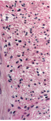
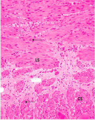
Describe bottom left and top right regions. |
Bottom left: normal cardiac muscle Top right: scar tissue cardiac muscle (after MI) = contains lymphocytes |
|
Describe. |
Skeletal muscle atrophy
(usually due to denervation) > shrinking skeletal muscle |
|
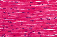
Describe. |
Cardiac muscle |
|
Describe. |
Cross section smooth muscle. |
|
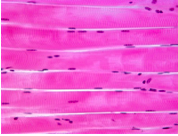
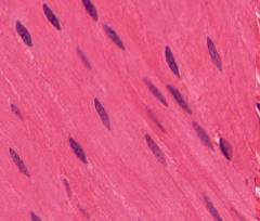
Describe. |
Longitudinal section smooth muscle |
|
Describe. |
Top left: cross section smooth muscle Bottom right: longitudinal section smooth muscle |
|
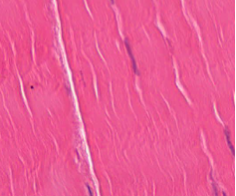
Describe. |
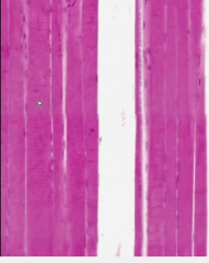
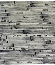
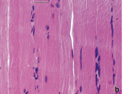
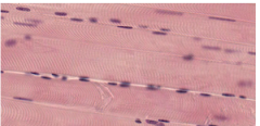
Skeletal muscle longitudinal section |
|
|
Properties skeletal muscle |
-Muscle cells formed by aggregate of myoblasts -Muscle fiber is a syncytium (multicellular) -Nuclei sit under plasma membrane (peripherally) -Muscle strength depends on # of muscle fibers |
|
Describe. |
Longitudinal section skeletal muscle |
|
Describe. |
Cross section skeletal muscle |
|

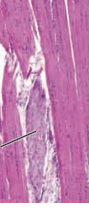
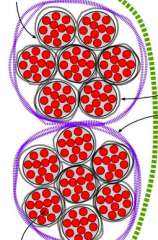
Describe.
Label arrows. Explain each. |
top left arrow: muscle cell/fiber (surrounded by endomysium) bottom left arrow: myofibril (several contained in 1 muscle fascicle) top right arrow: endomysium(surrounding muscle fiber) purple lining: perimysium (surrounds fascicle: containing several muscle cells (w/ myofibrils)) green lining: epimysium (surrounds entire muscle0 |
|
Describe. |
|
|
|
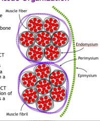
Different layers of skeletal muscle. |
Endomysium > Delicate layer that surrounds individual muscle fibers/cells Perimysium > Surrounds bundle of fibers to form a fascicle Epimysium > Sheath of DCT surrounding the collection of fascicles that comprise a muscle or simply surrounding a muscle (inherently contains several fascicles) |
|
Describe. |
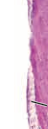
Muscle-tendon junction |
|
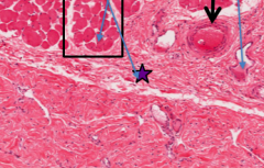
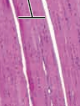
Describe. Label > Box > purple star > black arrow |
Box: Fascicle Purple star: Epimysium Black arrow: blood vessel |
|
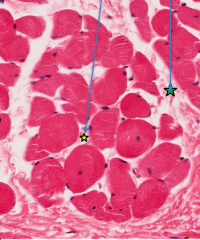
Describe. |
Yellow star: Endomysium (surrounds muscle fiber/cell ) Green star: Perimysium (surrounds muscle fascicle (several muscle fibers/cells)) |
|
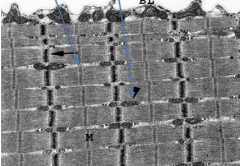
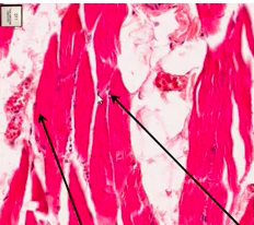
Describe. Label > left arrow > right arrow |
Left arrow: Glycogen Right arrow: Mitochondria |
|
|
Which of the following is the structurla and functional subunit of a muscle fiber? a. actin b. myosin c. entire muscle d. myofibril e. myofilaments |
d. Myofibril (composed of myofilaments) |
|
|
What are myofibrils composed of? |
Contractile myofilaments Functional subunit o myofibril: sarcomere |
|
|
What is the functional subunit of a myofibril? |
Sarcomere |
|
|
The sarcomere is the functional subunit of which of the following? a. myofilament b. entire muscle c. muscle fascicle d. myofibril e. myosin f. actin |
d. myofibril Myofibrils: > Sarcomeres: functional subunit of myofibril |
|
|
Myofilaments are principally located where? |
Myofibrils |
|
|
Which of the following is incorrect? a. red fibers have extensive blood supply b. white fibers have low mitochondria and myoglobin supplies. c. red fibers have the fastest enzymatic velocity d. intermediate fibers are fatigue resistant and fast twitching. e. red fibers are small, aerobic, slow-twitch f. White fibers have high concentrations of glycogen and glytolytic enzymes |
c. red fibers have the fastest enzymatic velocity Incorrect as red fibers have the slowest enzymatic velocity Differentiation: red fiber/white fiber: >Type I skeletal/type IIb >slow twitch, fatigue resistant/fast twitch, fatigue prone >high mitochondria, myoglobin, low glycogen/ low mitochondria, myoglobin, high glycogen and glycogen enzymes >slowest enzyme velocity/highest enzyme velocity |
|
|
Compare Red, White fibers |
Red/white: > Type 1/Type IIb > Lots mitochondria, myoglobin/Little mitochondria, myoglobin > extensive blood supply/low blood supply > Low glycogen/high glycogen > slowest enzymatic velocity / fastest enzymatic velocity > small fatigue resistant (aerobic) slow twitch/ large fatigue prone (anerobic) fast twitch |
|
|
Sheet of myomesin holds what in place? a. actin b. myosin c. titin d. desmin e. laminin |
b. myosin (thick filaments) |
|
Describe. |
Dystrophin shown. |
|
Describe. |
Duchenne muscular dystrophy |
|
|
Describe skeletal muscle contraction |
Overview steps: Attachment, Release, Bending, Force generation, reattachment Attachment: > normal myosin attached to actin (rigor state) = if no ATP as in death, 'rigor mortis' (stiffness) Release > when ATP binds to Myosin > conformational change at binding site > Myosin head uncoupled Bending > ATP hydrolysis to ADP + Pi > myosin head bends + displaces along thin filament Force generation: > Myosin head (w/ ADP-P) binds to Actin weakly, Pi released > Return to unbent position, movement > Power stroke (ADP released) Reattachment > myosin head itghtly bound to new actin |
|
|
What occurs when ATP is hydrolyzed in the myosin head during skeletal muscle contraction? a. detachment of myosin head from actin b. weak binding of myosin to actin c. power stroke d. bending of myosin head and displacement along actin e. reattachment of myosin head tightly bound to new actin f. attachment of myosin head to actin |
d. bending of myosin head and displacement along actin eliminations: a. incorrect, occurs when ATp binds to myosin head, causing a conformational cahnge detaching b. incorrect, occurs when Myosin-ADP-Pi complex binds to myosin, not in pure ATP state c. incorrect, occurs when myosin-adp complex bound to actin + ADP released (powerstroke d. correct e. incorrect, occurs after the powerstroke in reattachment f. incorrect, this occurs when there is no ATP present |
|
|
What occurs when there is no ATP to bind to myosin during skeletal muscle contraction? a. detachment of myosin head from actin b. weak binding of myosin to actin c. power stroke d. bending of myosin head and displacement along actin e. reattachment of myosin head tightly bound to new actin f. attachment of myosin head to actin |
f. attachment of myosin head to actin Elimination a. incorrect, occurs when ATP binds to myosin head, causing conformation change in myosin head b. incorrect. binding ADP-Pi-myosin complex to actin c. incorrect, occurs when Pi released movement, and then Power stroke (ADP released) when myosin is bound to actin d. incorrect, occurs when ATP hydrolysis occurs e. inccorects, occurs after poewrstroke (ADP, Pi released) f. correct |
|
|
What occurs when ATP binds to myosin head during skeletal muscle contraction? a. detachment of myosin head from actin b. weak binding of myosin to actin c. power stroke d. bending of myosin head and displacement along actin e. reattachment of myosin head tightly bound to new actin f. attachment of myosin head to actin |
a. detachment of myosin head from actin |
|
|
What occurs after ATP hydrolysis in myosin head and the myosin displaced along actin? a. detachment of myosin head from actin b. weak binding of myosin to actin c. power stroke d. bending of myosin head e. reattachment of myosin head tightly bound to new actin f. attachment of myosin head to actin |
b. weak binding of myosin to actin |
|
|
What occurs after the power stroke? a. detachment of myosin head from actin b. weak binding of myosin to actin c. movement of myosin along actin d. bending of myosin head e. reattachment of myosin head tightly bound to new actin f. attachment of myosin head to actin |
e. reattachment of myosin head tightly bound to new actin |
|
Explain. left dark region right light region |
left dark: cardiac right light: conduction tissue (Purkinje fibers) |
|
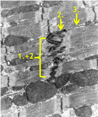
Describe each component. Describe. |
1. Fascia adherens > anchors thin filaments, make up transverse component 2. Macula adherens (desmosomes) > keep cells together during prolonged contraction 3. Gap junction |
|
|
Match. 1. This allows cells to communicate 2. Anchors thin filaments in smooth muscle 3. Anchors thin filaments in cardiac muscle 4. Anchors cells together, maintained in prolonged contraction 5. anchors thin filaments, make up transverse component. Describe thoroughly 6. Which tissue type contains caveoli associated with SR? 7. Which tissue contains t-tubule associated with Z line? 8. which tissue contains t-ubule associated with AI junction? 9. which tissue contains dyads? 10. Which tissue contains triads 11. Which of the following is innervated by alpha motor neurons to contract to generate force? 12. Which is smaller, specialized fibers for proprioception that sit within muscle spindles, innervated by gamma motor neurons? 13. What is an encapsulated receptor found at junction between muscle and tendon? 14. What contains intrafusal fibers? Options: Skeletal/cardiac/smooth a. gap junctions b. dense bodies c. Z lines d. AI junction e. fascia adherens f. macula adherens g. Muscle spindle h. Golgi tendon organ i. extrafusal fibers j. intrafusal fibers |
1. gap junction 2. dense bodies 3. Z lines 4. desmosomes (macula adherens) 5. Fascia adherens in intercalated discs or cardiac muscle 6. Smooth muscle 7. Cardiac muscle 8. Skeletal muscle 9. Cardiac muscle 10. Skeletal muscle 11. Extrafusal fibers 12. intrafusal fibers 13. Golgi tendon organ 14. Muscle spindle |
|
|
Muscle spindles -structure -function -innervation |
Structure: > intrafusal fibers Function: > Proprioception Innervation: > gamma motor neurons |
|
|
Golgi tendon organ -function -properties |
Structure: > monitors muscle load (force of contraction Properties: > encapsulated receptor found at junction of muscle and tendon > afferent nerve endings interwoven between collagen fibers |
|
|
Matching
1. Which is innervated by Gamma motor neurons? 2. Which is innervated by alpha motor neurons? 3. Which monitor muscle load (force of contraction? 4. Which responsible for proprioception? 5. Which is an encapsulated receptor found at junction between muscle and tendon? 6. What contains afferent nerve endings interwoven between collagen? 7. What contains nuclear bag (2-4) and nuclear chain (6-8) fibers? a. Extrafusal fibers b. intrafusal ibers c. Muscle spindle d. Golgi tendon organ |
1. intrafusal fibers, proprioception (in muscle spindles) 2 extrafusal fibers , contract to generate force 3. Golgi tendon organ 4. Intrafusal fibers (in muscle spindles), gamma motor neuron innervated 5. Golgi tendon organ 6. Golgi tendon organ 7. muscle spindle |
|
|
A certain structure is responsible for proprioception. Which of the following is a possible property? a. innervated by alpha motor neurons b. encapsulated receptor found at junction between muscle and tendon c. central region devoid of contractile proteins d. afferent nerve ending interwoven between collagen fibers e. monitors muscle load |
c. central region devoid of contractile proteins Explanation: This describes either intrafusal fibers or muscle spindle which both for proprioception but itnrafusal are found in muscle spindles. Select either one. Choices: a. incorrect, golgi tendon body b. incorrect, golgi tendon body c. correct, muscle spindle d. incorrect, golgi tendon body |
|
|
Which of the following is stimulated by alpha motor neurons? a. golgi tendon body b. intrafusal fibers c. extrafusal fibers d. muscle spindle |
c. extrafusal fibers > Contracts to gen. motor force > Responds to alpha motor neurons |
|
|
Which of the following is stimulated by gamma motor neurons and are small? a. golgi tendon body b. intrafusal fibers c. extrafusal fibers d. muscle spindle |
b. intrafusal fibers > found in muscle spindle |
|
|
Which of the following is specialized for proprioception and contains both afferent and efferent motor innervation. a. golgi tendon body b. intrafusal fibers c. extrafusal fibers d. muscle spindle |
d. muscle spindle |
|
|
Which of the following contains encapsulated intrafusal fibers in parallel with muscle belly? a. golgi tendon body b. intrafusal fibers c. extrafusal fibers d. muscle spindle |
d. muscle spindle |