![]()
![]()
![]()
Use LEFT and RIGHT arrow keys to navigate between flashcards;
Use UP and DOWN arrow keys to flip the card;
H to show hint;
A reads text to speech;
37 Cards in this Set
- Front
- Back
|
3 parts of Anterior elbow |
1. coronoid fossa 2. radial fossa 3. articular surfaces of radius, ulna & humerus |
|
|
Where are the 2 common locations for joint effusion in the elbow? |
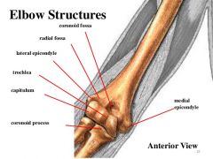
1. coronoid fossa 2. radial fossa |
|
|
3 parts of Medial elbow |
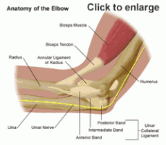
1. flexor tendons 2. ulnar nerve 3. ulnar collateral ligament |
|
|
3 parts of Posterior elbow |
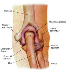
1. triceps tendon 2. olecranon bursa 3. olecranon fossa |
|
|
What is responsible for extension of the elbow? |
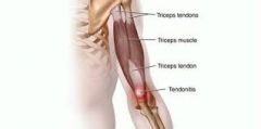
triceps tendon |
|
|
What is located on the Lateral elbow? |
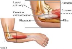
common extensor tendons |
|
|
What structure is responsible for extension of wrist, hand & digits? |
common extensor tendons |
|
|
What are the 2 parts in the Dorsal aspect of the wrist? |
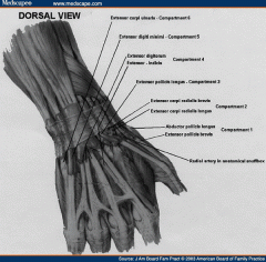
1. extensor mechanism 2. extensor tendons |
|
|
What is divided into 6 compartments and is responsible for the extension of the wrist, digits & thumb |
extensor mechanism |
|
|
Most common area for pathology in the wrist |
compartment 1 - on lateral aspect (thumb side) |
|
|
What functions to adduct, abduct & extend the wrist & digits? |
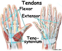
extensor tendons |
|
|
What does the Palmar / volar aspect of the wrist contain? |
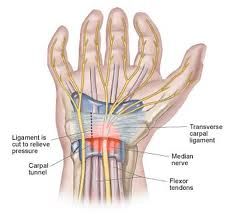
- flexor tendons - median, ulnar, and radial nerves - major blood vessels *carpal tunnel |
|
|
What is the canal through which the flexor tendons & median nerve pass? |
carpal tunnel |
|
|
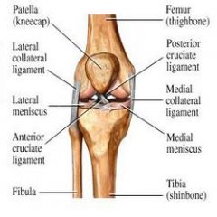
3 knee bones |
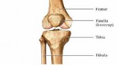
1. distal femur 2. proximal tibia 3. patella |
|
|
3 sections of Anterior knee |
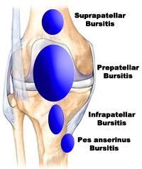
1. suprapatella 2. infrapatella 3. prepatella |
|
|
What is located in the suprapatella & is responsible for extension of lower extremity? |
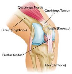
quadriceps tendon |
|
|
What attaches the patella to the tibia? |

patellar tendon |
|
|
What lies anterior to the patella & patellar tendon? |
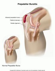
prepatellar bursa |
|
|
3 parts of Medial knee |
1. medial collateral ligament 2. external medial meniscus (cartilage) 3. pes anserine bursa |
|
|
What connects the medial condyle of the femur to the medial aspect of tibia? |
medial collateral ligament aka tibial collateral ligament |
|
|
What is a common location for fluid from overuse of the knee? |
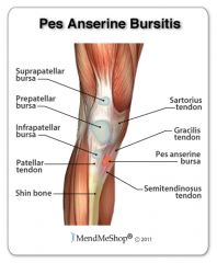
pes anserine bursa |
|
|
4 parts of Lateral knee |
1. lateral collateral ligament 2. iliotibial band 3. biceps femoris tendon 4. external lateral meniscus |
|
|
What connects the lateral condyle of the femur to the fibula? |
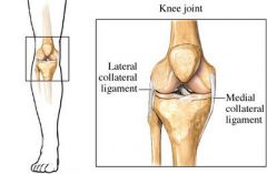
lateral collateral ligament aka fibular collateral ligament |
|
|
What attaches the lateral femur & muscles to the lateral tibia? |
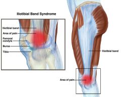
iliotibial band |
|
|
What attaches the hamstring muscles to the fibula and functions to flex the knee & extend the hip? |
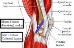
biceps femoris tendon |
|
|
4 parts of Posterior knee |
1. posterior femur 2. gastrocnemius muscles 3. gastrocnemius-semimembranous bursa 4. popliteal artery & vein |
|
|
What is the common location for fluid / Baker's cyst? |
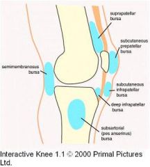
gastrocnemius-semimembranous bursa |
|
|
What vessels lie posterior & lateral to the gastrocnemius bursa? |
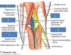
popliteal artery & vein |
|
|
4 bones of the ankle |
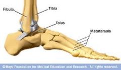
1. tibia (medial malleolus) 2. fibula (lateral malleolus) 3. 7 tarsal bones (calcaneus & talus most important) 4. 5 metatarsal bones |
|
|
2 parts of Anterior ankle |
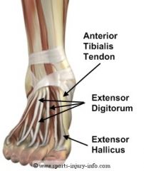
1. anterior tibialis tendons 2. extensor hallucis longus (EHL) & extensor digitorum longus (EDL) |
|
|
What originates on lateral condyle of tibia & attaches to metatarsal / tarsal bones? |
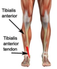
anterior tibialis tendons |
|
|
What functions in dorsiflexion of ankle & inversion of foot? |

anterior tibialis tendons |
|
|
What functions in dorsiflexion of toes? |

extensor hallucis longus (EHL) and extensor digitorum longus (EDL) |
|
|
2 parts of Medial ankle |
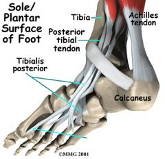
1. complex area of tendons, ligaments, vessels, nerves 2. posterior tibialis tendon (PTT) |
|
|
What attaches the tibia to most of the tarsal & metatarsal bones, and functions in support & stability of arch during weight bearing? |

posterior tibialis tendon |
|
|
3 common ligaments in Lateral ankle |
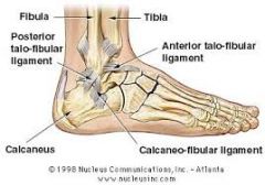
1. anterior talofibular ligament 2. calcaneal fibular ligament 3. anterior tibofibular ligament |
|
|
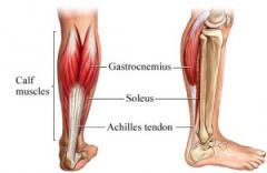
What is formed by the fusion of the soleus & gastrocnemius muscles & inserts into posterior surface of calcaneus? *common site for rupture |
achilles tendon |