Reading...
![]()
Play button
![]()
Play button
![]()
Use LEFT and RIGHT arrow keys to navigate between flashcards;
Use UP and DOWN arrow keys to flip the card;
H to show hint;
A reads text to speech;
16 Cards in this Set
- Front
- Back
|
What are venae comitantes and where are they found in the forearm?
|
- Venae commitantes = veins that run alongside arteries and use the pulsatile nature of the artery to aid their return flow. Often 2 veins straddle one artery
- In the arm, generally deep veins and arteries run together and superficial veins run independently - Therefore, the brachial veins, ulnar veins and radial veins are examples of these |
|
|
Describe the blood flow from the axilla into and from the heart
|
- The subclavian artery and vein run with the brachial plexus through the axilla
- The subclavian artery runs off the brachiocephalic trunk on the right, and from the aorta directly on the left - The left subclavian vein drains into the left brachiocephalic vein whereas the right subclavian drains directly into the superior vena cava (i.e. opposite to arteries) |
|
|
Why may an axillary vein thrombus occur and how can it be indicated?
|
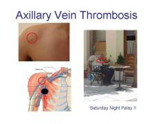
- May occur independently or be caused e.g. by putting a catheter in under the clavicle
- Pressure into the axilla e.g. using axillary crutches may also put pressure on the vein causing a thrombus - Cephalic vein swelling within the deltopectoral groove may indicate a thrombus |
|
|
Describe the 3 parts of the subclavian artery and vein
|
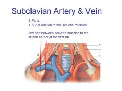
- 1st part arises from the brachiocephalic trunk (R) or from the arch of the aorta (L) at the level of T3/4 and travels to the medial border of scalenus anterior
- 2nd part lies posterior to scalenus anterior and in front of scalenus medius (vein lies anterior to scalenus anterior) - 3rd part begins at the lateral margin of scalenus anterior and runs to the lateral margin of the first rib |
|
|
What are the limits of the axillary artery and vein?
|
- Starts from the subclavian at the lateral border of the first rib
- Ends as the brachial artery at the lower margin of teres major - Closely related to the brachial plexus, and the cords are named according to their relationship |
|
|
What are the divisions and branches of the axillary artery?
|
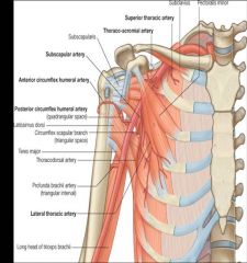
- Divided into 3 parts by pectoralis major
1. Runs between the lateral border of the 1st rib and the superior border of pectoralis minor and has 1 branch = Superior thoracic artery 2. Runs posterior to pectoralis major and gives off 2 branches = Thoraco-acromial = Lateral thoracic 3. Extends from the inferior border of pectoralis major to the inferior border of teres major and gives off 3 branches = Subscapular = Anterior circumflex humeral = Posterior circumflex humeral |
|
|
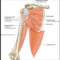
What nerves/vessels run from anterior to posterior through the quadrangular space, triangular space and the triangular interval?
|
|
|
|
Describe the features of the subscapular artery
|
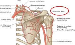
- Largest branch of the axillary artery (3rd part)
- Major source of blood to the posterior axilla - Runs down inferior margin of subscapularis - Gives off circumflex scapular and thoracodorsal arteries |
|
|
Describe the anterior and posterior circumflex humeral arteries and why they are significant
|

- Dislocations are more likely to affect nerves than arteries, but fractures are also common at the humeral surgical neck and sharp bits of bone can damage the arteries
|
|
|
Describe the limits of the brachial artery and its important branches
|
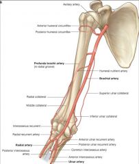
- STARTS – from Axillary Artery at lower border of Teres Major muscle.
- ENDS – at the distal margin of the Elbow Joint when it divides into Radial & Ulnar arteries. - Important branches = profunda brachii artery and the humeral nutrient artery which supplies the bone (muscle blood vessels will also help supply the bone) |
|
|
Describe the course of the profunda brachii artery
|
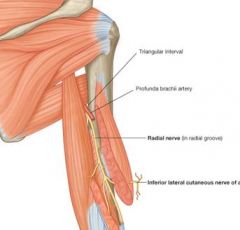
- Leaves the posteromedial aspect of the brachial artery and runs through the triangular interval with the radial nerve to travel in the radial groove
- Stuck to the bone, so very likely to be damaged in a mid-shaft fracture - Venae comitantes also run alongside it |
|
|
Describe where you would take pulses in the upper limb
|
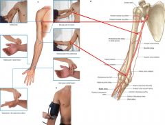
Note that all must be in places where can push the artery against a bone
|
|
|
Describe the course and supply of the radial artery
|
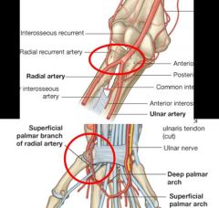
- STARTS- at the bifurcation of the Brachial artery at the distal margin of the elbow.
- ENDS – with deep palmar arch branches in the hand - No major branches in the forearm - Supplies the thenar muscles, the thumb and 1/2 of the 1st finger and gives off the deep palmar arch which supply the metacarpals |
|
|
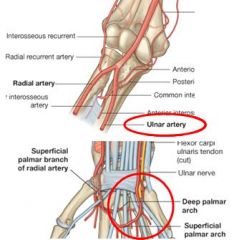
Describe the course and supply of the ulnar artery
|
- STARTS- at the bifurcation of the Brachial artery at the distal margin of the elbow.
- ENDS – with branches in the hand (superficial palmar arch). - Major source of blood to the forearm and hand - Gives off the common interosseous artery which divides into posterior and anterior and forms an anastomosis around the elbow joint with branches of the profunda brachii and the ulnar collateral artery - Superficial palmar arch supplies the hypothenar eminence and gives off palmar digital arteries to all of the fingers |
|
|
Describe the venous drainage of the upper limb
|
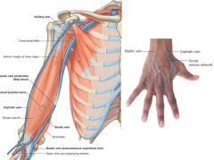
- Superficially Cephalic (lateral) and basilic veins (medial) form from the dorsal venous network of the hand
- Travel up the medial and lateral borders of the forearm, but anastomose in the cubital fossa, forming the median cubital vein - Deep ulnar and radial veins travel with their associated arteries and form the brachial vein, which joins with the basilic vein to form the axillary vein |