![]()
![]()
![]()
Use LEFT and RIGHT arrow keys to navigate between flashcards;
Use UP and DOWN arrow keys to flip the card;
H to show hint;
A reads text to speech;
64 Cards in this Set
- Front
- Back
|
Anatomy |
The study of internal and external body stuctures and their physical relationships among other body parts. |
|
|
Physiology |
Study of how living organisms perform their functions |
|
|
Medical Terminology |
Involves using word roots, prefixes, suffixes, and combining forms to build terms related to the body in health and disease. |
|
|
Cytology |
The study of the internal structure of individual cells. |
|
|
Cells |
The simplest units of life |
|
|
Histology |
The examination of tissues |
|
|
Tissues |
Groups of specialized cells and cell products that work together to perform specific functions |
|
|
Organs |
Tissues combine to form organs which carry out specific functions |
|
|
Scientific Method |
a system of advancing knowledge that begins by proposing a hypothesis to answer a question, and then testing that hypothesis with data collected through observation and experimentations. At the core of medical diagnosis. |
|
|
Organ System |
a group of organs interacting to perform a particular function |
|
|
Organism |
The highest level of organization that we consider |
|
|
Atoms |
the smallest stable units of matter |
|
|
Homeostasis |
(homeo=unchanging + stasis=standing) refers to the existence of a stable internal environment |
|
|
homeostatic regulation |
is the adjustment of physiological systems to preserve homeostasis |
|
|
autoregulation |
A process that occurs when a cell, tissue, an organ, or an organ system adjusts in response to some environmental change. For example: when oxygen levels decline in a tissue, the cells release chemicals that widen, or dilate, blood vessels. |
|
|
Extrinsic regulation |
Is a process that results from the activities of the nervous system or endocrine system |
|
|
receptor |
A sensor that is sensitive to a particular stimulus or environmental change. Example: thermometer, says what the temperature is |
|
|
Control center |
Receives and processes the information supplied by the receptor and sends out commands. Example: thermostat, if temperature goes above set-point the control center makes sure either the air or heat comes on to return to set-point. |
|
|
effector |
a cell organ that responds to the commands of the control center and whose activity either opposes or enhances the stimulus. Example: turns on the heat or the air |
|
|
set point |
The body's desired value. Example: the setting on the thermostat |
|
|
Negative feedback |
Opposes variations from normal and is a way of counteracting a change. It is the primary mechanism of homeostatic regulation, and it provides long-term control over the body's internal conditions and systems |
|
|
Positive feedback |
Is an initial stimulus which produces a response that exaggerates or enhances the original change in conditions, rather than opposing it. |
|
|
Disease |
When the body does not remain in a state of homeostasis, organ systems begin to malfunction which produces disease. |
|
|
state of equilibrium |
Exists when opposing processes or forces are in balance. In the case of body temperature, a state of equilibrium exists when the rate of heat loss equals the rate of heat production. |
|
|
Dynamic Equilibrium |
When each physiological system functions to maintain a state of equilibrium that keeps vital conditions within a normal range of values. Example: when muscles become more active, more heat is produced. |
|
|
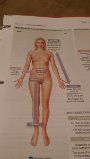
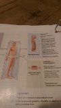
Superficial Anatomy |
involves locating structures on or near the body surface |
|
|
Anatomical position |
When the body is in this position, the hands are at the sides with the palms facing forward, and the feet are together. |
|
|
Supine |
A person lying down in the anatomical position face up |
|
|
Prone |
A person lying down in the anatomical position face down |
|
|
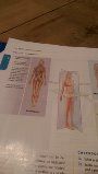
Abdominopelvic quadrants |
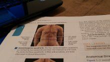
|
|
|
Abdominopelvic Regions |
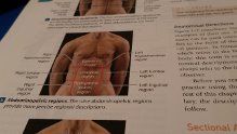
|
|
|
Abdominopelvic relationships |
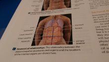
|
|
|
Proximal |
Toward the point of attachment of a limb to the trunk, ex: the shoulder is proximal to the wrist |
|
|
Distal |

Away from the point of attachment of a limb to the trunk. Ex: the fingers are distal to the wrist. |
|
|
Lateral |

Away from the midline. |
|
|
Medial |

Toward the midline |
|
|
Cranial or Cephalic |

Toward the head. The cranial nerves are in the head. |
|
|
Posterior or Dorsal |
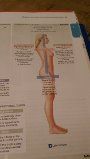
Posterior: The back surface Dorsal: the back Example: the scapula (shoulder blade) is located posterior to the rib cage. |
|
|
Caudal |

Toward the tail |
|
|
Anterior or Ventral |

Anterior: the front surface Example: the umbilicus (navel) is on the anterior (or ventral) surface of the trunk. Ventral: The belly side |
|
|
Transverse Plane |

aka horizontal plane, separates superior and inferior portions of the body |
|
|
Frontal Plane |
aka coronal plane, separates anterior and posterior portions of the body |
|
|
Sagittal Plane |
separates right and left portions of the body |
|
|
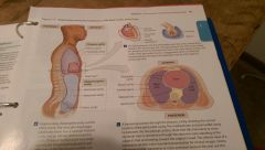
Thoracic Cavity |

Contains two pleural cavities, each surrounding a lung, the pericardial cavity of the heart, and a large tissue mass, the mediastinum, and the peritoneal cavity extends only partway into the pelvic cavity. |
|
|
Plueral cavity |
located in the thoracic cavity is subdivided into left and right pleural cavities (holding the lungs), separated by mediastinum. Each pleural cavity surrounds a lung and is lined by a shiny, slippery serous membrane that reduces friction. |
|
|
Pericardial Cavity |
inside the mediastinum. A small chamber that surrounds the heart. Resembles that of a fist pushing into a balloon. |
|
|
Pleura |
serous membrane lining the pleural cavity |
|
|
visceral pleura |
covers the outer surfaces of a lung |
|
|
parietal pleura |
covers the mediastinal surface and the inner body wall |
|
|
Abdominal Cavity |
Contained in the abdominopelvic cavity superior of the pelvic cavity. The abdominal cavity contains the liver, stomach, spleen, small intestine, and most of the large intestine |
|
|
Pelvic Cavity |
Contained in the abdominopelvic cavity and located inferior to the abdominal cavity. Contains the urinary bladder, various reproductive organs, and the distal (farthest) portion of the large intestines. |
|
|
Abdominopelvic Cavity |
Extends from the diaphragm to the pelvis. Contains peritoneal cavity, visceral peritoneum as well as the abdominal and pelvic cavities |
|
|
Diphragm |
A muscle that separates the thoracic cavity from the abdominopelvic cavity |
|
|
Viscera |
The internal organs enclosed by the trunk cavities (thoracic and abdominopelvic) |
|
|
Serous Membrane |
A fluid filled, thin tissue layer which surrounds the body cavities |
|
|
Serous Fluid |
Watery fluid, moistens serous membranes, coats opposing surfaces, and reduces friction |
|
|
Pericardium |
The serous membrane associated with the heart |
|
|
Mediastinum |
Consists of a mass of connective tissue that surrounds, stabilizes, and supports the esophagus, trachea, and thymus, as well as major blood vessels that originate or end at the heart |
|
|
Peritoneum |
A serous membrane lining the peritoneal cavity within the abdominal cavity |
|
|
X-Rays |
This technique uses high-energy radiation to create images of dense tissue such as when a dentist obtains images of the teeth |
|
|
CT Scans |
This technique X-Rays to create three-dimensional pictures which show soft tissue clearly |
|
|
MRI Scan |
This technique utilizes magnetic energy and is useful for imaging soft tissues |
|
|
PET Scan |
This technique images metabolic activity such as detecting a cancerous tumor in the brain |
|
|
Ultrasound |
This technique utilizes sound waves and can be used safely on fetuses |