![]()
![]()
![]()
Use LEFT and RIGHT arrow keys to navigate between flashcards;
Use UP and DOWN arrow keys to flip the card;
H to show hint;
A reads text to speech;
42 Cards in this Set
- Front
- Back
|
Three types of muscle tissue |
•Smooth •Cardiac •Skeletal |
|
|
Smooth Muscle |
•In the walls of organs •Not striated •Involuntary |
|
|
Skeletal Muscle |
•Attached to bones and skin •Striated •Voluntary |
|
|
Cardiac Muscle |
•Found only in the heart •Striated •involuntary |
|
|
Properties of Muscle Tissue |
•Excitability •Contractility •Extensibility •Elasticity |
|
|
Excitability |
Responds to chemicals released from nerve cells |
|
|
Contractility |
Ability to shorten and generate force |
|
|
Extensibility |
Ability to be stretched without damaging the tissue |
|
|
Elasticity |
Ability to return to original shape after being stretched |
|
|
Muscle Functions |
•Movement of bones or fluids •Maintaining posture and body position •Stabilizing joints •Heat generation (esp. Skeletal muscle) |
|
|
Connective tissue sheaths |
•Endomysium •Perimysium •Epimysium |
|
|
Endomysium |
The innermost sheath consisting of areolar connective tissue surrounds individual muscle fibers |
|
|
Perimysium |
A fibrous connective tissue surrounding groups of muscle fibers called fascicles |
|
|
Epimysium |
The outermost sheath called Epimysium wraps around the whole muscle and helps transfer the force generated by contraction of the muscle onto bones or skin. It consists of dense irregular connective tissue. |
|
|
Skeletal Muscle: Attachments |
Muscles attach: •Directly- Epimysium of muscle is fused to the periosteum of bone of perichondrium of cartilage •Indirectly- Connective tissue wrappings extend beyond the muscle as a rope like tendon or sheet like aponeurosis |
|
|
Profile of a skeletal muscle fiber |
•Long cells- fibers not visible without a Microscope •Multi nucleated •Sarcolemma- plasma membrane of the muscle •Sarcoplasm- muscle cytoplasm •Sarcoplasmic reticulum- muscle endoplasmic reticulum •Mitochondria- Many to make ATP •Glycosomes- For glycogen storage, myoglobin for O2 storage
|
|
|
What sets muscle tissue apart? |
•It's ability to convert chemical energy into mechanical •The structures that are designed to do just that are rod like structures called myofibrils that make up ~80% of the cell volume |
|
|
Sarcomere |
Smallest contractile or functional until of a muscle fiber is a sarcomere. It is defined as the region of a myofibril between two successive z discs. It is composed of thick and thin myofilaments. |
|
|
Muscle Hierarchy (Big-Small) |
1. Muscles 2. Fascicles 3. Muscle Fibers (cells) 4. Myofibrils (hundreds to thousands in a single muscle fiber) 5. Sarcomeres 6. Myofilaments (thick and thin) |
|
|
Sarcomere |
I band- LIGHT A band- DARK |
|
|
Z Disc |
Coin shaped sheet of proteins that anchors the thin filaments and connects myofibrils to one another |
|
|
Thick filament |
Runs the entire length of the A band |
|
|
M line |
Line of protein that holds adjacent thick filaments together |
|
|
H zone |
Lighter mid-region where filaments do not overlap |
|
|
Thin filament |
Runs the entire length of the I band and partway into the A band |
|
|
Sarcoplasmic Reticulum |
•Network of smooth endoplasmic reticulum surrounding each Myofibril •Pairs of terminal cisternae form perpendicular cross channels •Functions in the regulation of intercellular Ca2+ levels |
|
|
Intercellular calcium turns muscle contraction on and off |
Where does this calcium come from? •Terminal cisternae of Sarcoplasmic reticulum |
|
|
T Tubules |
•the Sarcolemma dips down as T tubules (Transverse tubules) at the A-I band junction •The T Tubules associate with the paired terminal cisternae to form triads that encircle each sarcomere |
|
|
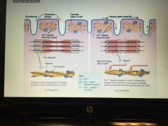
The Sarcolemma dips down as t tubules at the A-I band junction. The t tubules contain voltage sensors that open gated SR foot proteins which release Ca2+ and start contraction |
Back (Definition) |
|
|
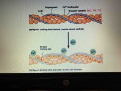
Once calcium is released it binds to troponin, tropomyosin moves and this exposes myosin binding sites on the actin |
|
|
|
Muscle contraction |
•The generation of force •Sarcomeres contract when the thick and thin myofilaments slide over each other |
|
|
Sliding Filament model of contraction |
•In the relaxed state, thin and thick filaments overlap only slightly •During contraction, myosin heads bind to actin, detach, and bind again, to propel the thin filaments toward the m line •As H zones shorten and disappear, Sarcomeres shorten, muscle cells shorten and the whole muscle shortens |
|
|
Requirements for skeletal muscle contraction |
•Skeletal muscle cells do not contract without a signal from their motor neuron in the central nervous system •Activation- neural stimulation at a neuromuscular junction •Excitation-contraction coupling: •Generation and propagation of an action potential along the Sarcolemma •Final trigger: A Brief rise in intracellular CA2+ levels |
|
|
Activation |
•Axons of motor neurons travel from the central nervous system via Nerves to skeletal muscles •Each axon forms several branches as it enters a muscle •Each axon ending forms a neuromuscular junction with a single muscle fiber |
|
|
Activation |
•Axons of motor neurons travel from the central nervous system via Nerves to skeletal muscles •Each axon forms several branches as it enters a muscle •Each axon ending forms a neuromuscular junction with a single muscle fiber |
|
|
Neuromuscular |
•Situated midway along the length of a muscle fibers •Axon terminal and muscle fiber are separated by a gel-filled space called the synaptic cleft •Synaptic vesicles- of axon terminal contain the neurotransmitter acetylcholine (ACh) •Junctional folds of the Sarcolemma contain ACh receptors |
|
|
Events at the neuromuscular junction |
•Nerve impulse arrives at axon terminal •ACh is released and binds with receptors on the Sarcolemma •Electrical events lead to the generation of an action potential •ACh effects are quickly terminated by the enzyme Acetylcholinesterase •Prevents continued muscle fiber contraction in the absence of additional stimulation |
|
|
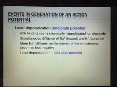
Events in generation of an action potential: 1. Local depolarization |
|
|
|
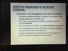
Events in generation of an action potential: 2. Generation and propagation of an action potential |
|
|
|
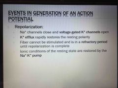
Events in generation of an action potential: 3. Repolarization |
|
|
|
Excitation-Contraction (E-C) Coupling |
• sequence of events by which transmission of an AP along the sarcolemma leads to sliding of the myofilaments • latent period: Time when EC coupling events occur • Time between AP initiation and the beginning of contraction • AP is propagated along sarcomere to T tubules •Voltage sensitive proteins stimulate Ca2+ release from SR • Ca2+ is necessary for contraction |
|
|
Cross bridge cycle |
• Crossbridge formation-high-energy myosin head attaches to thin filament • working (power) stroke- myosin head pivots and pulls thin filament toward M line • Crossbridge detachment: ATP attaches to myosin head and the cross bridge detaches •"Cocking" of the myosin head- energy from hydrolysis of ATP cocks the myosin head into the high energy state • continues as long as the calcium signal and adequate ATP are present |