![]()
![]()
![]()
Use LEFT and RIGHT arrow keys to navigate between flashcards;
Use UP and DOWN arrow keys to flip the card;
H to show hint;
A reads text to speech;
25 Cards in this Set
- Front
- Back
|
Matrix |
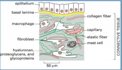
Connective tissue cells are surrounded by an extracellular matrix full of proteins and carbohydrate mixes
Ex. GAGs, proteoglycans, collagens/ elastin (fibers), fibronectin/ laminin (adhesion molecules)
Influences cell survival, development, shape, and function
Secreted by cells within it
|
|
|
Collagens |
Most abundant protein in animals
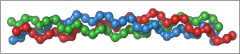
Long, stiff, triple-stranded helix (ropelike)
3 peptide α chains, rich in proline (stabilizes helix) and glycine (smallest amino acid, permits tight winding of the helix)
42 distinct α chains encoded by separate genes (thousands of molecules possible, but only ~40 collagens have been found) |
|
|
Types of collagen
types 1, 2, 3, and 4 |
Type I – bONE Type III - Lymph (reticular)
|
|
|
Synthesis of collagen |
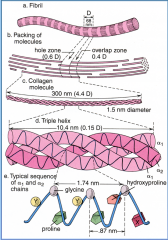
Collagen α- chains (3) form collagen molecules, molecules aggregate to form fibrils, and fibrils form fibers
Collagens are synthesized => ER assembly into triple helix=>N- and C-terminal telopeptides which prevent cross linking of the 3 chains=>secretion with procollagen peptidases=> propeptides (also called telopeptides) are removed=>lysyl oxidase crosslinks hydroxylysine making fibrils and fibers outside of the cell. |
|
|
Post-translational modifications of Collagens |
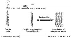
Post-translational hydroxylation of proline and lysine allows inter-chain H-bonds
One of the enzymes that cause linkages is Lysyl Oxidase, which acts extracellularly to cross-link collagen triple helices.
Hydroxylation events need Vitamin C
Hydroxylysine and hydroxyproline are postranslationally modified amino acids that are common in collagens.
Cross-links occur between certain hydroxylysines and lysines in short non-helical segments at the ends of collagen molecules.
Scurvy results from vitamin C deficiency (causes general weakness, anemia, gum disease, and skin hemorrhages)
|
|
|
Ehrlers Danlos |
Problems with propeptide collagenase (called ADAMTS2) and hence do not generate mature collagen fibrillar formation. |
|
|
Messenchym |
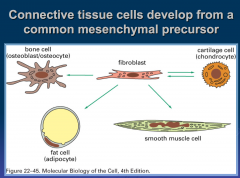
Meshwork of undifferentiated connective tissue cells
Mostly mesodermal
Give rise to connective tissues, blood, lymphatics, bone, and cartilage
Contact inhibited, so they come together after tissue damage to seal up cuts
Hyaluronic acid is the dominant GAG |
|
|
-blast cells vs -cyte cells |
-blast cells : make stuff
-cyte cells : fully matured cell, no longer making stuff
-blast refers to the committed, not terminally differentiated precursor cell in a given lineage of cells. -cyte refers to the terminally differentiated cell.
(ex.)
skeletal muscle differentiation - myoblasts fuse together to form multinucleated myocytes that become the individual fibers of the muscles.
epidermis - During wound healing, melanoblasts migrate out under epidermis where they differentiate into the melanocytes that give our skin color.
|
|
|
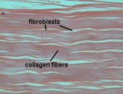
Fibroblasts |
a cell in connective tissue that produces collagen and other fibers
Most common cells of connective tissue in animals
Euchromatic nuclei and a lot ER
Plays a critical role in wound healing |
|
|
Fibrocytes |

An inactive mesenchymal cell
not synthesizing / secreting proteins
small amount of cytoplasm and rough ER
fibroblasts embedded in collagen fibers
|
|
|
Chondroblasts |
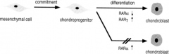
A cell of growing cartilage tissue
They lie in the space or lacunae present in groups of two or more
Euchromatic nuclei and a lot ER |
|
|
Chondrocytes |
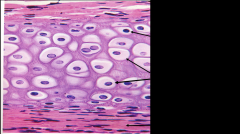
a cell that has secreted the matrix of cartilage and become embedded in it |
|
|
Osteoblasts |
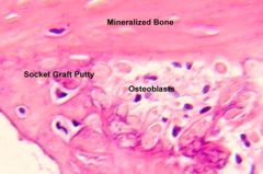
a cell that actively synthesizes and secretes the matrix for bone formation
Euchromatic nuclei and a lot ER
Found on the surface of the bone |
|
|
Osteocytes |
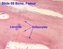
a bone cell, formed when an osteoblast becomes embedded in the matrix it has secreted |
|
|
Osteoclast |
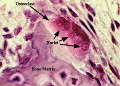
a large multinucleate bone cell that absorbs bone tissue during growth and healing
Derived from monocytes |
|
|
3 types of Loose Connective Tissue |
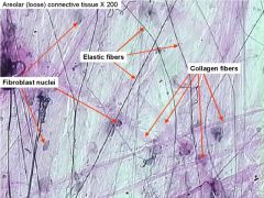
Lots of ECM matrix
Resist compression
Adipose - fat cells (white & brown)
Areolar - breasts, skin, gut, trachea |
|
|
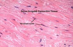
3 types of dense connective tissues
|
Lots of fibers
Fibrous - tendons/ligaments (regular), skin (irregular)
Elastic - aorta/arteries
Cartilege/Bone - next lecture
Dense connective tissue isn’t dense with cells but rather has a lot of ECM. Loose connective tissue (blood, lymph, fat) actually is much more cellular.
|
|
|
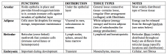
Loose Connective Tissue Chart:
Functions, distribution, tissue types |
|
|
|
Brown fat |
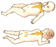
a dark-colored adipose tissue
On chest, sternum, and vertebral column of newborns
Involved in heat production in hibernating animals and human babies
Uncoupled oxidative phosphorylation
In contrast to white adipocytes (fat cells), which contain a single lipid droplet, brown adipocytes contain numerous smaller droplets and a much higher number of (iron-containing) mitochondria, which make it brown.
Many blood vessels
initiated by sensory afferent nerves in the skin which sense temperature (cold temperature stimulates sympathetic output) Sympathetic neurons stimulate beta oxidation of fatty acids in the brown fat tissue.
Rather than generate ATP, there is a mechanism in brown fat to bypass or short circuit ATPase synthetase. This results in a huge O2 utilization to dispose of the electrons.
|
|
|
White Fat |
used to store energy and acts as thermal insulator that help maintain body temperature.
Its cells have receptors for insulin, growth hormones, norepinephrine and glucocorticoids.
One large fat droplet |
|
|
Adipocytes |
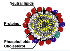
have a single lipid droplet and a thin surrounding cytoplasmic rim
The droplet consists of a core of neutral lipids (triglycerides or cholesterol esters)
Outer layer is a monolayer of amphipathic molecules (phospholipids, cholesterol and proteins)
The surrounding layer contains enzymes and other proteins needed for lipolysis |
|
|
White fat regulation |
Lipolysis upregulators: epinephrine, norepinephrine, natriuretic peptide.
Downregulator (fat storage): insulin
ob/ob mice lack leptin (anorexic factor produced by adipose tissue) and overeat.
Db/db mice make normal leptin but have mutant receptor and overeat.
|
|
|
Reticular connective tissue |
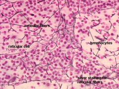
a type that is present in lymphoid organs (nodes, spleen etc).
Filled with thin collagen III fibers and associated cells. |
|
|
Elastic Fibers
Marfan syndrome |
Elastic fibers are made of elastin and fibrillin proteins; are irregularly arranged, distensible, and recoils;
in artery walls Marfan syndrome is due to a mutation in fibrillin-1, leading to elongated face, fingers, etc.
Also affects the heart and vasculature and so has life threatening risks involving heart problems and aortic aneurysm/dissection.
The lens of the eye dislocates and aorta is floppy and does not provide rebound (diastolic BP will be low). Marfan’s patients die of aortic rupture. |
|
|
Wharton’s jelly |
Mucus jelly surrounding umbilical cord. |