![]()
![]()
![]()
Use LEFT and RIGHT arrow keys to navigate between flashcards;
Use UP and DOWN arrow keys to flip the card;
H to show hint;
A reads text to speech;
14 Cards in this Set
- Front
- Back
|
Hyoid bone |
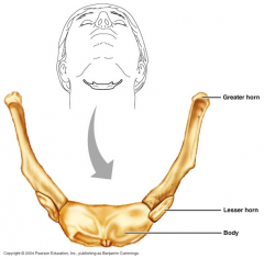
U-shaped bone superior to the thyroid cartilage and inferior to the chin
Parts - body, greater horn, lesser horn
It does not articulate with any other bones.
It is anchored in place by the suprahyoid and infrahyoid muscles.
Movements of the hyoid bone result in movements of the tongue and larynx. |
|
|
Laryngeal Skeleton |
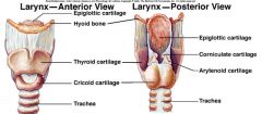
Protect the larynx (voice box)
Epiglottis and Cartileges:
1. Thyroid 2. Cricoid 3. Arytenoid 4. Corniculate |
|
|
Thyroid cartilage |
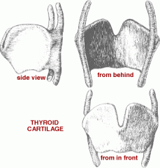
The "Adams Apple"
Attached superiorly to the hyoid bone via the thyrohyoid membrane and inferiorly with the the cricoid cartilage at its inferior horns.
The largest of the cartilages that make up the laryngeal skeleton |
|
|
Thyrohyoid membrane |
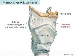
A thin, fibrous, membranous sheet filling the gap between the hyoid bone and the thyroid cartilage |
|
|
Cricoid cartilage |
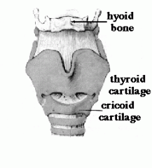
The only laryngeal cartilage that forms a complete ring around the airway.
It is narrow on top and wide at the bottom
Articulates with the inferior horns of the thyroid cartilage
This synovial joint that allows the angle between the cricoid and thyroid cartilages to change, thereby affecting the tension of the vocal ligament.
Inferiorly, the cricoid cartilage is attached to the first tracheal ring.
Excerpt From: Paulette Bernd, Ph.D. “Head & Neck.” iBooks. |
|
|
Tracheal rings |
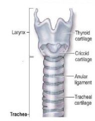
C-shaped cartilaginous rings that protect the trachea
There is no cartilage on the posterior surface of the trachea, instead there is smooth muscle.
This allows the esophagus to expand while swallowing. |
|
|
3 Salivary Glands |
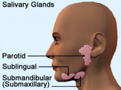
1. Parotid Gland 2. Submandibular Gland 3. Sublingual Gland |
|
|
Parotid Gland |
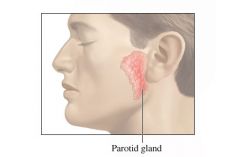
The largest of the three major salivary glands
Located just in front of and slightly inferior to each ear
The facial nerve (VII), external carotid artery and retromandibular vein all go through this gland.
The parotid gland produces saliva as a result of parasympathetic innervation from the lesser petrosal branch of the glossopharyngeal nerve (IX); the postsynaptic parasympathetic neurons are in the otic ganglion.
The saliva is delivered to the oral cavity via the parotid duct |
|
|
Parotid duct |
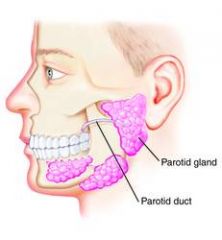
Carries salivary secretions from the parotid gland to the oral cavity.
The duct goes from the parotid gland lateral to the masseter muscle and the buccal fat pad.
At the anterior edge of the fat pad, the parotid duct goes deep and pierces the buccinator muscle to enter the oral cavity through a small orifice opposite the second maxillary molar tooth. |
|
|
Submandibular gland |
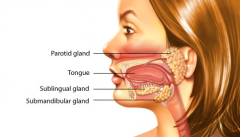
Major salivary glands located beneath the floor of the mouth
U-shaped
Its external portion fills most of the submandibular triangle while its deep portion is superior to the mylohyoid muscle.
Produces saliva as a result of parasympathetic innervation from the chorda tympani branch of the facial nerve (VII); the postsynaptic parasympathetic neurons are in the submandibular ganglion.
The submandibular duct (used to be called Wharton’s duct) opens into a sublingual caruncle (pink arrows) lateral to the frenulum of the tongue. |
|
|
Thyroid Gland |
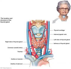
Located to the sides of the larynx and trachea (in the front of the neck).
Consists of right and left lobes, with a connecting isthmus in front of the trachea.
The thyroid gland’s blood supply comes from the superior and inferior thyroid arteries
It is drained by superior, middle, and inferior thyroid veins
The function of the thyroid is to regulate metabolism via hormones |
|
|
Carotid sinus |
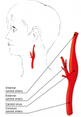
A slight dilation (widening) of the proximal part of the internal carotid artery.
The carotid sinus is sensitive to pressure changes in the arterial blood at this level. |
|
|
Buccal fat pad |
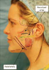
An accumulation of encapsulated fat superficial to the buccinator muscle.
The parotid duct lies superficial to the buccal fat pad and goes deep at its anterior border.
Loss of this fat pad gives the appearance of sunken cheeks. |
|
|
Articular disc of the temporomandibular joint |
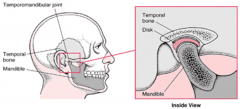
Divides the temporomandibular joint cavity into two separate synovial compartments |