![]()
![]()
![]()
Use LEFT and RIGHT arrow keys to navigate between flashcards;
Use UP and DOWN arrow keys to flip the card;
H to show hint;
A reads text to speech;
78 Cards in this Set
- Front
- Back
|
Def: Virus |
Genetic element that cannot replicate independently of a living (host) cell. A.k.a. - Obligate Intracellular Parasite |
|
|
Def: Virion |
Extracellular form of a virus. It exists outside host and facilitates transmission from one host cell to another. |
|
|
When do Viruses Replicate? |
Replication/reproduction occurs only upon infection (entry into host cell). |
|
|
Viral Components and Activities |
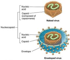
Capsid
Naked Virus
Enveloped Virus
Nucleocapsid |
|
|
Def: Capsid |
The protein shell that surrounds the genome of a virus particle. |
|
|
Def: Naked Virus |
(e.g., most bacterial viruses) have no other layers. |
|
|
Def: Enveloped Virus |
(e.g., many animal viruses) have an outer layer consisting of a phospholipid bilayer (from host cell membrane) and viral proteins. |
|
|
Def: Nucleocapsid |
Nucleic acid + protein in enveloped viruses. |
|
|
|
|
|
Def: Virulent (lytic) Infection |
Replicates and destroys host. |
|
|
Def: Lysogenic Infection |
Host cell genetically altered because viral genome becomes part of host genome. |
|
|
Viral Genomes |
Either DNA or RNA genomes.
Single-stranded or double-stranded Single-stranded may be plus sense (same as mRNA) or minus sense (complementary to mRNA). Either linear or circular.
Usually smaller than those of cells. |
|
|
Virus Classification |
Bacterial viruses (bacteriophages; model systems).
Archaeal viruses.
Animal viruses (extensively studied).
Plant viruses (less well studied).
other viruses. |
|
|
Average Size of Virus |
Most viruses are smaller than prokaryotic cells; range from 0.02 to 0.3 µm. |
|
|
Virion Structure: Capsomere |
Individual protein molecules arranged in a precise and highly repetitive pattern around the nucleic acid making up the capsid. Capsids can be put together through self-assembly (spontaneous) or require host cell folding assistance. |
|
|
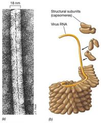
Virus Symmetry: Helical |
Rod-shaped viruses (e.g., tobacco mosaic virus or TMV).
Length of virus determined by length of nucleic acid. Width of virus determined by size and packaging of capsomeres. |
|
|
Virus Symmetry: Icosahedral |
Spherical viruses (e.g., human papillomavirus). Most efficient arrangement of subunits in a closed shell. Requires fewest capsomeres. |
|
|
Enveloped Viruses (Part 1) |
Have lipoprotein membrane surrounding nucleocapsid.
RNA or DNA genomes. Envelope proteins attach to and infect animal host cell.
|
|
|
Enveloped Viruses (Part 2) |
Relatively few enveloped plant or bacterial viruses because of cell walls surrounding cell membrane.
Entire virion enters animal cell during infection.
Enveloped viruses exit more easily. |
|
|
Enzymes Inside Virions: Lysozyme |
Makes hole in cell wall to allow nucleic acid entry. Also lyses bacterial cell to release new virions. |
|
|
Enzymes Inside Virions: Neuraminidases |
Destroy glycoproteins and glycolipids. Allows liberation of viruses from cell. |
|
|
Enzymes Inside Virions: Nucleic Acid Polymerases |
RNA replicases: RNA-dependent RNA polymerases. Reverse transcriptase: RNA-dependent DNA polymerase in retroviruses. |
|
|
Prokaryotic Viruses VS. Eukaryotic Viruses |
Nucleic acid entry in prokaryotes and virion entry in eukaryotes. |
|
|
Phases of Rep. In Permissive (supportive) Host Cell |
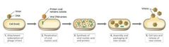
Attachment (adsorption) of the virion. Penetration (entry, injection) of the virion nucleic acid. Synthesis of virus nucleic acid and protein by host cell metabolism as redirected by virus. Assembly of capsids and Packaging of viral genomes into new virions. Release of mature virions from host cell. (or, A.P.S.A.P.R.) |
|
|
A.P.S.A.P.R. Visual |
|
|
|
Overview of Virus Life Cycle |
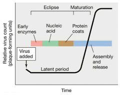
Eclipse: genome replicated and proteins translated. Maturation: packaging of nucleic acids in capsids. Latent Period: eclipse + maturation. Release: cell lysis, budding, or excretion. Burst Size: number of virions released. |
|
|
Def: One-Step Growth Curve |
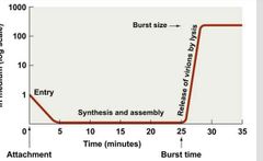
Increase occurs when cells burst. |
|
|
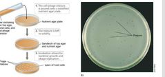
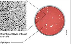
Culturing Viruses |
Bacterial viruses are easiest to grow (hosts in liquid medium or spread as “lawns” on agar and inoculated with virus). Animal viruses (and some plant viruses) can be cultivated in tissue cultures (from animal organ in culture medium). |
|
|
Culturing Viruses: Visual |
|
|
|
What is a Plaque Assay? |
Used for detecting and counting viruses. Plaques are clear zones that develop on lawns of host cells where successful viral infection occurs. |
|
|
Def: Titer |
Number of infectious units per volume of fluid. |
|
|
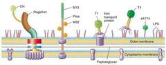
Attachment (Part 1) |
Major factor in host specificity
Requires complementary receptors on the surface of a susceptible host for its infecting virus.
Receptors include proteins, carbohydrates, glycoproteins, lipids, lipoproteins, or other cell structures. |
|
|
Attachment (Part 2) |
Receptors on host cell carry out normal functions for cell (e.g., uptake proteins, cell-to-cell interaction, flagella, pili). |
|
|
Attachment Visual |
|
|
|
Penetration (Part 1) |
Capsid left outside cell.
Viral genome and viral proteins (for some viruses) enter host cell. |
|
|
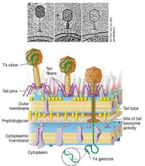
Penetration of T4 Bacteriophage (Part 1) |
Virions attach to cells via tail fibers that interact with polysaccharides on E. coli LPS layer.
Tail fibers retract, and tail pins contact cell wall.
|
|
|
Penetration of T4 Bacteriophage (Part 2) |
T4 lysozyme forms small pore in peptidoglycan.
Tail sheath contracts, and viral DNA passes into cytoplasm.
Capsid stays outside. |
|
|
Penetration Visual |
|
|
|
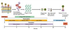
How long does Virion synthesis take? |
About 30 minutes and ends in release of new virions from lysed cell. |
|
|
How many parts of T4 Genome are there? |
3...
(1) Early proteins
(2) Middle proteins
(3) Late proteins |
|
|
T4 Early Proteins |
Enzymes needed for DNA replication and transcription.
Enzyme for the synthesis and glucosylation of the T4 base Hydroxymethylcytosine. Enzymes that function in T4 replisome. Proteins that modify host RNA polymerase. |
|
|
T4 Middle and Late Proteins |
Head and tail proteins and enzymes required to liberate mature phage particles. Additional RNA polymerase-modifying proteins. Viral head and tail proteins. Enzymes for liberating new virions from cell. |
|
|
Replication Chart |
|
|
|
Effect of T4 proteins on Host |
T4-specific proteins modify host RNA polymerase specificity to recognize only phage promoters. Host transcription shut down. |
|
|
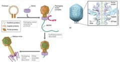
T4 Genome Packaging |
Genome is pumped into head under pressure using ATP. Packaging in three stages... - Proheads (bacteriophage head precursors) assembled. - Packaging motor assembled at opening. - Double-stranded linear genome pumped into prohead using ATP. |
|
|
T4 Genome Assembly |
After head is filled with DNA, T4 tail, tail fibers, and other components are self-assembled late enzymes break membrane and peptidoglycan.
Lysis occurs, virions released. |
|
|
Virus Replication Visual |
|
|
|
Basic Viral Replication Visual |
|
|
|
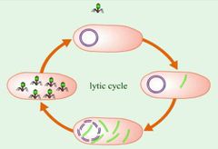
Def: Virulent Life Cycle |
A virus that always lyses, kills, their host after infection. |
|
|
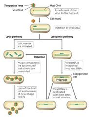
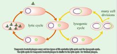
Def: Temperate Life Cycle |
Viruses that replicate their genomes in tandem with host genome and without killing host, establishing long-term, stable relationship.
Can be lytic/virulent.
Can enter lysogeny: most viral genes are not transcribed, viral genome is replicated with host chromosome and passed to daughter cells. |
|
|
Def: Lysogen |
lysogen: host cell that harbors temperate virus. Can result in lysogenic conversion with new properties (e.g., virulence in pathogens). |
|
|
Replication Cycle of Temperate Phage |
In lysogeny, genome is either integrated into bacterial chromosome (lambda) or exists as a plasmid (P1). Prophage: viral DNA. Lysogeny maintained by phage-encoded repressor protein. Inactivation of repressor induces lytic stage. Cell stress (e.g., DNA damage) induces lytic pathway. |
|
|
Temperate Phage Chart |
|
|
|
Bacteriophage Lambda (Part 1) |
Linear, dsDNA virus with head and tail.
Complementary, single-stranded “cohesive” regions 12 nucleotides long at the 5′ terminus of each strand.
Upon penetration, DNA ends base pair, forming the cos site, and the DNA ligates and forms double-stranded circle. |
|
|
Bacteriophage Lambda (Part 2) |
When it enters lytic pathway, lambda synthesizes long, linear concatemers of DNA by rolling circle replication.
Genome-sized lengths cut at cos sites; genomes packaged into phage heads.
After tails added, lysis occurs. |
|
|
Bacteriophage Lambda (Part 3) |
Transduction (packaging of host chromosomal genes and transfer to new host) can also occur. When lambda is lysogenic, its DNA integrates into E. coli chromosome at the lambda attachment site (attλ) using lambda integrase. |
|
|
What is regulates Lambda lysis/lysogeny |
Regulates the lambda lifestyle. Key elements are two repressor proteins.
CI protein (the lambda repressor): causes repression of lambda lytic events.
Cro repressor: controls activation of lytic events.
First repressor to accumulate controls infection outcome. |
|
|
Temperate Bacteriophage Viral Replication Visual |
|
|
|
An Overview of Animal Virus Infection (Part 1) |
Major tenets (capsid and DNA/RNA genome, infection and takeover of host, assembly and release) universal.
Classified by genomes. |
|
|
An Overview of Animal Virus Infection (Part 2) |
Most human viral diseases are caused by RNA viruses.
Two key differences.
Entire virion enters the animal cell.
Eukaryotic cells contain a nucleus, the site of replication for many animal viruses. |
|
|
|
|
|
An Overview of Animal Virus Infection (Part 3) |
Bind specific host cell receptors, typically used for cell-cell contact or immune function.
Different tissues and organs express different cell surface proteins. Often viruses only infect certain tissues.
Entry usually occurs by fusion with cytoplasmic membrane or endocytosis. |
|
|
An Overview of Animal Virus Infection (Part 4) |
Uncoating occurs at cytoplasmic membrane or cytoplasm.
Viral DNA genomes enter nucleus, most viral RNA is converted to DNA within nucleocapsid.
Bind specific host cell receptors, typically used for cell-cell contact or immune function. |
|
|
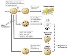
Def: Latent Infection |
Viral DNA exists in host genome and virions are not produced; host cell is unharmed unless/until virulence is triggered. |
|
|
Def: Persistent infections |
Release of virions from host cell by budding, does not result in cell lysis.
Infected cell remains alive and continues to produce virus. |
|
|
Def: Transformation |
Conversion of normal cell into tumor cell. |
|
|
Animal Virus Life Cycle Visual |
|
|
|
Animal Virus Developmental Chart |

|
|
|
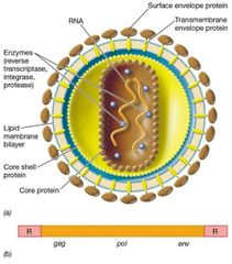
Retrovirus |
RNA viruses that replicate through a DNA intermediate (e.g., human immunodeficiency virus [HIV]).
Contain a reverse transcriptase (copies information from RNA to DNA), integrase, and protease.
Enveloped. |
|
|
Enveloped Animal Virus Visual |
|
|
|
Uniqueness of Retrovirus Genome |
Two identical ss(+)RNA molecules. Contains specific genes. gag: encode structural proteins. pol: encode reverse transcriptase and integrase. env: encode envelope proteins. |
|
|
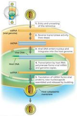
Retrovirus Replication (Part 1) |
Entrance into the cell with removal of envelope at the membrane.
Reverse transcription of one RNA genome begins in nucleocapsid.
Single DNA strand produced.
Reverse transcriptase uses this to make a complementary strand, forming dsDNA product.
|
|
|
Retrovirus Replication (Part 2) |
dsDNA enters nuclease with integrase, which incorporates retroviral DNA into host genome to form provirus, which remains indefinitely.
Transcription of retroviral DNA.
Assembly and packaging of genomic RNA.
Budding of enveloped virions and release from cell. |
|
|
Retrovirus Life Cycle Visual |
|
|
|
ICTV Criteria for Viral Classification - 5 Categories |
(1) Genome composition.
(2) Capsid Symmetry.
(3) Envelope.
(4) Size of the Virion.
(5) Host Range. |
|
|
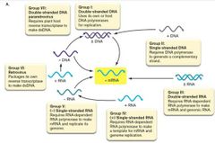
David Baltimore |
Proposed the main distinctions among classes of viruses be:
The genome composition (RNA or DNA).
The route used to express messenger RNA (mRNA). |
|
|
7 groups of Baltimore Virus Classification |
Group I: Double-stranded DNA viruses.
Group II: Single-stranded DNA viruses.
Group III: Double-stranded RNA viruses.
Group IV: (+) single-stranded RNA viruses.
Group V: (–) single-stranded RNA viruses.
Group VI: RNA retroviruses.
Group VII: DNA para-retroviruses. |
|
|
Baltimore Virus Classification Visual |
|