Reading...
![]()
Play button
![]()
Play button
![]()
Use LEFT and RIGHT arrow keys to navigate between flashcards;
Use UP and DOWN arrow keys to flip the card;
H to show hint;
A reads text to speech;
58 Cards in this Set
- Front
- Back
|
epithelium arrises from what 3 basic tissue layers
|
endoderm, mesoderm, ectoderm
|
|
|
General characteristics of epithelium
|
has a basement membrane, epithelial cells are tightly joined, many modifications to cell surface, form the fx part of certain glandular organs
|
|
|
Be able to describe difference b/w polarized and nonpolarized epithelial cells and give examples
|
polarized: intestinal cells, clear direction and distinction b/w lumen and basement membrane
Non-polarized: endocrine cells, less organized |
|
|
Properties of a basement membrane
|
Sheet-like structure , permeable- associated with reticular fibers (net-like)
|
|
|
Types of epithelium
|
simple columnar (SIntes)
Simple Columnar Ciliated (oviduct) Pseudostratified columnar ciliated (respiratory) Stratified epithelium (squamous cells) Transitional epithelium (mesh-like-urinary tract-needs to be flexible) |
|
|
Wedge shaped cells indicate what type of epithelium
|
transitional epithelium (mesh-like stretchability)
|
|
|
Multilayered cells that change from columnar cubical to flat indicate
|
stratified epithelium
|
|
|
What type of cells will you see al the cells attached to the basement membrane and ciliated (think breathing)
|
Pseudostratified columnar ciliated epithelium
|
|
|
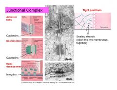
cell attachments
|
junctional complexs(terminal bars in LM) gap junctions, hemidesmosomes, tightt jnxt, adhesion belts, desmosomes
|
|
|
Tight junctions other name and functions
|
Zonula occludens - constant feature of jnxtional complexes
forms a zone or collar-like junction around cell prevents diffusion of molecules and ions b/w cells |
|
|
adhesion belts other name and function
|
zonula adherens
also forms a band around the cell cytoplasmic plaques of actin-binding proteins under the plasmalemma Actin microfilaments (anchored to the dense plaque) Transmembrane glycoprotein (E-cadherin) |
|
|
desmosomes
|
macula adherens or focal adhesions between cells
scattered along cel margins has cytoskeletal intermediate filaments anchored to plaques instead of actin |
|
|
gap junctions
|
macula communicans- gap jnxs can occur in cells other than epithelia to do cell-cell communication like in the cardiac cell
|
|
|
hemidesmosomes
|
attach cell to basement membrane
|
|
|
Gap junctions and how they communicate (aka nexus communicans)
|
Selective diffusion of some molecules- with membranes that are closesly applied to each other (almost fused) and where the channels and pores span both adjacent membranes - no specific direction depends on concentration gradient to move either direction- important to smooth and cardiac muscle
|
|
|
What is the major role of a tight junction and if it becomes loose what will happen- what kind of physiological conditions would have loose jnxs
|
role- keep material sealed in
loose-material can move more freely and diffuse easier physio conditions- stem cells in brain and new borns |
|
|
Two types of basal surface specializations
|
caveolae and pinocytotic vesicles ( types of infoldings and invaginations)
|
|
|
Apical surface specializations can be differentiated by their
|
motility and length
|
|
|
Microvilli definition and detection
|
a non-motile apical surface specialization
many finger like projections from cell surface can only detect individual microvillus with TEM |
|
|
Microvilli structure
|
contain actin microfilaments-which anchor at the tip of the microvillus and extend down the terminal web
|
|
|
Stereocilia
|
a type of microvilli (also non-motile) long modified microvili which contain actin microfilaments
|
|
|
Cilia definition and detection
|
a motile apical surface specialization
Resolvable by light microscopy |
|
|
Cilia structure
|
contain microtubules anchored to a basal body
9+2 arrangement of doublet microtubles in cilia 9 sets of triplet microtubles in the basal body (similar to centriole) |
|
|
Name an organ you'd be likely to encounter ciliated cells
|
GI tract
|
|
|
Ways to classify epithelial cells
|
number of layers (simple vs stratified)
Shape (squamous, cuboidal, columnar, pyramidal) Presence of surface specializations (cilia, microvilli, stereocilia, keratinization) |
|
|
Location/ Structure in the body where stereocilia cells could be found
|
male reproductive tract
|
|
|
How to differentiate simple cells
|
squamous- flattened cells with bulging nucleus, cytoplasm hard to distinguish
Cuboidal- cells appear square w/ central nucleus Columnar (columnar with basally placed and elongated nucleus) |
|
|
Where can simple squamous cells be found
|
lining lungs body cavities and blood vessels (as mesothelium and endothelium)
|
|
|
Pseudostratified columnar epithelia
|
cells actually contact the basement membrane (top layer of cells usually columnar and ciliated
|
|
|
Where is transitional epithelium found
|
only in teh urinary tract (urothelium)
|
|
|
Stratified squamous epithelia
|
many layers, the outermost of which is squamous in shape, can see cell division occuring
KERATINIZED (cornified) or non keratinized found |
|
|
Keratinized
|
lost nucelus where cells change from cuboidal to squamous and may be keratinized
|
|
|
Layers in the keratinized stratified squamous
|
stratum corneum
stratum lucidum stratum granulosum stratum spinosum stratum basale |
|
|
stratum corneum
|
soft keratin (tip of cell-near apical surface)
|
|
|
stratum spinosum
|
attached by desmosomes
|
|
|
stratum basale
|
next to basement membrane stem cells located in this layer and where cell division occurs
|
|
|
In a slide displaying an arteriole could you classify the epithelium on teh slide
|
vasco-epithelium and simple squamous (RBC has no nucleus and surrouned by WBC in the arteriole
|
|
|
Glandular epithelium and functional portion of a gland
|
develop from epithelial cells
usually simple cubodial columnar or pyramidal |
|
|
And glandular epithelium that retains connection to the surface is considered
|
exocrine (duct)
|
|
|
And glandular epithelium that is not connected to the surface is considered
|
endocrine
|
|
|
The secretory end piece of the glandular epithelial cell is called
|
adenomere
|
|
|
Types of glands
|
unicellular, simple, compound
|
|
|
unicellular glands
|
single secretory cells in teh epitheilial layer GOBLET cell
|
|
|
Simple gland (duct system in unbranched) would be found
|
usually in small glands which is another part of an organ like large intestine
|
|
|
compound gland
|
when the duct system branches (usually in the glands that are organs---pancreas, Brunner's gland in teh duodenum
|
|
|
Duct status in the endocrine
|
Not present -secretion into circulation
|
|
|
Duct status in the exocrine
|
duct is present to the surface of epithelum
|
|
|
Classification of glands (exocrine types)
|
merocrine (exocytosis secretion)
apocrine (pinches off apical part- mammary galnd some sweat) holocrine (entire cell is sloughed as secretory) |
|
|
Ways to classify glands besides endocrine vs exocrine
|
Shape (tubular or alveolar)
Type of secretion- serous (protien rich) or mucous (more viscous and glycoprotein rich) |
|
|
The more protein rich a cell or secretion the less or more intense the staining will be
|
more intense (dark) so a serous acini stains darker than a mucous acini
more protien-more ribosomes-darker stain |
|
|
Follicular endocrine gland
|
Found in thyroid and is poalrized where cells line up nicely
|
|
|
endocrine glands that are nonpolarized
|
pituitary, adrenal, pancreatic endocrine cells
|
|

caveolae
|

caveolae
|
|

|

|
|

|

|
|

|

|
|

|

|
|

|
|