Reading...
![]()
Play button
![]()
Play button
![]()
Use LEFT and RIGHT arrow keys to navigate between flashcards;
Use UP and DOWN arrow keys to flip the card;
H to show hint;
A reads text to speech;
108 Cards in this Set
- Front
- Back
|
1 µm = ? m = ? mm
|
10-6 m = 10-3 mm
|
|
|
1 nm = ? m = ? mm
|
10-9 m = 10-6 mm
|
|
|
1000 nm = ? µm
|
1000 nm = 1 µm
|
|
|
0.001 µm = ? nm
|
0.001 µm = 1 nm
|
|
|
Explain the importance of oil when using the oil immerion objective.
|
Oil reduces light loss between slide and the lens. Used to keep light from bending.
|
|
|
Identify a use for Dark field when the Bright field illumination is not sufficient.
|
Dark objects are visible against a bright background. Shows a light silhouette of an organism against a dark background. It is most useful for detecting the presence of extremely small organisms.
|
|
|
Identify a use for Phase Contrast when the Bright field illumination is not sufficient.
|
Accentuates diffraction of the light that passes through a specimen. Brings direct and reflected or diffracted light rays together (in phase) to form an image of the specimen on the ocular lens. Allows the detailed observation of living organisms.
|
|
|
Identify a use for DIC when the Bright field illumination is not sufficient.
|
Accentuates diffraction of the light that passes through a specimen; uses two beams of light.
-Provides a colored, three-dimensional image of hte object being observed. Allows detailed observations of livng cells. |
|
|
Identify a use for Florescence when the Bright field illumination is not sufficient.
|
-Uses UV light.
-Fluorescent substances absorb UV light and emit visible light. -Cells may be stained with fluorescent dyes (fluorochromes). Specimens are first stained with fluorochromes and then viewed through a compound microscope by using an ultraviolet light source. Microorganisms appear as bright objects against a dark background. Used primarily in a diagnostic procedure called fluorescent-antibody (FA) technique, or immunofluorescence. -absorb UV light and emit visible light |
|
|
Identify a use for Transmission Electron Microscop (TEM), when the Bright filed illumination (the one we use) is not sufficient
|
-Ultrathin sections of specimens.
-Light passes through specimen, then an electromagnetic lens, to a screen or film. -Specimens may be stained with heavy metal salts. -10,000-100,000; resolution 2.5 nm |
|
|
Identify a use for Scanning Electron Microscop (SEM), when the Bright filed illumination (the one we use) is not sufficient
|
-An electron gun produces a beam of electrons that scans the surface of a whole specimen.
-Secondary electrons emitted from the specimen produce the image. -Three-dimensional views of the surfaces of whole microorganisms can be obtained. Magnification: 1000–10,000×. Resolving power: 20 nm. |
|
|
-Uses electrons instead of light.
-The shorter wavelength of electrons gives greater resolution. |
Electron Microscopy
|
|
|
PEQ Explain how electron microscopic methods (TEM & SEM) differ from light microscopy
|
-TEM differs from light in that it uses electrons instead. The shorter wave lengths of electrons gives greater resolution.
-differ from Light Microscopes Uses a beam of electrons, instead of light. Electromagnets, instead of glass lenses, control focus, illumination, and magnification. Thin sections of organisms can be seen. Magnification: 10,000–100,000×. Resolving power: 2.5 nm. |
|
|
coloring a microorganism with dye to make some structures more visible
|
stain
|
|
|
-use of single basic dye
-A mordant may be used to hold the stain or coat the specimen to enlarge it. -An aqueous or alcohol solution of a single basic dye, used to make cellular shapes and arrangements visible. A mordant may be used to improve bonding between the stain and the specimen. |
simple stain
|
|
|
-The Gram stain classifies bacteria into gram-positive and gram-negative.
-Gram-positive bacteria tend to be killed by penicillin and detergents. -Gram-negative bacteria are more resistant to antibiotics. Decolorizer make gram negatives colorless. Safranin makes gram negative red. -Gram stain and acid-fast stain, differentiate bacteria according to their reactions to the stains. Gram stain procedure uses a purple stain (crystal violet), iodine as a mordant, an alcohol decolorizer, and a red counterstain. Gram-positive bacteria retain the purple stain after the decolorization step; gram-negative bacteria do not and thus appear pink from the counterstain. -Acid-fast microbes, such as members of the genera Mycobacterium and Nocardia, retain carbolfuchsin after acid-alcohol decolorization and appear red; non–acid-fast microbes take up the methylene blue counterstain and appear blue. |
differential stain
|
|
|
-negative, heat, and flagella
-Capsule staining provides a contrasting background, so the capsules of these bacteria show up as light areas surrounding the stained cells. |
special stain
|
|
|
procedure classifies bacteria into two large groups: gram-positive and gram-negative.
|
Gram stain
|
|
|
Reagents in gram stain
|
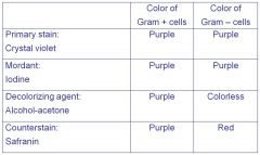
•Primary stain: crystal violet
•Mordant: iodine •Decolorizing agent: -alcohol-acetone •Counterstain: safranin |
|
|
PEQ Describe the order and names of the reagents in the Gram Stain, and be able to explain what is happening to differentiate the cells at each step.
|
1. A heat-fixed smear is covered with a basic purple dye, usually crystal violet. Because the purple stain imparts its color to all cells, it is referred to as a primary stain.
2. After a short time, the purple dye is washed off, and the smear is covered with iodine, a mordant. When the iodine is washed off, both gram-positive and gram-negative bacteria appear dark violet or purple. 3. Next, the slide is washed with alcohol or an alcohol-acetone solution. This solution is a decolorizing agent, which removes the purple from the cells of some species but not from others. 4. The alcohol is rinsed off, and the slide is then stained with safranin, a basic red dye. The smear is washed again, blotted dry, and examined microscopically. |
|
|
PEQ Describe the order and names of the reagents in the Gram Stain, and be able to explain what is happening to differentiate the cells at each step.
|
|
|
|
Understand the mechanism of these stains and give an example of something we might be looking for with each: Acid Fast, Endospore, Capsule Stains
|
-Acid fast: binds strongly only to bacteria that have a waxy material. We look for tuberculosis and leprosy microbes. Reddish color
-Endospore: special resistant structure that protects bacterium. Appear green -Capsule stains: determines the organism’s virulence. Stained with colored particles then with a simple stain. |
|
|
In the this staining procedure, the red dye carbolfuchsin is applied to a fixed smear, and the slide is gently heated for several minutes. (Heating enhances penetration and retention of the dye.) Then the slide is cooled and washed with water. The smear is next treated with acid- alcohol, a decolorizer, which removes the red stain from bacteria that are not acid-fast. The acid-fast microorganisms retain the red color because the carbolfuchsin is more soluble in the cell wall lipids than in the acid-alcohol One that differentiates bacteria into distinctive groups which binds strongly only to bacteria that have a waxy material in their cell walls. Microbiologists use this stain to identify all bacteria in the genus Mycobacterium, including the two important pathogens Mycobacterium tuberculosis, the causative agent of tuberculosis, and Mycobacterium leprae (lep′rī), the causative agent of leprosy
|
Acid Fast
|
|
|
Malachite green, the primary stain, is applied to a heat-fixed smear and heated to steaming for about 5 minutes. The heat helps the stain penetrate the _____ wall. Then the preparation is washed for about 30 seconds with water to remove the malachite green from all of the cells’ parts except the ______(s). Next, safranin, a counterstain, is applied to the smear to stain portions of the cell other than ________(s). In a properly prepared smear, the __________(s) appear green within red or pink cells. Because ________(s) are highly refractive, they can be detected under the light microscope when unstained, but they cannot be differentiated from inclusions of stored material without a special stain.
|
Endospore
|
|
|
Many microorganisms contain a gelatinous covering called a _______. In medical microbiology, demonstrating the presence of a _______ is a means of determining the organism’s virulence, the degree to which a pathogen can cause disease. It provides a contrasting background, so the _______(s) of these bacteria, Klebsiella pneumoniae, show up as light areas surrounding the stained cells.
|
Capsule Stains
|
|
|
standard unit of length
|
meter
|
|
|
1/100 m
|
centimeter
|
|
|
1/1000 m
|
millimeter
|
|
|
1/1,000,000 m
|
micrometer
|
|
|
1/1,000,000,000 m
|
nanometer
|
|
|
length of waves; light, gamma, radio
|
wavelength
|
|
|
centi c 10-2 = 0.01
Milli m 10-3 = 0.001 micro µ 10-6 = 0.000,001 nano n 10-9 = 0.000,000,001 |
centi c 10-2 = 0.01
Milli m 10-3 = 0.001 micro µ 10-6 = 0.000,001 nano n 10-9 = 0.000,000,001 |
|
|
the ability of the lenses to distinguish two points a specified distance apart.
(also called resolving power) is the ability of the lenses to distinguish fine detail and structure ~ distinguish between two points a specified distance apart. |
resolution
|
|
|
the same as resolution
|
resovling power
|
|
|
a constant based on the quality of the lens, the higher the numerical aperture the better the resolution
|
numerical aperture
|
|
|
the change in direction of a wavefront at an interface between
|
reflection
|
|
|
the bending of light when it passes from one medium to another; i.e. air to water.
|
refraction
|
|
|
transmitting waves or something.
|
transmission
|
|
|
the ability to absorb
|
absorption
|
|
|
to zoom in at an image
|
magnification
|
|
|
the bending or spreading of a beam of light when passing through a narrow opening
is the scattering of light rays as they “touch” a specimen’s edge. The diffracted rays are bent away from the parallel light rays that pass farther from the specimen. When the two sets of light rays– direct rays and reflected or diffracted rays–are brought together, they form an image of the specimen on the ocular lens, containing areas that are relatively light (in phase), through shades of gray, to black. |
diffraction
|
|
|
the positive ion, the chromophore is a cation
A salt in which the color is in the positive ion; used for bacterial stains. |
basic dyes
|
|
|
the negative ion, the chromophore is an anion
A salt in which the color is in the negative ion; used for negative staining. |
acidic dyes
|
|
|
aqueous or alcohol solution of a single basic dye
A method of staining microorganisms with a single basic dye. Used to make cellular shapes and arrangements visible. A mordant may be used to improve bonding between the stain and the specimen |
simple staining
|
|
|
react with different kind of bacteria and can be used to distinguish them.
A stain that distinguishes objects on the basis of reactions to the staining procedure. |
differential staining
|
|
|
-Staining the background instead of the cell
-preparing colorless bacteria against a colored background -A procedure that results in colorless bacteria against a stained background.. |
negative staining
|
|
|
uses fluorochromes and a laser light.
|
confocal microscopy
|
|
|
lens that stays in focus when the magnification is changed.
|
parfocal
|
|
|
the electrons pass through a thin section of the specimen
|
transmission electron microscope
|
|
|
visible light passes throught he specimen; uses separate objective and ocular lenses
|
compound light microscope
|
|
|
Details become visible because of differences in the refractive index of different parts of the cell
|
Phase-contrast microscope
|
|
|
visible light is scattered after striking the specimen, and the specimen is visible agains a darkened background
|
darkfield microscope
|
|
|
a special microscope using ultraviolet illumination
|
fluorescence microscope
|
|
|
the electrons strike the surface of the speciment, and secondary electrons leaving the surface are veiwed on a television-like screen
|
scanning electron microscope
|
|
|
makes use of relatively low-energy red light to excite fluorochromes; can track cellular activity in real tiem
|
two-photon microscopy
|
|
|
pertaning to the relative velocities of light through a substance
|
refractive index
|
|
|
involves the use of antibodies and ultraviolet light
|
immunofluorescence
|
|
|
one-millionth of a meter
|
resloving power
|
|
|
one ten-billionth of a meter
|
angstrom
|
|
|
the ability to sparate two points in a microscopy filed
|
reslvoing power
|
|
|
Adhere(s) best to bacteria, which have a negative charge, because the color molecule has a positive charge
|
basic dyes
|
|
|
used in diagnosis of tuberculosis
|
acid-fast stain
|
|
|
involves(s) the use of a negative stain made from India ink particles
|
capsule stain
|
|
|
schaeffer-fulton stain
|
endospore stain
|
|
|
uses carbolfuchsin dye
|
acid-fast stain
|
|
|
uses malachite green
|
endospore stain
|
|
|
relfect(s) a bsic difference between microbial cell walls: ethanol will not remove stain from bacteria
|
gram stain
|
|
|
a microscope that uses laser illumination
|
confocal
|
|
|
extremely thin microbes, for example the spirochete
|
darkfield
|
|
|
this type of electron microscope yields images with seemingly three-dimensional view of the specimen
|
scanning
|
|
|
light rays that pass through different portions of the specimen reach the eye with their wave peaks reinforced or cancelled, making structures of the specimen relatively light or dark
|
phase-contrast
|
|
|
a microscope that uses sound waves to form an image
|
scanning acoustic
|
|
|
formerly known as a micron
|
micrometer
|
|
|
formerly known as a millimicron
|
nanometer
|
|
|
this is 10 -10 of a meter
|
angstrom
|
|
|
a billionth of a meter
|
nanometer
|
|
|
did not do fill in the blanks
|
did not do fill in the blanks
|
|
|
used to distinguish mycobacterium species and some species of Nocardia. Once, stained with carbolfuchsin and treated with acid-alcohol, remain red because they retain the carbolfuchsin stain. Non-acid-fast bacteria, when stained and treated the same way and then stained with methylene blue, appear blue because they lose the carbolfuchsin stain and are then able to accept the methylene blue stain.
|
acid-fast
|
|
|
used to demonstrate the presence of capsules. Because capsules do not accept most stains the capsules appear as unstained halos around bacterial cells and stand out against a constrasting background
|
negative
|
|
|
used to detect the presence of endospores in bacteria. When malachite green is applied to a heat-fixed smear of bacterial cells, the stain penetrates the endospores and staines them green. When safranin (red) is then applied, it stains the remainder of the cells red or pink.
|
endospore
|
|
|
Tuberculoses
|
acid-fast
|
|
|
Be able to explain the importance of ioil when using the oil immersion objective
|
Immersion oil is used with the oil immersion lens to reduce light loss between the slide and the lens
|
|
|
capsule
|
Klebsiella pnemoniae
|
|
|
endospore
|
tetnus
|
|
|
the most common microscope used in microbiology is the
|
compound light microscope
|
|
|
the total magnification of an object is calculated by multiplying the magnification of the objective lens by the magnification of the ocular lens.
|
compound light microscopy
|
|
|
uses visible light
|
compound light microscope
|
|
|
the maximum resolution, or resolving power (the ability to distinguish two points) is 0.2 um; maximum magnification 2000x
|
compound light microscopy
|
|
|
brightfiled illumination is used for stained smears
|
compound light microscopy
|
|
|
unstained cells are more productively observed using darkfield, pahse-contrast, or DIC microscopy
|
compound light microscopy
|
|
|
shows a light silhouette of an organism against a dark background
|
darkfield microscope
|
|
|
it is most useful for detecting the presence of extremely small organisms
|
darkfiled microscope
|
|
|
birngs direct and reflected or diffracted ligth rays togehter (in phase) to form an image of the specimen on the ocular lens
|
phase-contrast microscopy
|
|
|
it allows the detailed observation of living organisms
|
Phase-contrast microscopy
|
|
|
provides a colored, three-dimensional image of the object being observed.
|
Differential Interference Contrast (DIC)
|
|
|
It allows detailed observations of living cells
|
Differential Interference contrast (DIC)
|
|
|
specimens are first stained with fluorochromes and then viewed through a compound microscope by using an ultraviolet light source
|
fluorescence microscopy
|
|
|
The microorganisms appear as bright objects against a dark background
|
fluorescence microscopy
|
|
|
used primarily in a diagnostic procedure called fluorescent-antibody (FA) technique, or immunofluorescence.
|
fluorescence microscopy
|
|
|
is an aqueous or alcohol solution of a single basic dye
|
simple stain
|
|
|
it is used to make cellular shapes and arrangments visible
|
simple stain
|
|
|
a mordant may be used to improve bonding between the stain and the specimen
|
simple stains
|
|
|
differentiate bactyeria according to their reactions to the stains; such as Gram stain and acid-fast stain
|
differential stains
|
|
|
The Gram stain proceudre uses a purple stain (crystal violet) iodine as a mordant, an alcohol decolorizer, and a red counterstain
|
differential stains
|
|
|
Gram-positive bacteria retaint he purple stain after the decoloriation step; gram-negative bacteria do not and thus appear pink from the counterstain
|
differential stains
|
|
|
acid-fast microbes, such as members of the genera Mycobacterium and Nocardia, retain carbolfuchsin after acid-alcohol decolorization and appear red; non acid -fast microbes take up the methylene blue counterstain and appear blue.
|
differential stains
|
|
|
negative staining is used to make microbial capsules visible
|
special stains
|
|
|
the endospore stain and flagella stain are special stains that color only certain parts of bacteria
|
special stains
|