Reading...
![]()
Play button
![]()
Play button
![]()
Use LEFT and RIGHT arrow keys to navigate between flashcards;
Use UP and DOWN arrow keys to flip the card;
H to show hint;
A reads text to speech;
75 Cards in this Set
- Front
- Back
|
True or false: Bone is a living, vital, responsive tissue
|
True
|
|
|
Like other connective tissue bone has what three components:
|
cells, fibers, and ground substance
|
|
|
Describe the organic matrix of bone:
|
The fibers and ground substance are called the organic matrix, or osteoid. This is at first unmineralized. Bone becomes rigid due to the mineralization of its osteoid, and its cells become trapped
|
|
|
Osteoblasts
produce: located: histology: |
produce-organic ECM
located- surface of bone tissue hist: basophilic, cuboidal if active, somewhat flattened if inactive |
|
|
Osteocytes
f(x)- location- cell processes- |
f(x)- help maintian bone matrix and control mineral homeostasis
osteocytes - cells w/in bone lacunae processes- gap junctions on their cell processes connect osteocytes |
|
|
Osteoclasts
f(x)- location- arrise from - example- histology- |
f(x)- remove bone
arrise from monocytes - multinucleated macrophages example of a synchtium histo- acidophili, ruffled border location- next to Howship's lacunae in the bone surface |
|
|
osteoid definition
components |
the unmineralized organic matrix of bone
90% collagen fibers (acidophilic) 10% ground substance-containing some GAGs |
|
|
Bone is not rigid until the osteoid becomes mineralized by the deposition of the ______amongst the collagen lattice
|
hydroxyapatitie crystals
|
|
|
The ECM of the organic matrix of bone contains _____produced by____________
|
collagen fibers produced by osteoblasts
|
|
|
Describe the periosteum
|
the outer covering for bone (thick)
has an outer fibrous layer made of dense CT and an inner cellular layer (composed of cells with osteogenic potential) |
|
|
_____fibers are collagen fibers which anchor the periosteum to bone
|
sharpey's fibers
|
|
|
desciribe the Endostem
|
a single thing, inner covering layer that is loosely organized
can be divided into several regions that are continuous wiht each other |
|
|
What are the regions of the endosteum?
|
diaphyseal endosteium (lining walls of marrow cavity)
Trabecular endosteum (covers trabeculae of spongy bone) Osteonal endosteum (goes into the canals that carry blood vessels w/in bone) |
|
|
Can osteoprogenitory cells (cells that will later be osteoblasts) arrise from the endosteum
|
yes, the endosteum is a potential source of osteoprogenitor cells
|
|
|
The osteon is also known as
|
A haversian system
|
|
|
Define the osteon
|
the basic unit of mature compact bone
|
|
|
What are osteons composed of
|
concentric lamellae or layers of mineralized bone matrix around a central canal, in which there is a blood vessel and CT
|
|
|
Most osteons are oriented _____to the long axis of the bone
|
parallel
|
|
|
Osteocytes are found in _____that are positioned b/w the lamellae of the osteons
|
osteocytes
|
|
|
What are the tiny channels that connect the lacunae called
|
canaliculi
|
|
|
volkmann's canals
|
the channels in bone that transmit blood vessles from the periosteum or endosteum into the bone, and communicate with the Haversian canals
|
|
|
intramemranous ossification process:
|
mesenchymal cells differentiate to osteoblasts, osteoblasts secrete osteoid, become trapped, bone spicules form and eventually fuse into solid bone. CT surrounding becomes endo and periosteum
|
|
|
When bone is formed in mesenchyme w/o a cartilage model it is called
|
intramembranous ossification
|
|
|
What kind of bones develop via intramembranous ossification
|
flat bones of the skull and other skull bones called membrane bones
and bone formation from the periosteum |
|
|
Spicule
|
small needlelike structure -one of hte silicate or calcium carbonate processes supporting the soft tissue
|
|
|
Trabecula
|
small bar, rod, bundle of fibers
--any of the intersecting osseous bars occuring in cancellous bone |
|
|
Two ways of classifying bones grossly are
|
compact and spongy bone
|
|
|
where can you find compact bone
|
it forms the outer shell of all bone and also the shafts in long bones
|
|
|
spongy bone is found where
|
at the expanded heads of long bones and fills most irregular bones
|
|
|
Is trabecula a micro or macroscopic way of discribing bone
|
it is a macroscopic structure (seen in the spongy part of bone)
|
|
|
A spicule is a macro or micro structure
|
a microscopic beam of bone
|
|
|
Endochondral bone growth arrises from
|
cartilage model
|
|
|
endochondral ossification occurs _____
|
in majority of bones in body from hyaline cartilage model
|
|
|
Is there hyaline cartilage in the endochondral ossified bone?
|
No, it was there as a model and was the orginal surface on which osteoid was deposited
|
|
|
Chondrification
|
the process of mesenchymal cells differentiated to chondroblasts
|
|
|
during chondrification cartliage is growing via
|
hypertrophy and then eventually will degenerate
|
|
|
what causes chondrocytes to degenerate?
|
a complex casscade of signals involving the periochondrium, chondrocytes, and the matrix mineralizing (becoming much more basophilic as this happens)
|
|
|
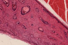
The bone collar is evidence of
|
bone development, one of the first signs -seen in the periostium area
|
|
|
During endochondral ossification, once the cartilage becomes calcified what happens?
|
blood vessels invade, bringing osteoprogenitor cells (can become osteoblasts in correct conditions). the large lacunae from the space chondrocytes were hanging out serve as the space which osteoblasts can start working- secreting osteoid on the calcified cartilage
|
|
|
Where do you usually find the primary center of ossification
|
the ends of the physes- interor of diaphysis (that has previously undergone endochondral ossification)
|
|
|
What is the secondary center of ossification/ where is usually found
|
one in each epiphysis, no bone collar
|
|
|
What is the bone collar formed from
|
from cells derived from the perichondrium/periosteum around the diaphysis – this is technically intramembranous ossification
|
|
|
what is the role of the bone collar?
|
will continue to add layers peripherally, and it will be remodeled to form the osteons of the compact bone of the diaphysis
|
|
|
The physes is synonymous with
|
growth or epiphyseal plate
|
|
|
the physes are remanants of the ______, where the _____remain the ability to divide
|
remnants of the cartilage model where chondrocytes retain ability to divide
|
|
|
In the physis area the chondrocyte proliferation is _____In orientation resulting in the width/length of the bone
|
linear, length
|
|
|
physes can be thought of as the ends of the ___-center of ossification
|
primary center, spreading toward either end of the bone and lengthening the bone
|
|
|
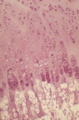
What are the five distinct zones of the physis
|
resting/ reserve
proliferation hypertrophy calcification ossification |
|
|
What occurs during the zone of ossification
|
capillary buds grow into tunnels previously occupied by dead chondrocytes
osteoblasts differentiate osteoid is deposited on tunnel walls |
|
|
explain how the ossification occurs in a 3D model
|
this deposition is seen as spicules with a thin core of cartilage ECM. This cartilage ECM is one key to identification of this process versus intramembranous ossification.
The spicules are later remodeled and cartilage ECM (and primary bone) is removed. |
|
|
in which zone does the cartilage matrix get mineralized
|
calcification
|
|
|
ossification zone is when
|
osteoid is laid down
|
|
|
chondrocytes in growth plate progress through a cycle of
|
division, hypertrophy and death
(do not become osteoprogeniotor cells) |
|
|
how do you achieve growth in width (expansion of bone)?
|
Osteoblasts in the inner layer of the periosteum deposit bone lamellae on the outer surface WHILE the osteoclasts in the diaphyseal endosteum remove bone from inner surface
NET result |
|
|
What growth process happens in the zone of proliferation
|
interstitial (growth from w/in -chondrocytes
|
|
|
The way that osteocytes proliferate is ___
|
appositional growth- tissue added to edge
|
|
|
Woven bone
|
bony tissue characteristic of the embryonal skeleton, in which the collagen fibers of the matrix are arranged irregularly in the form of interlacing networks
|
|
|
Through the process of remodeling woven bone is replaced by
|
lamellarbone
|
|
|
Lamellar or layer bone
|
the normal type of adult mammalian bone, whether cancellous or compact, composed of parallel lamellae in the former and concentric lamellae in the latter; lamellar organization reflects a repeating pattern of collagen fibroarchitecture.
|
|
|
Is lamellar bone ever remodeled
|
continuously throught life
|
|
|
how are osteons formed
|
new osteons form in a tunnel behind a cutting cone of osteoclats, and a blood vessel surrounded by osteoprogenitor cells grows into the , osteoblats differentiate and line edges of tunnel.
|
|
|
Spongy bone is what forms on the ____side of the growth plate and in most of the ___cavity of the epiphysis
|
metaphyseal side, in most of the marrow cavity
|
|
|
spongy bone remains in the form of ____even after remodeling
|
form of trabeculae
|
|
|
Relative to the formation of the lamellae in osteon, the reversal line is deposited
|
first
|
|
|
osteon/ haverian systems are evidence of
|
remodeling
|
|
|
reversal lines are deposited from outside in (one furthest out is the oldest)
|
so think of the deposition of osteon like rust building up on a pipe, the longer the osteon has been around the smaller the central canal will be
|
|
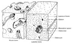
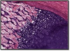
woven vs lamellar bone
|

woven vs lamellar
|
|
note 5 distinct zone
|

note 5 zones
|
|

note 5 distinct zone
|

note 5 zones
|
|

identify osteon being formed in vertebrae
|
osteon formation in IV
|
|

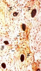
osteon (note dark lines are lacunae connected by canaliculi)
|
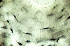
osteon (note dark lines are lacunae connected by canaliculi)
|
|
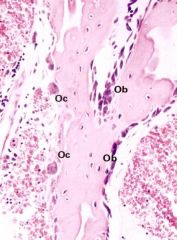
osteoclasts vs osteoblasts
|

osteoclasts vs osteoblasts
|
|

haverian system
|
Haverian system
|
|
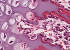
endochondrial ossification
|

endochondrial ossification
|
|
|

capillary growing into space chondrocyte was
|