Reading...
![]()
Play button
![]()
Play button
![]()
Use LEFT and RIGHT arrow keys to navigate between flashcards;
Use UP and DOWN arrow keys to flip the card;
H to show hint;
A reads text to speech;
45 Cards in this Set
- Front
- Back
|
Main difference between eukaryotes and fungi
|
fungi are Contain nucleus, mitochondria, 80S ribosomes
-Plasma membrane contains ergosterol instead of cholesterol. -Cell wall is composed of chitin and various glucans, mannans, and complex polysaccharides -Unicellular or multicellular depending on the species -lacking chlorophyll |
|
|
Effect of antibacterial on fungi?
|
None
|
|
|
Yeast are what kind of organism?
|
Yeast: Unicellular growth form of fungi, spherical/ellipsoid.
|
|
|
What is the reproduction of yeast?
|
Reproduction occurs by budding or fission
“Mother” cell pinches off to produce “daughter” cell Daughter cell elongates and forms a pseudohyphae. |
|
|
Mold are what kind of organism?
|
Multicellular organisms with tubular structures known as hyphae that grow at the tips by apical extension.
|
|
|
Mold reproduction
|
Hyphae can be septate or nonseptate (coenocytic)
Hyphae come together to produce mycelium (colony) Reproduction occurs by formation of spores |
|
|
the vegetative part of a fungus is known as
|
Mycelia consisting of a mass of branching, thread-like hyphae
|
|
|
Hyphae may initially develop from a
|
a GERM-TUBE (a short, immature hypha) that emerges from a germinating spore
|
|
|
The identification of yeast in the clinical lab
|
Colony morphology
Yeast cells, pseudohyphae or hyphae (grow on cornmeal agar) and biochemical tests (sugar assimilation, enzymatic activity). |
|
|
what is thermal dimorphism?
|
filamentous mold at 25oC, yeast at 37oC
|
|
|
Molds and yeast are not exclusive forms, some species may exist in
|
both yeast and mold forms (dimorphism).
|
|
|
Molds Vegetative hyphae
|
Hyphae that grow on or beneath culture medium surface
Form seen in tissue, few distinguishing features |
|
|
Molds Aerial hyphae
|
Hyphae that project above the surface of the media
Contain structures for production of spores (asexual propagules) usually only seen in culture |
|
|
Molds - identification based on
|
colony morphology (pigment, texture) and morphology of reproductive structures
Conidia - spores formed by budding (blastoconidia) or disarticulation of existing hypha (arthroconidia) Sporangiospores - produced by free-cell formation within sporangium in nonseptate molds |
|
|
1) fungal elements (appear violet in color)
2) Stains for chitin 3)For cryptococcus (deep red in appearance) 4)Fungi appear pinkish red 5)- Fungi including Pneumocystis; gray to black with green background |
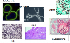
1)H&E- fungal elements
2)Calcofluor white 3)Mucicarmine 4)Periodic Acid Schiff (PAS) 5) Gomori Methenamine Silver (GMS) |
|
|
1) Molds with non-septate (coenocytic) hyphae
2)Most with separate filamentous form, but some are typical yeasts 3)Created just for Pneumocystis jiroveci (carinii) 4)Vegetative yeast cells that proliferate by budding or fission 5) Sexual reproduction with thin-walled sac (ascus); septate hyphae |
1)Zygomycetes
2)Basidiomycetes 3)Archiascomycetes: 4)Hemiascomycetes: 5)Euascomycetes |
|
|
Zygomycetes EX
|
Rhizopus spp.; Mucor spp.
|
|
|
Basidiomycetes ex
|
(Cryptococcus; Malassezia)
|
|
|
Hemiascomycetes: ex
|
(Candida)
|
|
|
Euascomycetes ex
|
Dermatophytes, Blastomyces, Histoplasma, Aspergillus
|
|
|
Fungi reproduce either
|
Asexual reproduction results in progeny that are genetically identical to the parent cell.
Sexual reproduction allows for recombination. Sexual reproduction is important for maintenance of pathogenicity. |
|
|
Primary fungi infection
|
Can initiate infection in a “normal”, immunocompetent host
Blastomyces, Coccidioides, Histoplasma, Paracoccidioides |
|
|
Opportunistic fungi infection
|
Fungal infections in immunocompromised (innate or acquired) individuals
Candida, Cryptococcus, Aspergillus, Pneumocystis |
|
|
Primary Fungal Pathogens All are
|
are agents of respiratory infections and none are obligate parasites
|
|
|
Primary Fungal Pathogens Saprobic vs. Parasitic Phases
|
Saprobic: Filamentous hyphae (in soil/decaying vegetation) that produce the airborne infectious cells (ie conidia)
Parasitic: Adapted to grow at 37C and to reproduce asexually in the host respiratory mucosa This “dimorphic” existence is a virulence factor in and of itself |
|
|
The Superficial Mycoses
|
Piedra (infection of the hair shaft)
Black piedra, white piedra and tinea nodosa Pityriasis versicolor: Tinea versicolor |
|
|
Piedra Growth of the fungus on the hair shaft
|
Small nodules/granules form sleeve/collar around shaft
Black piedra-scalp, penetrates shaft, breaks hair White piedra-beard (occ genital hair), soft, creamy |
|
|
Tan to brown, or non-colored scaly patches primarily on chest and back, but can occur on the extremities
Coincidence with regions rich in sebaceous glands (trunk area) |
Pityriasis (Tinea) Versicolor by M. furfur
|
|
|
Pityriasis (Tinea) Versicolor seen up to
|
Up to 3% of dermatology diagnoses in the summer months in temperate climates
Most often seen in tropical climates (up to 40% prevalence) |
|
|
Pityriasis (Tinea) Versicolor Diagnosis
|
Skin scraping -> KOH
Fungal elements with spaghetti & meatballs Rarely cultured Media needs fatty acids |
|
|
Pityriasis (Tinea) Versicolor tx
|
Topical antifungals (miconazole) or keratinolytic
|
|
|
M. furfur has been demonstrated to cause a life-threatening infection fungemia in
|
neonates that are receiving lipid-rich parenteral nutrition.
Cradle cap is seborrheic dermatitis in neonates |
|
|
Cutaneous Mycoses
|
1) Dermatophytoses
Tinea (ringworm) of various body parts 2) Onychomycosis Fungal infection of the nails |
|
|
Infections of keratinized tissues (keratinophilic and keratinolytic)
Erythematous outer ring -> crusting healing center Classified according to anatmoic site/structure: |
Dermatophytosis or Tinea (Ringworm)
Tinea barbae: Beard Tinea corporis: Body-ring in the body Tinea cruris: Groin (jock itch) spares penis/scrotum Tinea pedis: Foot (athlete’s foot) Tinea unguium: Nail (onychomycosis caused by dermatophytes) Tinea nigra: Palm (**non-dermatophytic Cladosporium werneckii) Tinea umbricata- Pacific islands, Amazon T. concentricum |
|
|
The dermatophytes causing tinea ringworm are
|
Microsporum spp, Epidermophyton spp, and Trichophyton spp
|
|
|
Tinea (Ringworm) dx
|
Wood’s lamp (UV)
Microsporum will fluoresce yellow (stars), KOH prep dissolves keratin, not chitin, see hyphae. Cx: Sabourauds agar |
|
|
Tinea (Ringworm Treatment:
|
Topical antifungals, though oral antifungals may be necessary in tinea capitis and severe cases of cruris and pedis.
|
|
|
Onychomycosis causative agents
|
Dermatophytes: Tinea Unguium. Mainly on toenails
Trichophyton rubrum Candida albicans, Candida parapsilosis – Mainly fingernails |
|
|
Subcutaneous Mycoses are due to
|
traumatic introduction into the subcutaneous tissues
ex Sporotrichosis |
|
|
Infection by direct implantation of spores into subcutaneous tissue from thorns or splinters
Lesions on hands and arms follow the lymphatics |
Rosegardener’s Disease”
Causative Agent: Sporothrix schenckii |
|
|
Sporothrix schenckii dissemination can occur in
|
in severely immunocompromised, and with an isolate that is capable of growth at body temperature.
|
|
|
Sporotrichosis Treatment:
|
Oral azole antifungals until lesions are clear, generally 3-6 months. (itra, vori, posa, or amphotericin)
|
|
|
Opportunistic pathogens are
|
Candida species-Most common
Cryptoccocus neoformans Aspergillus fumigatus Rhizopus species Mucor Pneumocystis Carinii |
|
|
what is the relationship between the morphology assumed by a fungus in tissue and the pathogenesis it causes?
|
Fungal morphology determines which component of ost defense is needed to fight infection
|
|
|
what 5 risks lead to systemic fungal infection?
|
1)HIV
2)Diabetes 3)Neutropenia-ex secondary to chemo 4)systemic corticosteroids 5)systemic broad-spectrum antibodies |