![]()
![]()
![]()
Use LEFT and RIGHT arrow keys to navigate between flashcards;
Use UP and DOWN arrow keys to flip the card;
H to show hint;
A reads text to speech;
10 Cards in this Set
- Front
- Back
- 3rd side (hint)
|
Most sensitive marker, gold standard for MI |
Troponin I |
|
|
|
Change in Troponin I levels in blood |
Rise 2-4hours after MI Peak at 24 hours Return to normal by 7-10 days |
|
|
|
CK-MB use |
Detects reinfarction. Levels rise 4-6 hours after MI. Peak at 24 hours. Return to normal by 72 hours. |
|
|
|
Treatment for MI |
Oxygen Beta blocker Aspirin Thrombolytic Morphine Ace inhibitor Nitrates |
O BATMAN |
|
|
< 4 hours from infarction |
No gross or microscopic changes. Complications: cardiogenic shock (massive infarction) Congestive HF Arrhythmia |
|
|
|
4-24 hours changes |
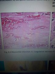
Gross: dark discoloration Microscopic: coagulative necrosis Complications: arrhythmia |
|
|
|
1-3 days changes |
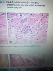
Gross: yellow pallor Microscopic: neutrophils Complications: Fibrinous pericarditis (chest pain w/ friction rub) |
|
|
|
4-7 days changes |
Gross: Yellow pallor (WBCs- acute inflammation) Microscopic: macrophages (eat dead necrotic debris) Complications: Rupture of: -ventricular free wall (leads to cardiac tamponade), -IV septum (leads to shunt), -Papillary muscle (leads to mitral insufficiency). |
|
|
|
1-3 weeks |
Gross: Red border emerges as granulation tissue enters from edge of infarct. (Blood vessels growing into it) Microscopic: Granulation tissue with plump fibroblasts, collagen and blood vessels. |
|
|
|
Months - changes |
Gross: white scar (scar not as strong as myocardium, weakened ventricular wall, increased risk of aneurysm, stasis along wall so thrombi can form) Microscopic: fibrosis Complications: Aneurysms, mural thrombi, dressler syndrome (antibodies formed against pericardium, 6-8 wks after phenomenon) |
|