Reading...
![]()
Play button
![]()
Play button
![]()
Use LEFT and RIGHT arrow keys to navigate between flashcards;
Use UP and DOWN arrow keys to flip the card;
H to show hint;
A reads text to speech;
34 Cards in this Set
- Front
- Back
|
What are the values of DEXA for osteopenia?
|
-1.0 to -2.5 SD
|
|
|
What does DEXA stand for?
|
Dual Energy x-ray absorptiometry
|
|
|
What are the DEXA values for Osteoporosis?
|
lower than -2.5
|
|
|
What is the caveat for Help I've Fallen and I can't Get up?
|
Most of the time the people break their hips first, then fall down, although it seems vice versa is most likely.
|
|
|
What are the two non-deficiency nor GI induced ostemalacia causes listed in lecture?
|
Hyperthyroid induced
Chronic use of anticonvulsant medication |
|
|
What is the one deficiency induced cause of osteomalacia?
|
Vitamin D deficiency
|
|
|
What are the two GI induced causes of osteopenia?
|
They are Gastrointestinal and biliary causes:
- renal osteodystrophy -fibrous dysplasia |
|
|
What are the three main phrases in lecture with adult-onset osteomalacia?
|
Osteoid is mineral deficient therefore Bone doesn't get mineralized from the get go
Looser lines or Osteoid Seams are present Individual Has "soft Bones" |
|
|
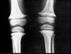
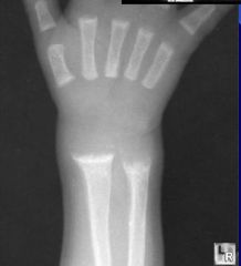
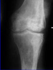
What are the six characteristics of Rickets in a radiograph?
|
-Cupping w/sclerosis and fraying of metaphysis
-Looser's lines (which are pseudofractures) -Poorly mineralized epiphyseal centers with delayed appearance -Widening osteoid seams -Osteopenia -BOWING- DuH |
|
|
what are parathyroid hormone disorders' effects on bone?
|
Existing bone is demineralized or hypermineralized.
|
|
|
What are the six characteristics of Hyperparathyroidism?
|
-medial side demineralization
-sub-periosteal bone resorption of distal tufts -Periosteal lifting -ground glass -lace-like appearance -Vascular calcifications (All of this points to the fact that we will see a combination of mineralization patterns) |
|
|
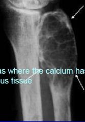
What is Osteitis Fibrosa?
|
Brown Tumors
-Areas where the calcium has been replaced with fibrous tissue "there has been so much leeching of the calcium of the bone that it has been replaced." |
|
|
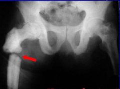
What causes Renal osteodystrophy?
What is a sign of it in the radiograph? |
Kidney pumps out Ca++, brings in PO4
Parathyroid kicks in to increase calcium in the blood Calcium is leeched out of the bone. Calcification of the meniscus |
|
|
What causes cretinism?
What are the symptoms and etiologies? |
Caused by congenital hypothyroidism
Deformed ossification centers with fragmented heterogenous appearance -long bones have short slender shafts -Metaphysis has dense transverse bands -metaphysis has wide cupping and osteoporosis |
|
|
What is Osteitis Deformans?
|
Paget's Disease- Chronic buildup and breakdown of bone
-over 40 male -bone pain with increasing hat size -has a genetic component -associated with CHF/ A-V shunts Can result in osteosarcoma |
|
|
What are the two types of bones mixed in Paget's disease?
|
Sclerotic bone mixed with lytic bone.
|
|
|
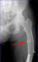
What type of fractures occur with Paget's disease?
|
Banana or chalk transverse fractures.
|
|
|
What lesions appear in the x-ray of a Pagets patient?
|
Flame shaped "blade of grass" lesions in long bones
Banana transverse fractures Picture frame vertebral body Osteolytic lesions |
|
|
What are the lesions in the skull of an individual with Paget's disease Called?
|
Osteoporosis Circumscripta
|
|
|
What disease is associated with Winber's sign? What is it?
|
Scurvy
Winberg's sign is very scloerotic rim around the epiphyseal area |
|
|
With Scurvy- What are the two items associated with the late, great Dr. Frankel?
|
Frankel line is an increased density at the metaphysis
A scurvy line is a lucent (decreased density) line on the shaft side of the Frankel line |
|
|
What is a "pelican" spur?
|
has to do with Scurvy, actually called Pelkan, but easier to associate with scurvy as pelican.
they are exostoses on the lateral border of the metaphysis |
|
|
What bacterial infection is associated with Sickle cell disease?
|
Salmonella osteomyelitis
|
|
|
What are the x-ray evidences of an individual with Sickle-cell disease?
|
-Decreased long bone density
-Trabecular coarsening -Cortical thickening -Destructive lesions- Infarcts -Mottling "moth eaten" areas - ** Bone within a bone** Brachymetatarsia from infarcts at growth centers Dactylitis with edema |
|
|
What are the three characteristics of Marfans?
|
Elongation and thinning of long bones
Arachnodactly Abnormal collagen |
|

|
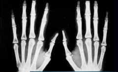
Marfans Arachnodactyly
|
|

|
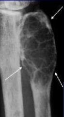
Brown Tumor
Osteitis Fibrosa |
|
|
Osteitis Fibrosa
|
|
|
Paget's Banana Fracture
|
|
|
Ricket cupping sclerosis and fraying of metaphysis
|
|
|
Rickets looser line
|
|
|
Rickets poorly mineralized epiphyseal centers and metaphysis deformities
|
|
|
Sickle Cell Bone within bone
|
|
|
Sickle Cell brachydactyly
|