Reading...
![]()
Play button
![]()
Play button
![]()
Use LEFT and RIGHT arrow keys to navigate between flashcards;
Use UP and DOWN arrow keys to flip the card;
H to show hint;
A reads text to speech;
79 Cards in this Set
- Front
- Back
|
Which axons are myelinated, 6-12uM in diameter, relay cutaneous tactile information and have a conduction velocity of 30-60M/s?
|
Abeta
|
|
|
Which axons are myelinated, 1-5uM in diameter, have a conduction velocity of 5-30M/s and relay painful modalities?
|
Adelta
|
|
|
Which axons are unmyelinated, .5-3uM in diameter, have a conduction velocity of <3M/s and are responsible for nociception?
|
C
|
|
|
Define sensory transduction
|
Converting mechanical, chemical, thermal or photic energy into electrical signals useful to the nervous system
|
|
|
Receptor and synaptic potentials are _________ (digital/analog)
|
analog
|
|
|
Action potentials are _________ (digital/analog)
|
digital
|
|
|
Intensity of a stimulus is coded as the ________ of the receptor potential.
|
Amplitude
|
|
|
Amplitude of the receptor potential is coded as the _________ of the action potentials.
|
Frequency
|
|
|
Frequency of action potentials is coded as the _________ of post-synaptic potentials
|
Amplitude
|
|
|
Sensory transduction is an exact transcription of a stimulus (True/False)
|
False (the stimulus is modified by the receptor and through CNS processing (think telephone game))
|
|
|
What is the purpose of auxilliary structures such as pacinian corpuscles and merkel's disks?
|
to modify the time course of a receptor potential (eg, pacinian corpuscles respond to a vibrating stimulus)
|
|
|
The excitation on a third order neuron has the same S/B ratio as the stimulus (True/False)
|
True
|
|
|
Receptors of multiple types can be found in a single neuron (True/False)
|
False (Line labeled coding segregates the information into exclusive channels so that an axon can only carry one type of signal)
|
|
|
If you were to stimulate an axon proximally to it's receptor, where would the brain perceive the location of the stimulus to be?
|
At the location of the receptor (explanation of phantom limb syndrome)
|
|
|
What are the four modalities of the epicritic (dorsal column) system?
|
fine tactile, 2-point discrimination, proprioception and vibration
|
|
|
The _________ is the structure in which the primary afferents of the epicritic (dorsal column) for the lower extremity ascend on the ________ (ipsilateral/contralateral) side of the spinal cord.
|
fasciculus gracilis, ipsilateral
|
|
|
The _________ is the structure in which the primary afferents of the epicritic (dorsal column) for the upper extremity ascend on the ________ (ipsilateral/contralateral) side of the spinal cord.
|
fasciculus cuneatus, ipsilateral
|
|
|
The primary afferents of the epicritic (dorsal column) system synapse at the ________ and cross the midline to ascend the_________. The secondary neurons then synapse in the ________.
|
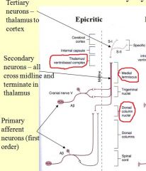
Dorsal Column Nuclei (nucleus fasciculus and nucleus cuneatus), Medial lemniscus, Thalamus/Ventrobasal complex
|
|
|
At what level of the brainstem do the internal arcuate fibers of the dorsal column nuclei begin crossing to the medial lemnisci?
|
|
|
|
Secondary epicritic afferents from the lower extremity travel along the _________ (medial/lateral) side of the medial lemniscus.
|
lateral (upper extremity travels along the medial side)
|
|
|
What are the modalities of the protopathic (anterolateral tract) system?
|
pain (nociception and nociperception), temperature and gross tactile
|
|
|
What is the primary stimulus of the protopathic (ALS) system?
|
K+
|
|
|
"Fast pain" fibers
|
Adelta
|
|
|
"Slow pain" fibers
|
C
|
|
|
The secondary protopathic (ALS) fibers from the lower extremity travel up the ________ (medial/lateral) side of the anterolateral tract.
|
lateral (body and arm are medial just like the medial lemniscus in the epicritic pathway)
|
|
|
Primary protopathic (ALS) fibers synapse and secondary fibers cross the midline before ascending the spinal cord (True/False)
|
True (in the epicratic pathway, the fibers ascend the spinal cord first and then synapse)
|
|
|
The trigeminal pathways carry both epicritic and protopathic fibers that synapse immediately upon entering the brainstem at the trigeminal nuclei. (True/False)
|
True
|
|
|
At what levels will you find the trigeminal tracts and/or nuclei?
|
Beginning at the spinal cord-medulla transition and ending at the ponto-medullary transition
|
|
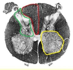
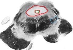
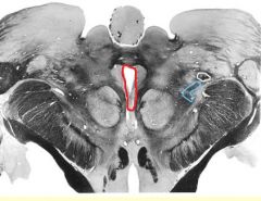
Name the structures circled and the level of the spinal cord.
|
Green - dorsal horn; Red - dorsal column (fasciculus gracilis); Yellow - ventral horn; White - Anterolateral tract; Lumbar Spinal Cord
|
|
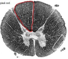
Name the circled structures and the level of the spinal cord.
|
Red - Dorsal column (fasciculus gracilis); White - anterolateral tract; Thoracic spinal cord
|
|
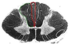
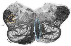
Name the circled structures and the level of the spinal cord.
|
Red - fasciculus gracilis; Green - fasciculus cuneatus; White - anterolateral tract; Cervical spinal cord
|
|
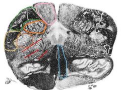
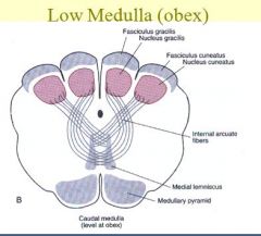
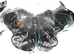
Name the circled structures and the level of the brain stem.
|
Pink - Nucleus gracilis; Green - fasciculus cuneatus; Orange - Nucleus cuneatus; Yellow - Spinal trigeminal tract; Brown - Spinal trigeminal tract; Red - Internal arcuate fibers; Blue - Medial lemniscus; White - anterolateral tract; Low medulla (obex)
|
|
Name the circled structures and the level of the brain stem.
|
Yellow - Spinal trigeminal tract; Blue - medial lemniscus; White - anterolateral tract; Rostral Medulla
|
|
Name the circled structures and the level of the brain stem.
|
Yellow - Spinal trigeminal tract; Red - spinal trigeminal nucleus; Blue - Medial lemniscus; White - anterolateral tract; Green - Infereior cerebral peduncle; Ponto-Medullary Junction
|
|

Name the circled structures and the level of the brain stem.
|
Green - Superior cerebral peduncle; Red - Fourth ventricle; Blue - medial lemniscus; White - anterolateral tract; Mid Pons
|
|
Name the circled structures and the level of the brain stem.
|
Green - Superior cerebral peduncle; Blue - medial lemniscus; White - anterolateral tract; Rostral Pons
|
|
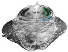
Name the circled structures and the level of the brain stem.
|
Green - Cerebral aqueduct; White - anterolateral system; Blue - medial lemniscus; Red - Periaqueductal gray; Midbrain
|
|
Name the circled structures and the level of the brain stem.
|
Red - third ventricle; Blue - medial lemniscus; White - anterolateral tract; Diencephalon
|
|
|
Secondary axons of the visceral pain pathway travel through the _________ where they synapse in the __________
|
dorsal columns, nucleus gracilis
|
|
|
What would result from lesions to the medial side of the fasciculus gracilis?
|
This would result in an abolishment of visceral pain signals from the ipsilateral side.
|
|
|
The anterior horn of the spinal cord is primarily supplied by what artery?
|
Anterior spinal artery
|
|
|
The dorsal columns are supplied by what artery?
|
Posterior spinal artery
|
|
|
What is known about the location of a lesion if the face is involved?
|
It is above the trigeminal nucleus
|
|
|
What artery supplies the medial lemnicus in the medulla?
|
Anterior Spinal artery
|
|
|
What artery supplies the spinal trigeminal artery and tract in the medulla?
|
Posterior spinal a.
|
|
|
What is area 3,1,2 on the Brodman map?
|
somatosensory area (postcentral gyrus)
|
|
|
Interneurons of the protopathic pathway in the spinal cord are ________ (inhibitory/excitatory) and are normally _________ (inhibited/activated).
|
Inhibitory, Inhibited (Gate control hypothesis. Tactile fibers activate the interneurons which in turn inhibit the protopathic pathway, decreasing pain sensation) (rub your hand when you smash it and the pain decreases)
|
|
|
Nucleus Raphe Magnus and Nucleus Reticularis Paragigantocellularis are found in the __________ and are involved in the _________ _________ ____________ pathway
|
Medulla, Descending Pain Modulation
|
|
|
Name the four nuclei of the descending pain pathways
|
PAG (periaqueductal gray), Locus cerulius, Nucleus Raphe Magnus, Nucleus Paragigantocellularis
|
|
|
What neurotransmitter is used by the PAG neurons?
|
Enkephalon
|
|
|
Which two descending pain modulation pathways use norepinephrine(noradrenaline) as their neurotransmitter?
|
Locus ceruleus and Nucleus Paragigantocellularis
|
|
|
Locus ceruleus and the Periaqueductal Gray (PAG) are located in the _________ and are involved in the _________ __________ _________ pathway
|
Midbrain, Descending Pain Modulation
|
|
|
Nucleus Raphe Magnus uses which neurotransmitter?
|
Serotonin
|
|
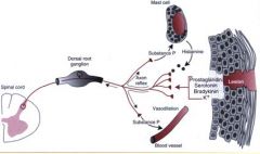
What is this cartoon illustrating?
|
Peripheral sensitization
|
|
|
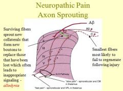
Axon sprouting can lead to what pathology?
|
Neuropathic pain - normal stimuli trigger severe pain (can occur post-amputation)
|
|
|
What Amendment provides the ability to confront adverse witnesses?
|
6th amendment provides the ability to confront adverse witnesses.
|
|
|
What happens when neuronal fibers fail to regenerate, leaving bare postsynaptic junctions?
|
|
|
|
Nipples are at what dermatome level?
|
T4
|
|
|
Umbilicus is at what dermatome level?
|
T10
|
|
|
The knee is at what dermatome level?
|
L4
|
|
|
Patient presents with sensory loss to the thumb. What dermatome is affected?
|
C6
|
|
|
Patient presents with sensory loss to the third digit. What dermatome is affected?
|
C7
|
|
|
Patient presents with sensory loss to the fifth digit. What dermatome is affected?
|
C8
|
|
|
Patient presents with sensory loss to the medial aspect of the elbow. What dermatome is affected?
|
T1
|
|
|
where is the substantia gelatinosa located?
|
The dorsal horn (lamina II)
|
|
|
Patient presents with sensory loss to the great toe. What dermatome is affected?
|
L4
|
|
|
Patient presents with sensory loss to the lateral foot and calf. What dermatome is affected?
|
S1
|
|
|
Patient presents with sensory loss to the middle of the dorsal foot and medial plantar foot. What dermatome is affected?
|
L5
|
|
|
The angle of the jaw receives sensory innervation from what nerve?
|
C2
|
|
|
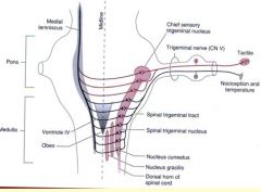
If a patient presents with a ringed pattern of sensory loss unilaterally on part of his face, where would a neuronal lesion be likely located?
|
The spinal trigeminal tract in the medulla
|
|
|
If a patient presents with V1-V3 patterned sensory loss, where is the neuronal lesion likely to be located?
|
The trigeminal nerve, peripheral to the spinal cord
|
|
|
How would you distinguish a peripheral nerve loss from a CNS lesion?
|
peripheral nerve loss follows a peripheral nerve pattern, a fragment of a dermatome (important in hands and feet)
|
|
|
Patient presents with sensory loss to the external acoustic meatus. What cranial nerve is likely involved?
|
CNX
|
|
|
A lesion in the medullary portion of the the spinal trigeminal nucleus would affect the fine tactile response of the face (True/False)
|
False (only protopathic pathways affected. a lesion would have to be at the level of the pons to affect the epicritic pathways)
|
|
|
How would a patient with peripheral neuropathy caused by diabetes likely present?
|
"Stocking Glove" sydrome. Loss of both sensory modalities in the hands and feet.
|
|
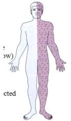
Patient presents with this pattern of sensory loss of both modalities. What is it called, where must the lesion be located and what could have caused it?
|
This is hemihypoesthesia. The lesion must be located rostral to the trigeminal nuclei (face is involved) and could have been caused by a stroke or neoplasm.
|
|
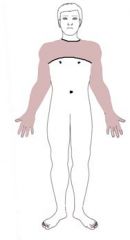
A patient presents with this pattern of sensory loss. What is it called and where must the lesion be located?
|
This is called crossed hypoesthesia. The lesion must be in the brain stem because the areas affected involve the face (trigeminal nucleus) and the contralateral side of the body (medial lemniscus).
|
|
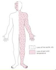
A patient presents with tactile sensory loss on his left lower extremity and pain/temp loss on his right lower extremity. What is this called, how could it have been caused, and on what side of the spinal cord is the lesion?
|
This is called the "Harlequin" or Brown-Sequard Syndrome. It is caused by a lesion/injury that hemisects the spinal cord. This can occur due to tumors, MS or trauma. Because the epicritic has been lost on the left side and the protopathic lost on the right, the lesion is on the left side (epicritic have not yet crossed the midline, protopathic have)
|
|
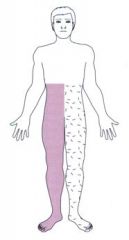
What would likely cause this pattern of pain/temp sensory loss and what is it called?
|
This is called syringomyelia. It is cause when a syrinx forms in the central canal of the cervical spinal cord, disrupting the protopathic secondaries as they cross from the dorsal horn to the ALS.
|