Reading...
![]()
Play button
![]()
Play button
![]()
Use LEFT and RIGHT arrow keys to navigate between flashcards;
Use UP and DOWN arrow keys to flip the card;
H to show hint;
A reads text to speech;
20 Cards in this Set
- Front
- Back
- 3rd side (hint)
|
Medial lemnisci
|
.
|
|
|
|
Spinocerabellar
|
Pathway for ventral and rostral spinocerebellar tracts[edit]
Some neurons of the ventral spinocerebellar tract instead form synapses with neurons in layer VII of L4-S3. Most of these fibers cross over to the contralateral lateral funiculus via the anterior white commissure and proceed up the spinal cord to synapse with neurons in the superior cerebellar peduncle. The fibers then often cross over again within the cerebellum to end on the ipsilateral side. For this reason the tract is sometimes termed the "double-crosser." The Rostral Tract synapses at the dorsal horn lamina (intermediate gray zone) of the spinal cord and ascends ipsilaterally to the cerebellum through the inferior cerebellar peduncle. |
Pathway for dorsal and spinocuneocerebellar tracts[edit]
The motor neurons synapse in an area known as Clarke's nucleus or "Clarke's column". This is a column of relay neuron cell bodies within the medial gray matter within the spinal cord in layer VII (just beneath the dorsal horn), specifically between T1-L3. These neurons then send axons up the spinal cord, and project ipsilaterally to medial zones of the cerebellum through the inferior cerebellar peduncle. Below L3, relevant neurons pass into the fasciculus gracilis (usually associated with the dorsal column-medial lemniscal system) until L3 where they synapse with Clarke's nucleus (leading to considerable caudal enlargement). The neurons in the accessory cuneate nucleus have axons leading to the ipsilateral cerebellum via the inferior cerebellar peduncle. |
|
|
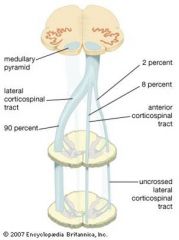
Pyramids
|
.The two pyramids contain the motor fibers that pass from the brain to the medulla oblongata and medulla spinalis, corticobulbar and corticospinal fibers.
When these pyramidal fibers are traced downward, it is found that some three-fifths or more of them leave the pyramids in successive bundles, and decussate in the anterior median fissure of the medulla oblongata, forming what is termed the pyramidal decussation or motor decussation. Having crossed the middle line, they pass down in the posterior part of the lateral funiculus as the lateral corticospinal tract. |
|
|
|
Spinothalamic
|
Immediately decussates in Spinal cord
|
|
|
|
Superior cerabellar peduncles
|
.
|
|
|
|
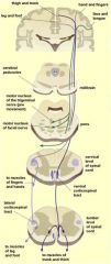
Corticospinal tract
|
Travels from the cerebral cortex down to the spinal cord.
CST actually consists of two separate tracts in the spinal cord: the lateral corticospinal tract and the anterior corticospinal tract. Contains mostly motor axons. Referred to as a pyramidal tract as when the tract passes the medulla, it forms a dense bundle of nerve fibres that is shaped somewhat like a pyramid |
|
|
The corticospinal tract is concerned specifically with discrete voluntary skilled movements, especially of the distal parts of the limbs.
|
Some motor fibres from the motor cortex do not decussate, but instead descend in the anterior corticospinal tract. These then cross in the anterior commissure of the cord at the level where fibres synapse with the anterior horn cell – they terminate in the anterior gray column of the spinal cord segments in the cervical and upper thoracic regions.
|

|
|
|
The upper motor neuron has its cell body in the cerebral cortex. Its axon descends to synapse on the
LMN, (an inter neuron, situated in the anterior gray column of the spinal cord. The axon is short ) and synapses with the LMN, in the anterior gray column. The axon then innervates the skeletal muscle through the anterior root and spinal nerve. |

|
|
|
|
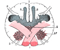
Section of the ____ ____ at the level of the decussation of the pyramids.
|
medulla oblongata
|
|
|
|
it leaves cortex it will decussate on contralateral XII nucleus.
tongue going away from lesion is ____ |
Hypoglossal
UMN |
|
|
|
it leaves cortex it will decussate on contralateral XII nucleus.
if it goes toward lesion you know you're dealing with ____ |
Hypoglossal
LMN lesion. |
|
|
|
.
|
.
|
|
|
|
.
|
.
|
|
|
|
.
|
.
|
|
|
|
.
|
.
|
|
|
|
.
|
.
|
|
|
|
.
|
.
|
|
|
|
.
|
.
|
|
|
|
.
|
.
|
|
|
|
.
|
.
|
|