Reading...
![]()
Play button
![]()
Play button
![]()
Use LEFT and RIGHT arrow keys to navigate between flashcards;
Use UP and DOWN arrow keys to flip the card;
H to show hint;
A reads text to speech;
43 Cards in this Set
- Front
- Back
- 3rd side (hint)
|
primitive gut tube is formed during
|
4th week
|
|
|
|
primitive gut tube is formed from incorporation of the dorsal part of the
|
yolk sac
into the embryo as a result of the = |
craniocaudal and lateral folding of the embryo
|
|
|
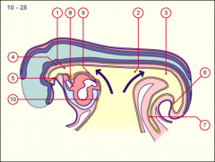
The primitive gut tube extends from the
|
oropharyngeal membrane to the cloacal membrane
and is divided into the foregut, midgut, and hindgut. |
|
|
***
Know it all fore mid hind |
Early in development, the epithelial lining of the gut tube proliferates rapidly and obliterates the lumen. Later, recanalization occurs.
|
|
|
|
Early in development, the epithelial lining of the gut tube proliferates rapidly and obliterates the lumen. Later
|
recanalization occurs.
|
|
|
|
Mucosa Epithelial Lining of glands Derived from =
|

Endoderm
|
|
|
|
Mesoderm Derives what =
5ct |
..
|
|
|
|
Oropharyngeal membrane / stomodeum
6.Cloacal membrane / proctodeum Come from |
Ectoderm (an Exception)
|
|
|
|
FOREGUT gives rise to:****
|
pharynx
lower respiratory system esophagus and stomach duodenum (proximal liver * pancreas * biliary apparatus * |
|
|
|
Esophagus develops from the ________ immediately caudal to the pharynx .
|
foregut
+ |
The foregut is divided into the esophagus dorsally and the trachea ventrally by the tracheoesophageal folds, which fuse to form the tracheoesophageal septum.
|
|
|
diverticulum becomes separated from the primitive pharynx by trachoesophageal folds which fuse to form the
|
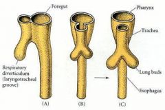
trachoesophageal septum,
dividing the foregut into laryngotracheal tube esophagus |
|
|
|
stratified squamous epithelium
mucosal glands submucosal glands of esophagus are derived from |
endoderm
|
|
|
|
lamina propria
muscularis mucosae submucosa skeletal muscle smooth muscle of muscularis externa adventitia of the esophagus derived from visceral |
mesoderm
|
|
|
|
Esophageal atresia :
occurs when the |
tracheoesophageal septum deviates too far dorsally
causing the esophagus to end as a closed tube. + |
The esophagus is divided into two pouch blind, an upper and lower, which may or may not communicate with the tracheobronchial tree.
|
|
|
About 33% of patients with esophageal atresia have other congenital defects associated with the
|
VATER
VACTERL |
VATER (vertebral defects, anal atresia, tracheoesophageal fistula, and renal defects) or
VACTERL (similar to VATER but includes cardiovascular defects and upper limb defects) syndromes. |
|
|
Esophageal atresia is associated clinically with =
|
tracheoesophageal fistula.
|
|
|
|
esophageal atresia S/S:
|
unable to swallow its own saliva
gastric distention cough, apnea/SOB tachypnea cyanosis |
|
|
|
#1 Incomplete Recanalization
#2 Myo Extra Hypertrophy #3 Membronous Diapragm |
Reasons for Esophogeal Atresia
|
|
|
|
esophageal atresia
Diagnosis |
This condition is visible, after about
___ weeks T/F a feeding tube will not pass through the esophagus. |
26 weeks
True |
|
|
esophageal atresia
Complications |
-aspiration pneumonia
-fistula between the lower esophagus and trachea |
|
|
|
If associated with TEF---Abdominal distention after crying
.Reflux of gastric contents into lungs, causing pneumonitis. Diagnostic feature is = |
Diagnostic features include inability to pass a catheter into the infant's stomach
|
|
|
|
Esophageal stenosis :
occurs when the = |
lumen of the esophagus is narrowed and usually involves the midesophagus.
|
|
|
|
Esophageal stenosis
stenosis may be caused by |
-submucosal/muscularis externa hypertrophy,
-emnants of the tracheal cartilaginous ring within the wall of the esophagu -#1 Cause incomplete recanalization... (membranous diaphragm obstructing the lumen ) |
|
|
|
Esophageal diverticulum:
|
a pouch that protrudes outward in a weak portion of the esophageal lining.
|
|
|
|
Esophageal diverticulum:
Esophageal diverticula are classified by their location within the esophagus: |
Zenker’s
Midthoracic Epiphrenic |
|
|
|
Esophageal diverticulum:
Zenker’s diverticula |
most common
located in the back of the throat above the esophagus |
|
|
|
Esophageal diverticulum:
Midthoracic diverticula |
mid-chest
|
|
|
|
Esophageal diverticulum:
Epiphrenic diverticula |
above the diaphragm
|
|
|
|
. Esophageal hiatal hernia :
|
herniation
into the pleural cavity caused by an abnormally large esophageal hiatus. |
|
|
|
An esophageal hiatal hernia renders the esophagogastric sphincter =
|
incompetent
|
|
|
|
►Omentum : fold of peritoneum extending from the
|
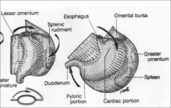
stomach to adjacent abdominal organs.
|
|
|
|
EnteroEndocrine Hormones
are from Endoderm the exception is = 2ct |
Enterochromaffin Cells
Enterochromaffin Like Cells (ELC) Which come from ___________ Cells Derived from ___ = |
Ectoderm (NCC)
|
|
|
The primitive gut tube extends from the
|
oropharyngeal mem =
3ctbrane to the cloacal membrane |
foregut
, midgut, hindgut. |
|
|
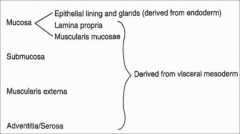
Embryologically, the epithelial lining and glands of the mucosa are derived from
|
endoderm
whereas the other components are derived from - |
visceral mesoderm.
|
|
|
____________ an invagination of the surface ectoderm of the embryo, at the point where later the mouth is formed.
|
Stomodeum
|
|
|
|
: is the back ectodermal part of an alimentary canal.
It is created during embryogenesis by a folding of the outer body wall |
Proctodeum
|
|
|
|
the trachoesophageal septum, dividing the foregut into =
|
laryngotracheal tube
esophagus |
|
|
|
The stratified squamous epithelium, mucosal glands, and submucosal glands of the esophagus are derived from
|
endoderm
|
|
|
|
The lamina propria, muscularis mucosae, submucosa, skeletal muscle and smooth muscle of muscularis externa, and adventitia of the esophagus are derived from visceral
|
mesoderm
|
|
|
|
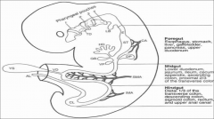
Name the structures comes from the Primordial
Forgut Midgut Hind gut |
Forgut
esophagus stomach liver gallbladder pancreas upper duodenum Midgut lower duodenum jejunum Ilium Secom appendix ascending: proximal 2\3 transverse colon Hind gut Distal1/3 transverse colon Descending Sigmoid Rectum upper anal canal |
|
|
|
Name the structures comes from the Primordial Hindgut
5 |
Distal1/3 transverse colon
Descending Sigmoid Rectum upper anal canal |
|
|
|
Name the structures comes from the Primordial Midgut
7 |
lower duodenum
jejunum Ilium Cecum appendix ascending: proximal 2\3 transverse colon |
|
|
|
Name the structures comes from the Primordial Forgut
6 |
esophagus
stomach liver gallbladder pancreas upper duodenum |
|