![]()
![]()
![]()
Use LEFT and RIGHT arrow keys to navigate between flashcards;
Use UP and DOWN arrow keys to flip the card;
H to show hint;
A reads text to speech;
123 Cards in this Set
- Front
- Back
|
Gram -ve vs gram +ve transport (1) |
Gram -ve have to transport across outer memb, periplasm, and cytoplasmic memb. (2 membranes)
Gram +ve only transport across single membrane |
|
|
OM permeability + source |
Lipopolysaccharides in OM form permeability barrer for water soluble and hydrophobic molecules - Transporters required |
|
|
Classes of OM transporters (3) |
Import: 1. Porins (channels) 2. TonB-dependent transporters Export: 3. Export channels |
|
|
Porins (overall) - F/restrictions/type of transport General porins - Basic S (2 *) - F: 1 (+ 3 things) - Crystal structure (2) - Example (3 + detail/significance) Specific porins example - F (1) + substrates (2) - S (2) |
Porins: Channels
- Substrates under ~600 Da pass through via facilitated diffusion (no energy) General porins S: 1. Integral outer membrane proteins 2. beta-barrel fold: first strand interacts with last strand - beta-barrel strands are amphipathic: inside of barrel = hydrophilic, outside = hydrophobic F: Allows diffusion of sugars, ions, AAs down a concentration gradient Crystal S: - 16 stranded b-barrels with 8 extracellular loops - Form trimers: 3 barrels -> 3 pores Ex: E. coli general porins: OmpF, OmpC, PhoE - 60% identical AA sequence - Different substrate preferences Specific porins - LamB: uses facilitated diffusion to transport narrow range of substrates: maltose, malto-oligosaccharides S: 1. Also trimer of beta-barrels 2. Aromatic AAs lining channel provide specificity for sugars by aiding facilitated diffusion through hydrophobic interactions |
|
|
TonB-dependent transporters - Why these are needed? (2) - Proteins involved (3) - L (1) - F/mechanism (1*) |
1. Bacteria need to acquire scarce nutrients (iron, B12), but facilitated diffusion is insufficient 2. Transport of these against a concentration gradient requires energy input, but OM has no energy source TonB, ExbB, ExbD = energy transducing proteins (TonB reaches from cytoplasmic membrane to OMP) - L: cytoplasmic membrane - TonB couples PMF to uptake of solutes: Entry of protons through channel ExbBD drives conformational changes that are transmitted via TonBto the OMP, which allows it to take up the molecules |
|
|
Exporter example in E. coli - Names/involved proteins/L (3) - F - important characteristic |
TolC-AcrA-AcrB in E. coli L: TolC = OM, AcrA = periplasm, AcrB = inner membrane F: efflux of organic solvents, detergents, antibiotics Energy-dependent - Needs to use energy (PMF or ATP) to transport substrates out of cell |
|
|
Solute uptake: diffusion vs active transport - What affects rate of uptake - Curve appearance - Maximums? - Km |
Diffusion - Through non-specific porins - Initial rate of uptake (Vi) of solute (S) proportional to external solute concentration (linear) - Does not approach maximum even at very high solute concentrations Transporter-driven (active transport) - Curve is hyperbolic, approaches Vmax - Transport of solute limited by number of transporters - At Vmax = all transporters occupied Km = [S] at 1/2 Vmax - Represents affinity for solute (low Km = higher affinity: low Km = less [S] needed to reach Vmax = high affinity) |
|
|
Primary vs secondary transport |
Primary transport: - driven by energy producing metabolic event (ATP hydrolysis, substrate-level phosphorylation) Secondary transport: - Driven by electrochemical gradient |
|
|
Types of transporters (3) |
1. Simple transport 2. Group translocation 3. ABC transporter |
|
|
Simple transporters - Types/def (3) - example/type it is/characteristic |
1. Uniporter: transport in one direction (down concentration gradient) across membrane
2. Symporter: Co-transport substrate with (ion gradient/PMF transport) in same direction 3. Antiporter: transport mol. across membrane while simultaneously transporting another molecules in the opposite direction Lac permease (E. coli) - Symporter (with proton) - Energy-dependent (PMF) |
|
|
Group Translocation - def - Types of molecules transported (3) Example from class - Type of transport? (primary/secondary) - Basic mechanism |
- Substance is chemically modified during transport across the membrane - Transports glucose, fructose, mannose Phosphotransferase system (PTS) in E. coli - Primary transport: energy is hydrolysis of high-energy P bond in phosphoenol pyruvate Mech: phosphate relay - PEP transfers P to nonspecific enzymes -> sugar-specific enzymes -> specific membrane transporter -> glucose (to make glucose 6-P) |
|
|
ABC transporters - type of transport (1 or 2nd) - what it transports (3+) - Specificity - Gram -ve vs gram +ve |
ABC = ATP Binding Cassete - Primary transport (ATP) - Transports organic compounds, inorganic nutrients, and trace metals - High substrate specificity Gram -ve: employ periplasmic-binding proteins and ATP-driven transport proteins Gram +ve: employ substrate-binding proteins and membrane transport proteins (no periplasm) |
|
|
ABC transporter structure - All ABCs have a minimum of? - Structures (2) + S/F - ESR? |
All ABCs have a minimum of 2 subunits (total 4 domains): 1. ATP binding subunit - Dimer of nucleotide binding domain proteins - Cytoplasmic - Each monomer binds a single ATP, provides energy for transport of substrates 2. Permease - dimer of integral membrane proteins Some ABC transporters (importers) have additional subunit: 3. ESR: extracytoplasmic solute receptor - Determines specificity of transporter - Soluble protein in periplasm, two domains joined by hinge region - Venus flytrap mechanism of binding to substrate, then brings to specific permease |
|
|
[T/F] ABC proteins are only found in gram -ve bacteria because there is no periplasm in gram +ve bacteria for the ESR to function
|
F, gram +ve bacteria have their ESR tethered to outside surface of membrane
|
|
|
Chemotaxis - def - what kind of bacteria can perform chemotaxis? |
Def: process by which bacteria move in a directed way toward attractands or away from repellents - Bacteria with swimming motility mediated by flagella can perform chemotaxis (other types of motility not discussed) |
|
|
Motility in bacteria with peritrichous flagella - characteristic of flagella - Motility (2 parts) |
Flagella can rotate in either direction CCW rotation of all flagella: flagella bundle together, produces smooth swimming Reversing flagella = CW rotation: tumbling |
|
|
Motor proteins of flagella - L (1+) - F (1) |
In cytoplasmic membrane, in contact with MS and C rings of basal body F: Responsible for switching direction of flagellar rotation |
|
|
Basic signal transduction components + F(3) |
Two component system 1. Histidine Kinase (HK) - senses signal (binding) - autophosphorylates on histidine residue - in response to signal, autophosphorylation ability modified - Transfers P to response regulator 2. Response regulator (RR) - receives P from HK, modifies the RR's activity 3. Phosphatase - Cleaves P from RR - Resets system to pre-stimulus state |
|
|
Chemotaxis signal transduction components +F (6) |
1. Methyl-accepting chemotaxis protein (MCP) - Receptors (integral memb. proteins) - Directly binds attractants/repellents if present 2. CheA - Histidine kindase (HK) - uses ATP to autophosphorylate his residue - Phosphorylates either CheY or CheB 3. CheY - Response Regulator, receives P from CheA - CheY-P binds flagellar motor and promotes CW rotation (tumbling) 4. CheZ = phosphatase - removes P from CheY, resets flagellar motor to CCW rotation 5. CheB - Response regulator, receives P from CheA - Methyl-esterase, removes methyl groups from MCP (receptors), making them more senssitive to chemoeffectors 6. CheR - Methyl-transferase, methylates chemoreceptors, making them less sensitive to chemoeffectors |
|
|
Chemotaxis signal transduction mechanism A. No chemoeffectors B. Attractant present (4 steps+) |
A. No chemoeffectors - Equilibrium between: a. CheY > CheY-P = swimming b. CheY < CheY-P = tumbling B. Attractants 1. Attractants bind to MCP and inhibits autophosphorylation of CheA 2. Shifts equilibrium to CheY (less CheA-P = less CheY-P) - CheY > CheY-P = swimming 3. less CheA-P = less CheB-P - low methyl-esterase activity, but CheR still active [CheR activity > CheB-P activity] -> methyl-transferase = More methyl groups = less sensitive - Receptors must sense higher [attractant] to continue to be stimulated/active 4. Adaptation (reset to pre-stimulus state) - MCPs no longer respond to same [attractant] - CheA autophosphorylation resumes -> CheY-P and CheB-P increases -> system resets to CheY/CheY-P equilibrium (swim/tumble) |
|
|
Swim or Tumble? 1. MCP mutant: constitutively on 2. CheA mutant: does not bind ATP 3. CheY mutant: cannot be phosphorylated 4. Mutant: lacks CheZ |
1.Swim 2. Swim 3. Swim 4. Tumble |
|
|
What parts of a pathway are regulatory enzymes usually found? (2 p) 2 Main ways of controlling activity of regulatory enzymes |
Found:
1. Branch points 2. Catalyze physiologically irreversible reactions Control: 1. Binding of small molecule effectors (allosteric effectors) 2. Covalent modification (methylation) |
|
|
Feedback inhibition - def/mech - what kind of pathways is it often found in - physiological function/reason? |
End-product of pathway inhibits regulatory enzyme of pathway, slows down production of end-product Biosynthetic pathways Reason: - So cell can maintain steady state where end product is utilized as it is synthesized |
|
|
Allosteric enzymes - basic mechanism - effect on Km |
Effector binds to allosteric site -> conformational change -> alters Km of enzyme - +ve effector: decrease Km (tighter binding/higher affinity) - -ve effector: increase Km |
|
|
Glutamine synthase activity regulation |
Regulated by covalent modification - Addition of AMP groups onto subunits - No AMP groups = fully active - 12 AMP groups added = inactive |
|
|
Bacterial mechanism for glucose intake/preparation
|
Glucose -> Glucose-6-P - using PEP PTS (phosphoenolpyruvate phosphotransferase system) - PEP -> pyruvate |
|
|
Glycolysis AKA |
Embden-Meyerhof-Parnas pathway |
|
|
Central metabolic pathways provide ___________ _________ for all other pathways. |
precursor metabolites |
|
|
Central metabolic pathways - categories (2) + what's in them (2, |
A. Carbohydrate metabolism 1 - EMP pathway/glycolysis 2 - Pentose-Phosphate pathway B. Carboxylic acid metabolism |
|
|
Stage 1 (3) and 2 (3) of glycolysis - include #s - similarities w/ PPP |
Stage 1 1. Glycolysis uses hexokinase + ATP (or PEP PTS) as first step 2. 6-Carbon glucose split into 2 (3C) PGALD 3. Total of 4 reactions, consumes 2 ATP/glucose Stage 2 (common with PPP) - Both convert glucose to phosphoglyceraldehyde - PGALD oxidized to pyruvate by 5 common reactions - Generates 4 ATP/glucose: Net +2ATP |
|
|
Which reactions in glycolysis generate ATP via substrate-level phosphorylation? |
The reactions from PGALD to pyruvate |
|
|
Glycolysis products per 1 mol glucose |
- 2 mol pyruvate - Net 2 mol ATP - 2 NADH + H+ (reduced cofactor) |
|
|
PPP - Overall steps (4) - similarities/interconnections with glycolysis (marked with *) - Products of each steps - Overall reaction (incl #s) |
Pentose Phosphate Pathway 1. ***starts with glucose 6-P from hexokinase or PTS 2. Glucose-6-P -> Ribulose 5-phosphate (5C pentose sugar) + 2 NADPH + CO2 - NADPH produced here 3. -> isomerization to 5-C sugars (used for nucleic acid synthesis) 4. -> transketolase/transaldolase Rxns -> 3-C PGALD *** - C6 pdt = Fructose-6-P*** (gluconeogenesis) Overall Rxn G6P + 6 NADP+ -> 3 CO2 + 1 PGALD + 6 NADPH + 6 H+ |
|
|
Precursors from EMP, PPP |
1. G6P -> polysaccharides, PPP, aromatic AAs 2. F6P -> amino sugars (cell wall), 3. PGALD <-> DHAP -> phospholipids (cell membranes) 4. 3-PGA -> serine, glycine, cysteine 5. PEP -> pyruvate, aromatic AAs, muramic acid (cell wall) |
|
|
AA biosynthesis - origin of precursors - precursors (5) + AAs formed from them (2,2,3,3,5) |
Glycolysis produces: 1. Pyruvate - Alanine family (valine, leucine) 2. 3-PGA - Serine family (glycine, cysteine) 3. PEP + Erythrose-4-P from PPP = Chorismate - Aromatic family (Phenylalanine, tyrosine, tryptophan) TCA cycle produces: 4. a-ketoglutarate - Glutamate family (proline, gln, arg) 5. Oxaloacetate - Aspartate family (Asn, Lys, Met, Thr, Ile) |
|
|
Ribose-5-P -> - 2 ways, difference? |
Ribose 5-P -> Ribonucleotides
1. -> RNA 2. --NADPH--> DNA - NADPH-dependent ribonucleotide reductase forms deoxyribonucleotides |
|
|
Importance of PPP for biosynthesis (3 products)
- + importance of PPP relationship to glycolysis (2) |
1. Produces pentose-P precursors to ribose/deoxyribose in nucleic acids 2. Provides erythrose-P = precursor to aromatic AAs 3. NADPH = major source of e- in reduction pathways Importance of interconnections: - PPP and glycolysis interconnect at F6P and PGALD 1. PPP allows organisms growing on pentoses to make hexose phosphates (F6P) 2. Transaldolase/transketolase rxns reversible, pentose phosphates/F6P can be synthesized from PGALD |
|
|
Gluconeogenesis importance |
can form 6-C compounds from 2-5-C compounds by running glycolysis in reverse |
|
|
Fates of pyruvate (2) - incl any important reactions/enzymes etc |
Determined by if cell is doing respiration or fermentation A. Respiration - pyruvate oxidized to acetyl-CoA by pyruvate dehydrogenase - CO2 + NADH produced (decarboxylation) - acetyl-CoA enters TCA cycle |
|
|
TCA cycle - Overall reaction - 4 very basic parts of TCA cycle |
Acetyl-CoA + 2H2O + ADP + Pi + FAD + NADP+ + 2 NAD+ ---> 2 CO2 + ATP + FADH2 + NADPH + 2 NADH + 3H+ + CoA-SH 1. [2C] Acetyl-CoA + [4C] Oxaloacetate -> [6C] citrate ->...... 2. ----> NADPH + CO2 + [5C] a-Ketoglutarate 3. -> NADH + CO2 + [4C] Succinyl-CoA ->...... 4. -----> Oxaloacetate |
|
|
___________ _________ replenish TCA cycle intermediates such as _________ that have been extracted for biosynthesis. - How is this accomplished? |
Anapleurotic reactions oxaloacetate CO2 is added to either PEP or Pyruvate (3-C molecules) to form [4-C] oxaloacetate |
|
|
E. coli is a _________________, and a _________ __________. (metabolic terms) |
chemoorganotroph facultative anaerobe |
|
|
Metabolic classes - 3 divisions - 8 total classes |
Determined by where they get their electrons (electron source) 1. Chemoorganotrophs - organic chemicals (glucose) 2. Chemolithotrophs - inorganic chemicals (H2, Fe2+, etc) 3. Phototrophs - light Determined by where they get their carbon (carbon source) 4. Heterotrophs - organic compounds - All chemoorganotrophs = heterotrophs 5. Autotrophs - CO2 - AKA primary producers because they synthesize new organic matter from CO2 Does cell need oxygen to obtain energy? 6. Aerobes: yes 7. Anaerobes: No, and they cannot obtain energy in presence of O2 8. Facultative aerobe: can use O2 if it's there, but can still grow without it - most chemolithotrophs/phototrophs |
|
|
Autotrophy - def - Pathway, purpose/F, requirements (6+ names) |
- Use of CO2 as sole carbon source Calvin Cycle - F: assimilate CO2 into organic carbon for cellular material Req: *need 6x everything per hexose to regenerate RuBP* 1. CO2 2. CO2 acceptor molecule: ribulose bisphosphate 3. NADPH 4. ATP 5. Ribulose bisphosphate carboxylase (RubisCO) 6. Phosphoribulokinase |
|
|
Calvin Cycle - Steps (6) include # of C's, inputs, byproducts, etc |
6x cycle required for one hexose 1. RubisCO catalyzes formation of 6 6-C molecules that rapidly split into 12 3-PGA [36C] from 6 ribulose bisphosphate [30C] and 6 CO2 [6C] 2. 12 ATP energy input -> 12 BPGA [36C] 3. 12 NADPH input -> 12 G3P [36C] 4. 2 G3P [6C] removed for biosynthesis via gluconeogenesis as F6P -> 10 G3P [30C] - F6P -> DNA/RNA (PPP), cell wall sugars, aromatic AAs 5. Sugar rearrangements (like PPP) -> 6 Ribulose 5-P [30C] 6. 6 ATP input -> phosphoribulokinase catalyzes reformation of ribulose bisphosphate (RuBP) |
|
|
Overall Rxn of Calvin Cycle |
6 CO2 + 12 NADPH + 18 ATP -> C6H12O6(PO3H2) + 12 NADP+ + 18 ADP + 17 Pi |
|
|
•What pathway provides the ribulosephosphate required for the Calvin Cycle in autotrophs? •What intermediate in the Calvin Cycle isdrawn off for hexose biosynthesis? •Why do autotrophs need to synthesizehexoses? |
|
|
|
What bacteria do not have the Calvin Cycle? - What do they do instead? |
Green bacteria Use reverse TCA cycle: - Use Oxaloacetate as CO2 acceptor -> reverse rxn to G3P |
|
|
Reduction potential of NAD+ and FAD+ |
NAD+: -0.32 V FAD+: -0.22 V |
|
|
Energy is released during redox rxns, how is it used/stored? - (2 + example(s)) |
A. Energy-rich storage compounds - e.g. ATP, PEP, CoA B. Electrochemical gradient (PMF) - used directly or used to make ATP |
|
|
Ways to make ATP (2)
- Processes (3) that use those ways |
A. Substrate level phosphorylation - Central metabolism - Fermentation B. Oxidative phosphorylation - Respiration |
|
|
Group transfer potential - def - ranges |
Energy released when chemical group (e.g. P) transferred - Can be used for substrate level phosphorylation to make ATP High energy => 29 kJ/mol |
|
|
Mitchell's Chemiosmotic Theory - 3 principles |
1. Cytoplasmic membrane impermeable to -OH and H+ 2. ETC localized in membrane so pH gradient and membrane potential are formed by excretion of protons during electron transport 3. ATP synthase takes advantage of pH gradient and membrane potential for ATP synthesis from ADP and Pi |
|
|
Electron carriers - 5 types + what they carry - |
Some carry H as well as e-
1. Flavoproteins (H and e-) 2. Quinones (H and e-) - Quinones are lipids, mobile in membrane, act as shuttle 3. Iron-Sulfur proteins (e- only) 4. Cytochromes (e- only) 5. Some e- carried in prosthetic group bound to protein in multiprotein complexes: oxidoreductases |
|
|
General bacterial e- transport pathway - How order is determined - steps/pathway/structures in pathway (3) |
e- carriers arranged in membrane in order of increasing reduction potential 1. e- donor passes its e- to a dehydrogenase complex (flavoprotein + Fe-S protein) 2. Dehydrogenase complex passes e- to quinone 3. Quinone passes e- to cytochrome -> terminal e- acceptor - If it is the last cytochrome in the chain = terminal oxidase complex (if O2 is terminal e- acceptor) - if O2 not terminal e- acceptor, = reductases |
|
|
Complex I-IV
- Where are coupling sites? (3) (not sure if need to know) |
1. Complex I (NADH:quinone oxidoreductase ) - NADH donates e- to FAD (in Complex I) - FADH donates e- to quinone 2. Complex II (Succinate dehydrogenase complex) - bypasses Complex I - Feeds e- and H+ from FADH to quinone pool 3. Complex III (cytochrome bc1 complex) - transfers e- from quinones to cytochrome c - cytochrome c shuttles e- to cytochromes a and a3 4. Complex IV (Cytochromes a and a3) - terminal oxidase; reduces O2 to H2O Coupling sites: places in ETC where redox rxns are coupled to H+ translocation 1. NADH:quinone oxidoreductase 2. Quinones (Q cycle) 3. Terminal oxidase |
|
|
ATP synthase - Structures (2) - directionality? |
Domains F0, F1 1. F0: proton channel that spans cytoplasmic membrane 2. F1: catalytic subunit for ATP synthesis/hydrolysis (on inner surface of membrane) Can run in either direction: - Use PMF to make ATP or - Use ATP to make PMF |
|
|
How many protons must flow through ATP synthase to form 1 molecule of ATP? |
3 |
|
|
Why does anaerobic respiration yields less energythan aerobic respiration? |
O2 has highest reduction potential, most energy released if O2 is terminal e- acceptor |
|
|
Anaerobic respiration is simply: |
•Use of electron acceptors other than O2 |
|
|
Aerobic vs Anaerobic respiration - only/main difference? - PMF result |
- Only difference is terminal electron acceptor, in both, organic substrates are oxidized by standard pathways to produce NADH and FADH2 for the ETC - Less PMF generated from anaerobic respiration (e.g. nitrate respiration) |
|
|
Assimilative metabolism vs Dissimilative metabolism - def - amount of substrates used - example: Nitrate |
Assimilative metabolism: use of inorganic compounds (NO3-, SO42-, CO2) and reduced compounds in biosynthesis - Only use enough to satisfy biosynthetic needs Dissimilative (bacteria only): Reduction of inorganic compounds for purposes of energy generation - Large amount of e- acceptor reduced, waste product is excreted Nitrate: Assimilation end result: organic N (R-NH2) Dissimilation end result: N gases (N2, NO, N2O) |
|
|
Most common anaerobic respiration = ? - products? - Different processes? (2 p) |
Most common: reduction of inorganic nitrogen compounds
1. Nitrate reduction (E. coli) - NO3- to NO2- 2. Denitrification - Reduction of nitrate to gaseous waste products (NO, N2O, N2) - Major biological source of N2 |
|
|
Denitrification in agriculture vs aquatic systems |
Agriculture: detrimental, removes fixed nitrogen from soil so it is unavailable to plants
Aquatic systems: good, removal of fixed nitrogen from waste water, prevents algal blooms in lakes |
|
|
Iron Respiration - Definition*** important distinction First example of iron respiration - origin/most basic example/overall reaction |
Use of Fe3+**(not 2+) as terminal e- acceptor in ETC - Fe3+ reduced to Fe2+ (iron reduction) Life emerged on Fe2+ rich earth - When light reacts with 2 Fe2+ and 2 H+ -> 2 Fe3+[e- acceptor] and H2 gas [e- donor] 2Fe(II) + 2H+ --hv(light)--> 2Fe(III) + H2 |
|
|
Iron reduction - Problem? - Mechanisms to counteract problem (2) - movement of electrons in iron reduction (4+2 steps) |
Problem: Fe3+ is insoluble at neutral pH - How to get e- to Fe3+ (TEA) ppt on outside of cell? Strategies: 1. Direct contact via "nanowires" - electrically conductive pili, mediate e- transfer 2. Soluble e- shuttle (cofactors) -a. shuttle reduced in cell (metabolic rxns) -b. reduced e- shuttle donates e- to Fe3+ -> oxidized shuttle + Fe2+ e- movement: 1. e- -> shuttle/quinone 2. -> cytochromes 3. -> periplasmic e- carriers 4. -> OMPs - a. OMP, directly reduces Fe3+ [shewanella] - b. OMPs directly reduce Fe3+, or pass e- to other carriers or pili (nanowires) [geobacter] |
|
|
Rank the terminal e- acceptors in most energy released when NADH is the e- donor - Fe3+ - NO3- - (SO4)2- - O2 Importance/significance (to fitness)? (3 parts + main concept) |
O2> NO3- > Fe3+ > (SO4)2- More energy released = Higher ATP yield - It takes the same amount of ATP to make a cell from the same substrate - Thus, higher ATP yield per mol substrate = higher cell yield, higher cell yield outcompetes cells with lower cell yield ATP yield per mol substrate (and ATP synthesis rate) is a critical selective force in microbial competition and evolution |
|
|
Chemolithotroph - def - common in nature? why/why not? Most chemolithotrophs are ____________ - Some are __________. <- Definition? |
Def: Obtains energy from oxidation of inorganic compounds (inorganic molecules used as e- donors) - Common type of metabolism because many types of reduced inorganic molecules in environment (e.g. pdts of anaerobic respiration) Autotrophs (carbon from CO2) - Mixotrophs: use inorganic molecule as e- donor, need organic carbon source |
|
|
Chemolithotrophs vs chemolithoautotrophs vs chemolithoautotrophs/mixotrophs |
Chemolithotroph: use inorganic mol. as e- donor (obtain energy from its oxidation) Chemolithoautotroph: use inorganic mol. as e- donor and C source = CO2 Chemolithoheterotroph: use inorganic mol. as e- donor and C source = organic carbon |
|
|
Aerobic hydrogen oxidizers - source of substrate - pathway (3 overall steps) enzymes/compounds/Fs - autotrophy problem (2p) + solution (+concept) |
Source: H2 is common product of fermentation - H2 used as e- donor H2 oxidized by membrane-integrated hydrogenase -> passes its e- to ETC (-> quinone -> cytochromes -> O2 [TEA]) -> creates PMF for ATP synthase Chemolithoautotrophs - Need NADH/reducing power for Calvin cycle - Not metabolizing glucose, no NADH produced from glycolysis/TCA cycle Solution: cytoplasmic hydrogenase - H2 can directly reduce NAD+ -> NADH because the reduction potential for H2 is very low |
|
|
Difference between autotrophic hydrogen oxidizers and other chemolithoautotrophs? + reason why - How other chemolithoautotrophs deal with it? (Requirement + 2 p of solution) - example(s) |
H2 (E' less than NAD+'s) can directly donate its e-'s to NAD+, but most other reduced inorganic compounds are poor e- donors (E' > E' of NAD+), can't directly donate e- to make reduced cofactors
Solution: - Energy input in the form of PMF required to generate NADH - Forward e- transport generates PMF - Reverse e- transport consumes PMF to produce NADH Example: sulfur oxidizers, iron oxidizers (all other chemolithoautotrophs) |
|
|
Fe2+ AKA ______ Fe3+ AKA ______ |
Fe2+: ferrous Fe3+: ferric |
|
|
Iron oxidation - Sign/niche - ^importance/reason for niche (3p) - amount of energy produced (1p) + importance/effects (1p) - e- flow (autotrophs) |
Ferrous (Fe2+) iron is e- donor, oxidized to Fe3+ - Fe3+ forms ferric hydroxide ppt in water - Many iron oxidizers grow at low pH (<1) Reason: Ferrous (Fe2+) iron is only stable at acid pH: - Rapidly oxidized and forms ppt at neutral pH, bacteria unable to use - Stable/available for use at acid pH Small difference between reduction potentials of Fe2+ and O2 at acidic pH - Large amts of Fe2+ oxidation still yield little energy/few cells Autotrophs also requires reverse e- flow to generate NADH for Calvin cycle |
|
|
How is growth of iron oxidizers monitored? |
Monitor production of Fe3+ ppt |
|
|
Respiration - (what is it) (2p) - relation to PMF - PMF def/F - aerobic vs anaerobic resp |
Redox rxns in ETC in cytoplasmic membrane release energy as e- pass from donors with lower E' to acceptor with higher E' - Energy release coupled to proton translocation to outside of membrane -> PMF PMF = electro (+outside, -inside) chemical ([H+out]>[H+in]) gradient - PMF used to generate ATP through ATP synthase (3 H+/ATP) Aerobic: terminal e- acceptor is O2 Anaerobic: TEA is something else e.g. NO3-, SO4-, Fe3+ |
|
|
Fermentation - def - differences between respiring organisms (energy generation, energy yield, |
Use of organic substrates as both e- donor and e- acceptor Differences 1. Respiring organisms generate PMF to drive ATP synthesis - Fermenting organisms use substrate level phosphorylation as major route for ATP synthesis 2. Fermenting organisms have lower ATP yield, and thus often have very high metabolic rates to allow competition |
|
|
Fermentation PW - Why fermentation needed (2p) + solution (1) Lactic acid bacteria PWs (2) - explain PW discussed in class (2p) |
- 2 NADH produced at end of stage 1 of glycolysis - For glycolysis to continue, NAD+ needs to be regenerated - Accomplished by using NADH to reduce pyruvate to fermentation end products (redox balance), which are excreted Lactic acid fermentation - Homofermentative or heterofermentative Homofermentative PW - Stages 1/2 of glycolytic pw the same, pdts = 2 pyruvate, 2 ATP, 2 NADH - 2 pyruvate reduced to 2 lactate + 2 NAD+ |
|
|
Possible fermentation end products (6) |
1. Acetate + H2 2. Lactate 3. Ethanol + CO2 4. butyric acid 5. butanol 6. isopropanol |
|
|
Classifications of Phototrophic Organisms (1->2p) Distinction between photosynthesis and phototrophy |
Phototrophs: use light as energy source - a. Photoautotrophs: use CO2 as carbon source - b. Photoheterotrophs: use organic C as C source (don't do photosynthesis) Phototrophy (ATP synthesis) != photosynthesis (CO2 fixation) - Photosynthesis: use of light to fix CO2 into biomass |
|
|
2 Classes of photosynthesis - differences/similarities (3p) - Organisms in each class (4,1) |
A. Anoxygenic photosynthesis - Use inorganic e- donors (S, Fe2+, H2, NO2-) for CO2 fixation - ATP from light (phototrophy) - O2 not produced 1. Purple sulfur 2. Purple non-sulfur 3. Green sulfur 4. Green non-sulfur B. Oxygenic - Use H2O as e- donor for CO2 fixation - ATP from light (phototrophy) - O2 produced 1. Cyanobacteria ("blue-green algae") |
|
|
Light-sensitive pigments - Function* - Pigments (2) and what organisms have them (2,3) - Structure (2p) |
F: Required for capture of light energy and conversion to ATP Pigments: 1. Bacteriochlorophyll - Purple and green bacteria (anoxygenic) 2. Chlorophyll - Cyanobacteria, algae, green plants (oxygenic) S: - similar to pyrolle/heme - Mg in center (instead of Fe in heme) = e- carrier |
|
|
Sulfur globules - def - found in: (2) + specific locations? |
Elemental sulfur storage (e- donor) - Intracellular in purple bacteria - extracellular for green bacteria |
|
|
Purple phototrophic bacteria - Characteristics of ALL purple phototrophic bac (4) |
All 1. Carry out anoxygenic PS 2. Contain bacteriochlorophylls and carotenoid pigments 3. Intracytoplasmic photosynthetic membranes (membranes req. for PMF/ETC of PS) 4. Purple non-sulfur bac. can use H2 as e- donor (sulfur bac uses S compounds) |
|
|
Purple Sulfur Bacteria - e- donors + their use - Locations (2) |
H2S used as e- donor -> So (elemental sulfur) - So stored as globules So -> oxidized to sulfate (SO42-) L: found in illuminated anoxic zones of lakes/aquatic habitats where H2S accumulates |
|
|
ATP synthesis in all purple bac - name of mechanism: + overall result - Structures involved (4) - Mechanism (4 steps) + molecules involved/Fs+ important concepts |
Photo-phosphorylation - cyclic electron flow (no net mvmt of e-) creates PMF Structures 1. Bchl = bacteriochlorophyll - Pair of Bchl in membrane forms reaction center P870 2. Quinones 3. Cytochromes 4. ATP synthase Steps: 1. Rxn center P870 abs photon and becomes excited P870* (E' becomes -1.0 V[strong e- donor] from +0.5 in P870) 2. e- from P870* -> Quinone pool 3. -> Cytochromes/ETC (e- transport generates PMF) 4. -> mobile cytochrome carries e- back to Rxn center, re-reduced to P870 |
|
|
Steps involved in PS but not req. for phototrophy (Purple bac vs green bac) - Reasons/solutions |
Purple bac: NADPH synthesis requires input of energy (in form of reverse e- flow) - Quinones in quinone pool have high E', can't donate e- to NADP+ for reducing power - Solution: use reverse e- flow (consume PMF) (external e- donors) to generate NADPH Green Bac do not require additional energy input for CO2 fixation - Fd from ETC has low E' ( - Fd reduces NAD+ to NADH directly (no energy req) |
|
|
Green sulfur bac - what kind of metabolism? - e- donor =? - Difference in autotrophy? - Special structure = ? - L? |
Anoxygenic photorophs - Use H2S as e- donor, oxidize to SO42- - Autotrophy uses reverse TCA cycle (not Calvin Cycle) - Have chlorosomes: oblong Bchl rich bodies bound by thin membrane L: anoxic environments rich in H2S |
|
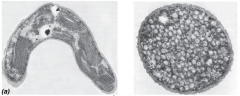
What kind of bacteria are these found in? F? Name left/right |
Purple bacteria (increase light absorbing area) Left: Lamellar membranes RT: Intracellular vesicle membranes |
|
|
Light-harvesting complex of green bacteria: - name - L: - Structures/Fs (2) - Why green bacteria have these |
Chlorosome L: cytoplasm, up against cytoplasmic membrane S: specialized compartments 1. Many Bchl c, d, e molecules inside chlorosomes (not protein associated) - Capture energy from light, pass to reaction center in membrane 2. Reaction Center = Bchl a - receives energy from antennae Why: efficient light gathering so green bacteria can grow at lowest light intensities of all known phototrophs |
|
|
Cyanobacteria - type of metabolism - potential byproduct - L |
- Most species are obligate O2 producing phototrophs - many produce neurotoxins L: widely distributed (terrestrial, freshwater, marine habitats) |
|
|
Oxygenic phototroph light harvesting - overall steps (3) + imp structures/pdts |
2 Photosystems in "Z scheme" Start: RC of PSII 1. e- from H2O (very +ve E') donated to PSII (even more +ve E') 2. Excited PSII passes e- through ETC to RC of PSI - e- flow similar to purple bacteria, but not cyclic - noncyclic e- flow generates PMF -> ATP synthesis PSI 3. PSI RC receives e-, becomes excited, passes e- down ETC directly to NAD(P)+ -> NAD(P)H (reducing power for CO2 assimilation) - e- flow similar to green bacteria |
|
|
Most abundant phototrophs on earth: (2) - type of bacteria? - % of global photosynthesis? |
1. Synechococcus 2. Prochlorococcus Cyanobacteria they carry out 35% of global PS |
|
|
Response to Stress by bacteria - 2 types + def + examples if possible - general F of response |
1. General stress response - common broad resp. to many varied stress conditions - rapid/emergency response - F: enhance survival under stress conditions, provides long-term adaptation to starvation and other stresses (most changes prevent damage e.g. smaller size of E. coli) 2. Specific response - Targeted response to particular stress in addition to general response - F: repair damage - e.g. oxidative stress, heat shock, acid stress |
|
|
General stress response - Triggers (4) - how it is regulated? |
Triggers: 1. Starvation (stationary phase) 2. High osmolarity 3. Temp 4. Acid pH Regulated by σS |
|
|
σS - what is it? alternate name? - gene? - pattern of expression? (2p) - Function in general stress response |
σS (AKA sigma factor S/RpoS) = master regulator of general stress response - coded by rpoS gene Expression: σS produced at high levels in stationary phase or in response to various stresses - low RpoS in exponential phase F: σS = sigma factor for RNAP, regulates >100 genes (oxidative stress genes, osmoprotection, morphology, metabolism, acid resistance, virulence) |
|
|
σS mutants - effects on virulence - Effect on stress response/results? (1+3p) |
Virulence: mutants lacking σS are more susceptible to stresses in host (oxidative burst of macrophages) Mutants lack normal stress response properties: 1. Remain rod-shaped in stationary phase (WT become rounded) 2. Sensitive to many stresses 3. Decreased survival rates in stationary phase (die sooner) |
|
|
σS induction by one stress (e.g. starvation) can do what?
|
provide cross-protection against other stresses (e.g. osmotic shock) because rpoS already induced |
|
|
Regulation of σS
- patterns of expression (unstressed vs stressed) - howσS is regulated in response to so many different stresses? (concept) |
Barely detectable in exponentially growing cells (unstressed) - Stresses/stationary phase cause σS to rapidly accumulate σS is regulated at transcription and translational levels (rpoS gene/rpoS mRNA) as well as σS protein stability |
|
|
Translational regulation of rpoS - regular pattern of expression (2p)(H: structures) - mechanism of regulation (2) - stresses that affect translational regulation + how? (2 examples, 1 main one) |
rpoS mRNA has long 5'-UTR - forms hairpin secondary structure which prevents translation (repression) in exponential phase/non-stress conditions Small RNA molecules produced under stress or stationary phase: - binds rpoS mRNA and opens secondary structure -> allows translation and production of RpoS 1. DsrA - only produced in resp. to low temperatures (20*C) - RpoS production increased in response to low temperature stress 2. RprA |
|
|
Regulation of RpoS protein stability - General pattern of expression in stress vs no stress conditions - Specific example from class (Molecule involved/F/responses to 2 specific stresses) |
IraP = small protein that stabilizes σS under some stress conditions - no effect on σS levels without stress A. Glucose Starvation - when cells starved for glucose, σS half-life increases dramatically, with no difference in WT and IraP mutants (IraP does not impactσS stability under glucose starvation conditions) B. Phosphate starvation - In IraP mutant, σS much less stable (half-life much lower than WT) - IraP stabilizes σS during phosphate starvation |
|
|
[T/F]During anaerobic growth,the cell does not need to use the TCA cycle |
F. Intermediates of TCA cycle always needed for biosynthesis |
|
|
[T/F]During aerobic respiration, reduced cofactors are reoxidized by donating electrons to organic molecule acceptors (e.g., resulting in generation of lactate) |
F. Aerobic resp = donate e- to O2 Anaerobic in place of aerobic would be correct |
|
|
Mechanism by which bacteria talk to each other = ? - what other important functions does this mechanism control? - Bacteria studied for this = ? |
Quorum sensing - also controls virulence/pathogenicity and other group behaviors (activated when high cell density) - Vibrio fischeri |
|
|
Bioluminescence of V. fischeri is dependent on _______ ___________? |
cell density |
|
|
General mechanism of quorum sensing (2p + details) |
A. Intra-species communication - Produces signal molecule specific to that bacteria ("me" signal) - Has receptors to that molecule in its membrane - When molecule increases to certain amount (cell density), receptor binds signal -> signal transduction -> turns on genes B. Inter-species communication - Universal communication molecule/enzymes - Every bacteria has the same molecule/enzyme, tells each other how many other bacteria there are |
|
|
QS experiment + what it proves - set up/control - experiment - what it proves/significance |
V. fischeri grown in culture, luminescence does not occur until a certain/sufficient cell density is reached.
- V. fischeri then grown in spent supernatant: luminescence occurs along with growth Proves: the signal molecule (AI = autoinducer) is a diffusible small molcecule |
|
|
Quorum sensing in V. fischeri
- Specific proteins/their F/L/genes/etc - Mechanism (3p +2) |
Two proteins control expression of lux operon (luxICDABE), which encodes gene products required for light production 1. LuxR = cytoplasmic AI receptor and transcriptional activator 2. LuxI = AI (autoinducer) synthase Mechanism: 1. AI produced by LuxI, can diffuse in/out of cell 2. At threshold concentration, AI binds LuxR -> activating gene expression 3. LuxR-AI induces luxI and luxCDABE genes - a. luxI: codes LuxI (AI synthase), more LuxI produced -> positive feedback loop, signal amplification -> whole population produces light - b. luxCDABE: luciferase enzyme - oxidizes long chain aldehyde and FMNH2, free energy released as blue-green light |
|
|
Autoinducers - _________ bacteria have ________-type proteins for quorum sensing - Structure of AIs produced by ^ bacteria - characteristics of AIs (2p+) |
- Gram -ve bacteria have LuxIR-type proteins for quorum sensing - the AIs produced by gram -ve bacteria are acyl-homoserine lactones (AHLs) Characteristics of AIs/AHLs - R group varies for specificity (LuxR-like proteins specific for a single AHL), but they are all hydrophobic - ^ makes the AIs freely diffusible through the membrane |
|
|
In a multi-species environment, each individual species will respond only to _____________________? |
A buildup of its own signal |
|
|
Vibrio harveyi - Significance V. harveyi QS mechanism - components (3 parts, +3 molecules in each part + Fs) - Mechanism (low cell density vs high) [4 steps each, incl. proteins/molecules involved] - important note about final molecule in this system |
First example of multiple/parallel QS system in single organism - 3 AIs/receptors function in parallel, channel info into shared regulatory PW Components of Parallel QS circuits A. HAI-1 (AI/AHL signal) - Synthase: LuxM - Sensor/receptor: LuxN (histidine kinase, pass phosphate downstream) B. AI-2 (furanosyl borate diester) = interspecies communication molecule - Synthase: LuxS - Sensor: LuxQ C. CAI-1 - Synthase: CqsA - Sensor: CqsS Mechanism: A. At low cell density (low [AI]): 1. HKs (receptors) autophosphorylate, transfer P to LuxU 2. LuxU-P phosphorylates LuxO (response regulator) 3. LuxO-P (withσ54) activates expression of 5 small RNAs (Qrr1-5) 4. Qrr sRNAs base pair with luxR mRNA, destabilizing it = no luminescence because no LuxR Important: LuxR in this system is NOT the AI receptor B. High cell density (high [AI]) 1. HKs act as phosphatases, drain P from LuxU 2. Less LuxU-P = less LuxO-P 3. Less LuxO-P = Qrr sRNAs not produced 4. No Qrr sRNAs = luxR mRNA not inhibited -> bioluminescence produced |
|
|
Quorum quenching - def - In competition - In plants |
Def: mechanisms to interfere with communication by QS systems Competition for limited resources: ability to disrupt other species' QS systems may be advantageous Plants: Disruption of pathogen QS systems by the host (plant) to combat infection |
|
|
Difference between antibiotic vs quorum quenching + significance |
QS does not control essential functions for growth, but control virulence genes - Can inhibit bacterial virulence without selection pressure (antibiotics) for resistant mutants |
|
|
[T/F]Quorum sensing is only used by a few species in specialized symbiotic relationships |
Used by ALL BACTERIA |
|
|
Bacterial development - vs eukaryotic development (2 differences) - result of bac dev.? - common bacterial development scheme? |
Bac vs Euk Dev 1. Plant/animal development leads to morphological complexity, e.g. tissue differentiation. Morphological changes are not the primary outcome of bac dev 2. Bacterial development usually induced in response to environmental change (Euk not strongly influenced by environment) Result: Cell type more adapted to environment Common scheme: resting state induced by nutritional stress |
|
|
Sporulation - def/effects/cause - imp distinction between conditions that induce vs other adverse conditions - disadvantage (1+2p) - example/basic overview of mechanism |
Resting state only induced by nutrient limitation and high population density. - Spores can survive under adverse conditions (UV/gamma rad, reactive oxygen, extreme temp, strong acids/bases, organic solvents) ***imp: exposure to these does not induce sporulation. (mentioned above) Disadvantage: committed pathway - give up growth for a significant period of time, even if conditions change - Germination and resuming growth are slow (require special conditions) Ex. Endospore development in B. subtilis 1. Endospore formed by asymmetric cell division and engulfment of prespore by mother cell 2. Mother cell provides energy and materials to build prespore into mature spore w/ protective outer layers |
|
|
Sporulation in Bacillus subtillus under: A. Normal conditions (activities of cell/proteins involved + F (2+)) B. __________ conditions (conditions that induce sporulation) - Mechanism (4 steps [3,3,4,2], + imp structures/chemicals) |
A. Vegetative (actively growing) cells have proteins that help divide via binary fission: 1. FtsZ - F: recruits other cell division proteins, etc (Z-ring) - [Z-ring] positioned at mid-cell by: nucleoid occlusion and polar localization of MinCD 2. MinCD - MinC: inhibits Z ring at poles B. Starvation conditions or cell population too dense -> switch to sporulation: 1. Asymmetric cell division - Increase in FtsZ concentration and pdtn of SpoIIE protein - Medial Z-ring spirals out/repositions towards poles - one polar Z-ring constricts -> polar division, forming septum separating prespore (AKA forespore) and mother cell 2. Engulfment - Mother cell engulfs prespore -> prespore surrounded by double layer membrane Steps: - a. cell wall material of septum degraded - b. Edges of septal membrane migrate around prespore cytosol - c. membranes fuse around prespore - prespore = protoplast, completely enclosed by mother cell cytoplasm 3. Spore morphogenesis/endospore maturation - a. Proteins coat DNA (protection from dmg, broken down during germination and serve as source of AAs) - b. mother cell synthesizes dipicolinic acid: taken up by prespore along with divalent cations -> leads to dehydration of spore - c. Spore cortex (modified cell wall) synthesized outside spore protoplast membrane - d. Multilayered protein coat (spore coat) assembled outside cortex 4. Mother cell lyses and endospore released - when conditions become favorable, endospore germinates -> produce new vegetative cells |
|
|
Myxobacteria - def/imp characteristics (Requirements/caveats/behaviors/motility/structures) |
Soil microbes, feed on organic matter and colonies of other bacteria - Require secreted enzymes for breakdown of nutrients - Enzymes only effective when secreted by large # of cells Myxobacteria are social: coordinated behaviors of populations - Social gliding motility - form fruiting bodies |
|
|
Life cycle of myxobacteria - 2 major stages (2) + conditions where they occur (1,3) |
A. Feeding, growth, division - occurs when B. Development - Fruiting body formation - 3 conditions: 1. nutrient depletion 2. Cells on solid surface 3. High cell density |
|
|
Myxococcus xanthus motility - type of motility (1) + pattern of movement (2p+) - What does ^ not require - 2 Types of engines for motility+ any conditions/req/S (5p, 3p+) |
Gliding motility (does not require flagella or pili) - Pattern: cells intermittently stop and reverse direction; no U-turns, the cell polarity is reversed Engines: A. "S" (social) motility - requires type IV pili (motility via retraction of pili) - Unipolar: only extend from one end of cell at a time - Retraction/disassembly of pilus = pulling - ATPase motor in inner membrane for depolymerization of pilus - No S motility for cells more than one pilus-length apart (from fibrils) B. "A" (adventurous) motility - Mutants lacking pili still have A-motility (pili not req.) - Slime jets: thick-walled rings clustered at either end of cell that secrete slime (only from one end at a time) - Slime ribbon secretion = pushing |
|
|
Motility is required for: (4) |
swarming, predation, fruiting body formation, sporulation |
|
|
Myxobacteria development - Steps(4)/important stimuli/structures/events |
1. Vegetative growth cycle and swarming Starvation induces development of fruiting bodies 2. Movement of cells into aggregation centers -> mounds -> fruiting bodies - Autolysis of up to 90% of population as cells aggregate 3. Remaining cells differentiate into myxospores a. Morphologically different (smaller, round) b. Metabolically dormant c. somewhat resistant to environmental stresses 4. Under appropritate conditions, myxospores germinate and begin life cycle again |
|
|
Advantages(3)/disadvantages(1+) of fruiting body formation |
Disadvantages 1. Energetically expensive and long (2-3 day) process Advs 1. Size (macroscopic) and bright color (pigments that protect fruiting body from photooxidative dmg) may attract insects/animals, aiding dispersal 2. Fruiting body may provide protection: cells on outside protect those on inside 3. Ensure future life cycles are started by community of bacteria rather than single cells (likely because social behavior is important for feeding strategy of myxobacteria) |
|
|
Both ___________ motility and _________ motility are required for _____________, a required process for development of myxobacteria. |
Social; adventurous Swarming |