Reading...
![]()
Play button
![]()
Play button
![]()
Use LEFT and RIGHT arrow keys to navigate between flashcards;
Use UP and DOWN arrow keys to flip the card;
H to show hint;
A reads text to speech;
58 Cards in this Set
- Front
- Back
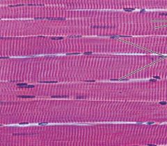
What specific tissue type is shown in the picture?
|
skeletal muscle tissue
|
|
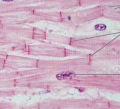
What specific tissue type is shown?
|
Cardiac muscle tissue
|
|
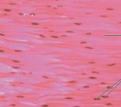
What specific tissue type is shown?
|
smooth muscle tissue
|
|
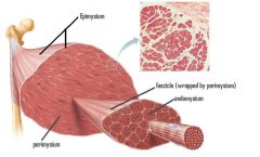
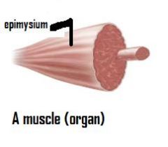
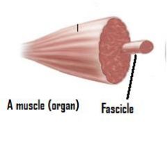
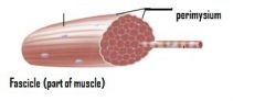
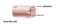
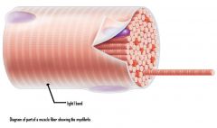
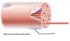
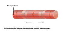
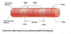
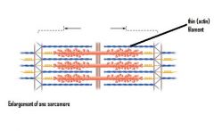
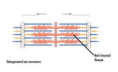
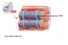
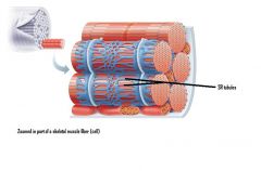
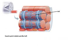
What term is labeled?
|

|
|
|

|
|
|
|
|
|

|
|
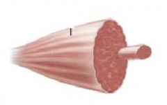
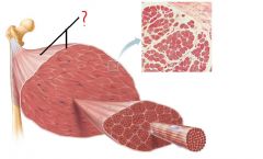
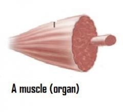
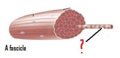
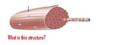
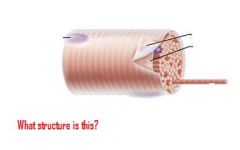
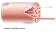
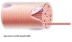
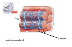
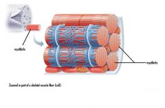
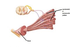
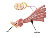
What is shown in the picture?
|
|
|
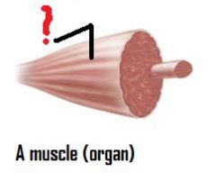
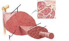
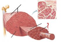
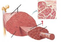
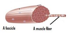
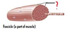
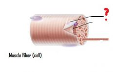
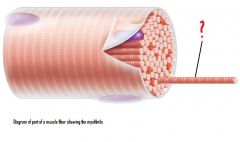
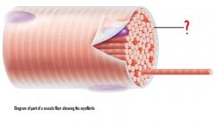
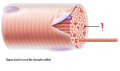
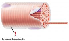
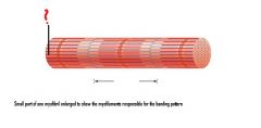
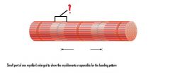
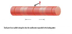
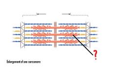
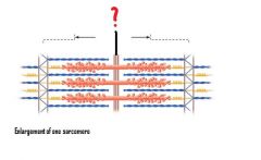
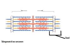
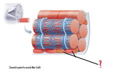
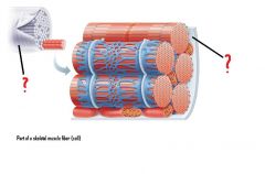
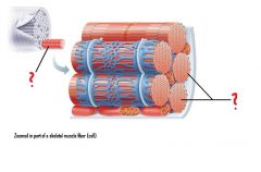
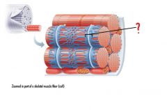
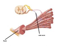
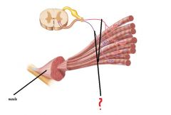
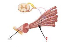
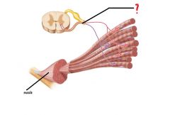
Label the structure indicated by the red question mark.
|
|
|
|

|
|
|
|
|
|
|
|
|
|
|
|
|
|
|
|
|
|

|
|
|
|
|
|

|
|
|

|
|

|
|
|
|

|
|
|

|
|

|
|
|
|
|
|
|
|
|
|
|
|
|
|
|
|
|
|

|
|
|
|
|
|
|
|
|
|
|
|
|
|
|
|
|
|
|
|
|
|
|
|
|
|
|
|
|
|
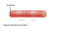
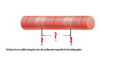
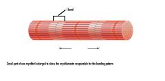
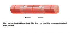
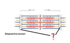
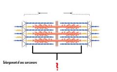
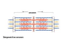
The picture shows what region of the sarcomere? What type of filaments are in this region?
|
I band shown ; thin filaments only
|
|
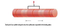
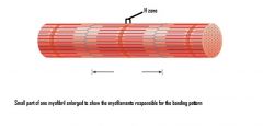
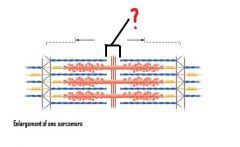
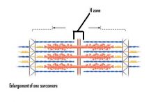
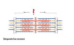
What region of the sarcomere is shown? What type of filaments are in this region?
|
H zone shown ; thick filaments only
|
|
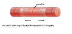
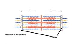
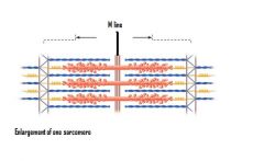
What region of the sarcomere is shown in the picture? What filaments are present in this region?
|
M line shown ; thick filaments linked by accessory proteins
|
|

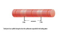
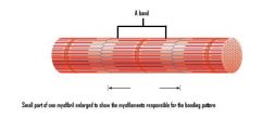
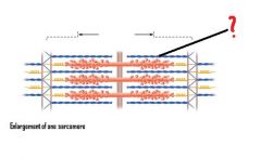
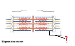
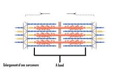
What region of the sarcomere is shown? What filaments are present in this region?
|
Outer edge of the A band shown ; thick and thin filaments overlap
|
|
|
|
|
|

|
|

|
|
|

|
|
|
|
|
|
|
|
|

|

|
|

|

|
|

|
|
|

|
|
|
|

|
|
|
|
|
|

|
|
|
|
|
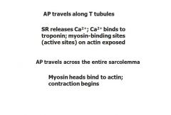
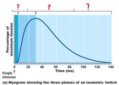
What's the correct order of steps of excitation-contraction coupling?
|

|
|
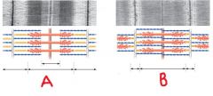
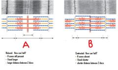
Which picture, A or B, shows a contracted muscle? Which picture, A or B shows a relaxed muscle? How can you tell?
|
|