Reading...
![]()
Play button
![]()
Play button
![]()
Use LEFT and RIGHT arrow keys to navigate between flashcards;
Use UP and DOWN arrow keys to flip the card;
H to show hint;
A reads text to speech;
57 Cards in this Set
- Front
- Back
|
Spinal cord cat
|
Whats seen here?
|
|
|
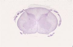
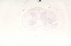
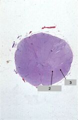
1. Spinal cord cat
2. Gray matter 3. White matter |
1. Whats seen here?
|
|
|
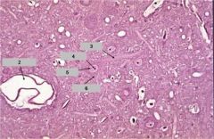
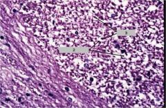
1. Spinal cord cat (gray matter)
2. Blood vessel 3. Process 4. Nucleolus 5. Nucleus 6. Cell body |
1. Whats seen here?
|
|
|
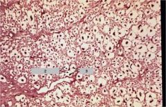
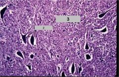
1. Spinal cord cat (white matter)
2. Myelin sheath 3. Axons |
1. Whats seen here?
|
|
|
Nissl stain cat spinal cord
|
1. Whats seen here?
|
|
|
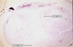
1. Nissl stain (cat spinal cord)
2. White matter 3. Gray matter |
1. Whats seen here?
|
|
|
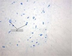
1. Nissl stain (cat spinal cord)
2. Cell bodies |
1. Whats seen here?
|
|
|
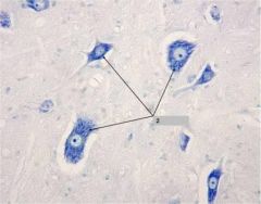
1. Nissl stain (cat spinal cord)
2. Cell bodies |
1. Whats seen here?
|
|
|
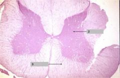
1. Spinal cord
2. Gray matter 3. White matter |
1. Whats seen here?
|
|
|
1. Spinal cord Grey matter
2. Cell bodies 3. Neuropil |
1. Whats seen here?
|
|
|
1. White matter spinal cord
2. Myelin Sheath 3. Axons |
1. Whats seen here?
|
|
|
1. Fetal spinal cord
2. Vertebral artery 3. Spinal cord surrounded by meninges 4. Dorsal root ganglion and ventral root |

1. Whats seen here?
|
|
|
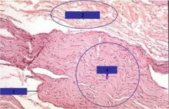
1. Fetal spinal cord surrounded by meninges
2. Pia 3. Arachnoid 4. Dura 5. Dorsal root 6. Ventral root |

1. Whats seen here?
|
|
|
1. Fetal spinal cord dorsal/ventral root
2. Ventral root 3. Dorsal root 4. Dorsal root ganglion 5. Vertebral artery 6. Spinal nerve 7. Vertebral body. |

1. Whats seen here?
|
|
|
1. Fetal spinal cord
2. Dorsal root ganglion cells |

1. Whats seen here?
|
|
|
1. Cerebral cortex
2. Cortex with meninges 3. White matter |

1. Whats seen here?
|
|
|
1. Cerebral cortex
2. Columns of neurons |

1. Whats seen here?
|
|
|
1. Cerebral cortex
2. Pyramidal cells 3. Blood vessel |

1. Whats seen here?
|
|
|
1. Cerebral cortex
2. Pyramidal cells |

1. Whats seen here?
|
|
|
1. Cerebral cortex
2. Pyramidal cells |

1. Whats seen here?
|
|
|
Cerebral cortex (golgi stain)
|

1. Whats seen here?
|
|
|
Cortex gray matter (golgi stain)
|

1. Whats seen here?
|
|
|
1. Golgi Cerebral cortex
2. Pyramidal cells |

1. Whats seen here?
|
|
|
1. Cerebellum
2. Cortex |

1. Whats seen here?
|
|
|
1. Cerebellum
2. White matter 3. Granule cell layer 4. Molecular layer 5. Purkinje cell layer |

1. Whats seen here?
|
|
|
1. Cerebellum
2. Granule cell layer 3. Purkinje cell layer 4. Molecular layer |

1. Whats seen here?
|
|
|
Ox smear
|

Whats seen here?
|
|
|
1. Ox smear
2. Neurons |

1. Whats seen here?
|
|
|
1. Ox smear
2. Neuron |

1. Whats seen here?
|
|
|
1. Nerve fibers
|

1. Whats seen here?
|
|
|
Peripheral nerve
|

?
|
|
|
1. Peripheral nerve
2. Nerve fascilcles |

1. Whats seen here?
|
|
|
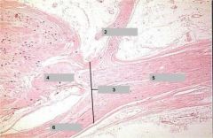
1. Peripheral nerve
2. Perineurium 3. Epineurium 4. Fascilcles |
1. Whats seen here?
|
|
|
1. Peripheral nerve
2. Axons 3. Schwann cell nucleus |

1. Whats seen here?
|
|
|
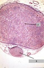
Peripheral nerve cross section
|

?
|
|
|
1. Peripheral nerve cross section
2. Perineurium 3. Epineurium 4. Fascicles |

1. Whats seen here?
|
|
|
1. Peripheral nerve cross section
2. Axon and myelin sheath |

1. Whats seen here?
|
|
|
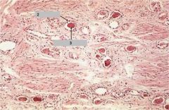
1. Lesser Omentum
2. Nerves |

1. Whats seen here?
|
|
|
1. Lesser Omentum
2. Nerve fascicles |

1. Whats seen here?
|
|
|
1. Lesser Omentum nerves
2. Nerve fascicles |

1. Whats seen here?
|
|
|
1. Lesser Omentum
2. Perineurium |
1. Whats seen here?
|
|
|
1. Lesser Omentum
2. Perinuerium |

1. Whats seen here?
|
|
|
1. Jejunum
2. Mucosa 3. Submucosa 4. Muscularis |

1. Whats seen here?
|
|
|
1. Jejunum
2. Submucosa 3. Muscularis 4. Inner cell layer 5. Outer longitudinal layer |

1. Whats seen here?
|
|
|
1. Jejunum
2. Cross section of SM 3. Myeenteric plexus 4. Longitudinal section of SM |

1. Whats seen here?
|
|
|
1. Jejunum
2. Inner Circular layer 3. Ganglion cells of myenteric plexus 4. Outer longitudinal layer |

1. Whats seen here?
|
|
|
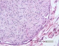
1. Dorsal root ganglion
2. Dorsal root ganglion 3. Ventral root 4. Spinal nerve 5. Dorsal ramus 6. Ventral ramus |

1. Whats seen here?
|
|
|
Dorsal root ganglion
|

1. Whats seen here?
|
|
|
1. Dorsal root ganglion
2. Unipolar cells 3. Satellite cells |
1. Whats seen here?
|
|
|
1. Dorsal root ganglion
2. Dorsal ramus 3. Spinal nerve 4. Dorsal root 5. Ventral ramus 6. Ventral root |
1. Whats seen here?
|
|
|
Dorsal root ganglion (trichrome)
|

1. Whats seen here?
|
|
|
1. Dorsal root ganglion (trichrome)
2. Dorsal root ganglion 3. Ventral root |
1. Whats seen here?
|
|
|
1. Dorsal root ganglion
2. Satellite cells 3. Unipolar ganglion cell 4. Myelinated axons |

1. Whats seen here?
|
|
|
Ventral root
|
1. Whats seen here?
|
|
|
1. Autonomic ganglion
2. Postganglic neurons |

1. Whats seen here?
|
|
|
1. Autonomic ganglia
2. Lightly myelinated or unmyelinated axons. 3. Multipolar ganglion cells |

1. Whats seen here?
|
|
|
1. Autonomic ganglia
2. Lightly myelinated or unmyelinated axons 3. Satellite cells 4. Multipolar neurons |

1. Whats seen here?
|