Reading...
![]()
Play button
![]()
Play button
![]()
Use LEFT and RIGHT arrow keys to navigate between flashcards;
Use UP and DOWN arrow keys to flip the card;
H to show hint;
A reads text to speech;
105 Cards in this Set
- Front
- Back
|
The sciatic nerve branches into what two major nerves in the leg?
|
Tibial nerve
Common fibular (peroneal) nerve |
|
|
Where does the lateral sural cutaneous branch from?
|
Common fibular nerve
|
|
|
Where does the Superficial fibular nerve branch from?
|
Common fibular nerve
|
|
|
1. Lateral Sural Cutaneous nerve
2. Superficial Fibular nerve 3. Sural nerve 4. Deep Fibular nerve |
Cutaneous innervation
|
|
|
Where does the sural nerve enter the foot?
|
Posterior to the lateral malleolus
|
|
|
What is the relationship of the superficial fibular nerve to the inferior extensor retinaculum?
|
It is superficial to it.
|
|
|
What is the major vein in the leg?
|
Great Saphenous vein.
|
|
|
Where does the great saphenous vein begin?
|
At the dorsal venous plexus
|
|
|
Where does the great saphenous vein end?
|
Femoral vein.
|
|
|
Where does the saphenous nerve begin?
|
Begins at the terminal branch of the femoral nerve.
(near the proximal anteriomedial portion of the leg) |
|
|
Where is the relation of the saphenous nerve to the medial malleolus?
|
Anterior to malleolus
|
|
|
What attaches at Gerdy's tubercle?
|
Illiotibial tract
|
|
|
What are the muscles of the Pes anserinus
|
Sartorius
Gracilis Semitendinosus |
|
|
The sural nerve branches from what two nerves?
|
Common fibular
Tibial |
|
|
What nerve supplies medial portion of foot?
|
Saphenous nerve
|
|
|
What nerve supplies the lateral portion of foot?
|
Sural nerve
|
|
|
What nerve supplies the posterior side of foot?
|
Tibial nerve
|
|
|
What nerve supplies anterior muscles of leg?
|
Deep Fibular nerve
|
|
|
What is the relationship of superficial veins and nerves to the extensor retinaculum?
|
Superficial to it.
|
|
|
1. S1
2. L5 3. L4 |

Origin of dermatone innervation
|
|
|
1. Iliotibial tract
2. Biceps Femoris 3. Fibularis Longus muscle 4. Extensor Digitorum Longus 5. Extensor Hallucis Longus 6. Fibularis Brevis 7. Fibularis Tertius 8. Extensor Digitorum Longus 9. Quadriceps Femoris muscles via patellor ligament 10. Pes Anserinus 11. Tibialis Anterior muscle 12. Extensor Hallucis Longus muscle |

Name the attachments
|
|
|
Fibularis Longus muscle inserts where?
|
Medial cuneiform
Base of 1st metatarsal (plantarly) |
|
|
Fibularis Brevis muscle inserts where?
|
Base of 5th metatarsal
|
|
|
Fibularis Tertius muscle inserts where?
|
Base of 5th metatarsal
|
|
|
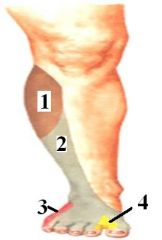
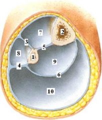
What are the borders of the anterior compartment of the leg?
|
1. Anterior Intermuscular Septum
2. Interosseus membrane 3. Deep fascia of the leg 4. Tibia |
|
|
What are the borders of the lateral compartment of the leg?
|
1. Anterior Intermuscular Septum
2. Posterior Intermuscular Septum 3. Deep fascia of the leg 4. Fibula |
|
|
What are the borders of the Deep Posterior compartment?
|
1. Interosseus membrane
2. Transverse Intermuscular septum 3. Tibia 4. Fibula |
|
|
What are the borders of the Superficial Posterior Compartment?
|
1. Posterior Intermuscular Septum
2. Transverse Intermuscular Septum 3. Deep Fascia of the leg |
|
|
1. Fibula
2. Tibia 3. Anterior Intermuscular Septum 4. Posterior Intermuscular Septum 5. Interosseus membrane 6. Transverse Intermuscular Septum 7. Anterior Compartment 8. Lateral Compartment 9. Deep Posterior Compartment 10. Superficial Posterior Compartment |
Name
|
|
|
In the Anterior compartment, what is the innervating nerve and action of the muscles?
|
Deep Fibular Nerve
Dorsiflex Ankle Extend Toes |
|
|
In the Lateral compartment, what is the innervating nerve and action of the muscles?
|
Superficial Fibular Nerve
Evert Foot Weakly Plantarflex Ankle |
|
|
In the Deep Posterior compartment, what is the innervating nerve and action of the muscles?
|
Tibial Nerve
Plantarflex Ankle Invert Foot Flex Toes |
|
|
In the Superficial Posterior compartment, what is the innervating nerve and action of the muscles?
|
Tibial Nerve
Plantarflexes Ankle |
|
|
What structures contribute to the capsule of the knee? x4
|
1. Lateral/Medial Patellar Retinacula
2. Paterllar Tendon 3. Iliotibial tract 4. Quadriceps tendon |
|
|
What are the muscles of the anterior leg?
|
1. Tibialis Anterior
2. Extensor Digitorum Longus 3. Extensor Hallucis Longus 4. Peroneus Tertius |
|
|
What muscles insert into the foot POSTERIOR to the transverse tarsal joint?
|
1. Gastrocnemius
2. Plantaris 3. Soleus |
|
|
Where would you note a common fibular nerve damage?
|
By the fibula head, right before it branches into deep and superficial fibular nerves.
|
|
|
Loss of dorsiflexion and eversion indicates what nerve damage?
|
Common fibular nerve
|
|
|
Loss of eversion indicates what type of nerve damage?
|
Superficial fibular nerve.
|
|
|
Describe the course of the ANTERIOR TIBIAL ARTERY and the DEEP FIBULAR NERVE in the leg:
- Proximal leg - Distal Leg - Ankle |
Between Tibularis Anterior and Extensor Digitorum Longus
Between Tibularis Anterior and Extensor Hallucis Longus Extensor Hallucis longus will cross OVER the artery and nerve. |
|
|
Describe the location and attachments of the Superior Extensor Retinaculum.
|
Anterior ankle, just above malleoli
Extends from fibula to tibia |
|
|
Describe the shape and attachment(s) of the inferior extensor retinaculum.
|
Y-shaped
Superior band attaches from calcaneous to medial malleolus Interior band blends with plantar aponeurosis |
|
|
What does the tendon extensor hallucis longus do at the ankle region?
|
Crosses over
Deep Fibular nerve and Dorsalis Pedis artery (continuation of anterior tibial artery) |
|
|
Where on the foot is a good location for taking a pulse? What vessel is this?
|
Between the tendons of extensor digitorum longus AND extensor hallucis longus.
Dorsalis Pedis artery |
|
|
Short extensors of foot:
- where are they - what are they called - which toe does not have one |
They lie deep to the extensor tendons.
Extensor hallucis brevis Extensor digitorum brevis 5th toe does not have one |
|
|
Where would you find the arcuate artery?
|
Runs transverse, under the extensor tendons, near PROXIMAL ends of metatarsals.
|
|
|
What nerves supply the 1st and 2nd toes?
|
Deep Fibular nerve - btw toes
Superficial Fibular nerves - outer edges of both toes. |
|
|
Inferior Fibular Retinaculum is located where?
|
Entirely on the calcaneous
|
|
|
Inferior Fibular Retinaculum holds what?
|
Tendons of Fibularis longus and Fibularis brevis muscles
|
|
|
How do the tendons of the fibularis longus and fibularis brevis enter the foot?
|
Posterior to the lateral malleolus.
|
|
|
The superior fibular retinaculum is located where?
|
Calcaneous to lateral malleolus
|
|
|
Dorsal Venous Arch gives rise to what?
|
Great Saphenous vein
Small Saphenous vein Saphenous vein |
|
|
1. Saphenous nerve
4. Medial Plantar nerve 5. Lateral Plantar nerve 6. Sural nerve 7. Calcaneal branches of (medial) tibial nerve and (lateral) sural nerve. |

Name the innvervating nerves
|
|
|
1. Superficial fibular nerve
2. Superior extensor retinaculum 3. Lateral Malleolus 4. Venous network of lateral malleolus and tributaries of small saphenous vein 5. Lateral Dorsal Cutaneous Nerve (branch of sural nerve) 6. Intermediate dorsal cutaneous nerve (branch of superficial fibular nerve) 7. Tendons of extensor digitorum longus 8. Dorsal digital nerves 9. Tendon of tibialis anterior 10. Saphenous nerve 11. Venous network of medial malleolus and tributaries of great saphenous vein. 12. Medial malleolus 13. Medial Dorsal Cutaneous Nerves (branches of superficial nerve) 14. Dorsal Venous Arch 15. Dorsal Digital Nerve (Continuation of Deep Fibular Nerve btw 1st and 2nd digits) 16. Tendon of extensor hallucis longus 17. Dorsal Digital Arteries 18. Fibularis Longus and Brevis 20. Extensor Digitorum Longus 21. Extensor Hallucis Longus 22. Inferior Extensor Retinaculum 23. Extensor Hallucis Brevis |

Name all
|
|
|
What holds down the tendons of the fibularis longus and fibularis brevis?
|
Extensor Retinaculum
and Fibular Retinaculum |
|
|
Tendon sheaths provide what?
|
Lubrication
|
|
|
Fibularis Tertius tendons runs how in terms of the malleolus?
|
Anterior to Lateral Malleolus
|
|
|
What is the action of the Fibularis Tertius?
|
Dorsiflex and Evert foot
|
|
|
What are the actions of the Fibularis Longus and Fibularis Brevis?
|
Evert Foot mostly
|
|
|
Where is the Inferior Fibular Retinaculum?
|
Completely on the Calcaneus
|
|
|
Where is the Superior Fibular Retinaculum?
|
Extends from the calcaneus (lateral side) to lateral malleolus.
|
|
|
What type of fascia is over the anterior compartment of the leg?
|
Crural fascia.
|
|
|
What is the medical terminology for shin splints and describe the etiology.
|
Anterior Compartment Syndrome
The crural fascia and injury (possibly due to strenuous exercise) cuts off blood supply and resulting in further damage to muscles. (Anoxic Necrosis) |
|
|
Describe the insertions of all muscles posterior to the transverse tarsal joint.
|
The GASTROCNEMIUS and SOLEUS join to end with the calcaneal tendon.
The PLANTARIS tendon also attaches to the calcaneus but as its own tendon. |
|

Answer this shit
|

Shit answered
|
|
|
What are the muscles of the deep posterior compartment of the leg?
|
1. Flexor Digitorum Longus
2. Flexor Hallucis Longus 3. Tibialis Posterior 4. Popliteus |
|
|
What are the muscles of the superficial posterior compartment of the leg?
|
1. Gastrocnemius
2. Plantaris 3. Soleus |
|
|
Fibularis longus and fibularis brevis is held posterior to the lateral malleolus by what? Identify which overlap each other.
|
Inferior Fibular Retinaculum AND
Inferior Extensor Retinaculum overlap. Superior Fibular Retinaculum |
|
|
1. Medial head of Gastrocnemius
2. Plantaris 3. Insert for Semimembranosus 4. Lateral head of Gastrocnemius 5. Popliteus muscle 6. Insert for Popliteus 7. Soleus 8. Soleus 9. Flexor Digitorum Longus 10. Tibialis Posterior 11. Flexor Hallucis Longus 12. Fibularis Brevis 13. Insert for Gastrocnemius, Soleus. Also for Plantaris 14. Insert for Tibialis Posterior 15. Insert for Tibialis Anterior 16. Insert for Fibularis Longus 17. Insert for Flexor Hallucis Longus 18. Insert for Flexor Digitorum Longus |

Posterior Leg Origins and Inserts
|
|
|
What is the superiomedial boundary for the Popliteal Fossa?
|
Semitendinosis
Semimembranosis |
|
|
What is the superiolateral boundary for the Popliteal Fossa?
|
Biceps Femoris
|
|
|
What is the inferiolateral boundary for the Popliteal Fossa?
|
Lateral Head of Gastrocnemius
|
|
|
What is the inferiomedial boundary for the Popliteal Fossa?
|
Medial Head of Gastrocnemius
|
|
|
What is the floor of the popliteal fossa?
|
Popliteus, Femur, and Oblique Popliteal ligament
|
|
|
Describe the location of the Inferior lateral and medial genicular arteries.
|
In the popliteal fossa, inferior to the knee joint.
|
|
|
1. Small Saphenous vein
2. Sural nerve |

ID
|
|
|
1. Medial sural cutaneous nerve
2. Sural nerve |

ID
|
|
|
1. Lateral Sural Cutaneous
2. Medial Sural Cutaneous 3. Sural Nerve 4. Tibial nerve (via medial calcaneal branch) |

ID
|
|
|
Lymph from posterior leg drains where?
|
Popliteal lymph nodes
|
|
|
Describe Small saphenous vein:
- origin - respect to malleolus - empties where? |
Dorsal Veous arch on LATERAL SIDE
Passes posterior to lateral malleolus with sural nerve Popliteal vein |
|
|
How does the fibular artery travel in the leg?
|
Between the FHL and Fibula
|
|
|
How does the Posterior tibial artery travel in the leg?
|
Between the FDL and the Tibularis Posterior
|
|
|
What does the posterior tibial artery run with?
|
Tibial nerve
|
|
|
Describe the tendons, nerves, and blood supply behind the medial malleolus.
|
From most medial to lateral:
Tibularis Posterior Flexor Digitorum Longus Posterior Tibial Artery Tibial Nerve Flexor Hallucis Longus |
|
|
Describe insertion of Tibularis Anterior.
|
1st metatarsal on medial cuneiform
|
|
|
Describe insertion of Tibularis Posterior.
|
Posterior on navicular
|
|
|
Actions of TA.
|
Dorsiflex and Invert foot
|
|
|
Action of EHL
|
Extends 1st toe and dorsiflex foot
|
|
|
Action of EDL
|
Extends lateral 4 toes and dorsiflex foot
|
|
|
Action of Fibularis Longus
|
Evert foot and plantar flexes foot
|
|
|
Actions of Gastrocnemius
|
Flexes knee
Plantar flexes foot |
|
|
Actions of Soleus
|
Plantar flexes foot
|
|
|
Actions of TP
|
Inverts foot and Plantar flexes foot
|
|
|
Actions of FDL
|
Flexes lateral four toes
and Plantar flexes foot |
|
|
Actions of FHL
|
Flexes 1st toe
and Plantar flexes foot |
|
|
Innervation for dorsum of foot?
|
Deep Fibular nerve
|
|
|
Innervation for sole of foot?
|
Medial and Lateral Plantar nerves
|
|
|
What are the branches of the popliteal artery?
|
Superior lateral genicular
Superior medial genicular Middle Genicular Inferior lateral genicular Infereior medial genicular |
|
|
What are the terminal branches of the popliteal artery.
|
Anterior Tibial Artery
Posterior Tibial Artery |
|
|
What are the branches of the Posterior Tibial Artery?
|
Fibular artery
Branches to bone and muscle |
|
|
What are the branches of the Anterior tibial artery?
|
Anterior tibial recurrent
Anteior medial malleolar artery Anterior lateral malleolar artery Branches to muscles |
|
|
What are the terminal branches of the posterior tibial artery?
|
Medial Plantar artery
Lateral Plantar artery |
|
|
What are the terminal branches of the anterior tibial artery?
|
Become dorsalis pedis in ankle.
|
|
|
What are the branches of dorsalis pedis artery?
|
Lateral tarsal
Medial tarsal Arcuate artery |
|
|
What is the terminal branch of dorsalis pedis artery?
|
Deep plantar artery
|