Reading...
![]()
Play button
![]()
Play button
![]()
Use LEFT and RIGHT arrow keys to navigate between flashcards;
Use UP and DOWN arrow keys to flip the card;
H to show hint;
A reads text to speech;
56 Cards in this Set
- Front
- Back
- 3rd side (hint)
|
Name some sites where CT is located.
|
o Padding organs and other regions of body
o Fascia throughout body o Underneath epithelium o Can be fluid-like or gel-like o Wide distribution |
|
|
|
Which of the following is NOT a role of CT?
A. Structural B. Metabolic C. Defensive D. Energetic |
D. Energetic is incorrect
|
|
|
|
What is the origin of CT?
|
mesodermal and specific neural crest cells that proliferate to form a primitive CT called mesenchyme in the early embryo
|
|
|
|
What are the two general components of CT?
|
Cells and ECM
Break down ECM further. |
1. Ground Substance - composed of GAGs (hydrophilic), proteoglycans (like condroitin sulfate), proteoglycan aggregates (hyaluronic acid), adhesive glycoproteins (involved in cell communication. ex. fibronectin, laminin)
2. Tissue Fluid 3. Fibers (1. Collagen and its four types, 2. Elastic fibers, 3. Reticular fibers) |
|
|
What is ground substance a component of and what are its components?
|
Ground substance is a component of ECM.
Ground substance's components are: GAGs (hydrophilic), proteoglycans (ex. chondroitin sulfate), proteglycan aggregate (hyaluronic acid), adhesive glycoproteins (fibronectin and laminin) Other components of ECM are tissue fluid and fibers. |
|
|
|
Which component of ground substance are very negatively charged structures that form a difficult to compress gel?
A. GAGs B. Proteoglycan C. Proteoglycan Aggregate D. Adhesive glycoprotein |
A. GAGs
|
|
|
|
What component of ground substance does hyaluronic acid fall under?
A. GAGs B. Proteoglycan C. Proteoglycan Aggregate D. Adhesive glycoprotein |
C. Proteoglycan Aggregate
makes a huge tree-like branched structure. This is what fills the ground substance and forms the physical barrier to bacteria that try to tunnel in. Some bacteria can produce hyaluronuase to degrade these, though. |
|
|
|
What component of ground substance does chondroitin sulfate, dermatin sulfate, keratin sulfate and heparin sulfate fall under?
A. GAGs B. Proteoglycan C. Proteoglycan Aggregate D. Adhesive glycoprotein |
B. Proteoglycan
|
|
|
|
What component of ground substance does fibronectin and laminin fall under? What are fibronectin and laminin important for function wise?
A. GAGs B. Proteoglycan C. Proteoglycan Aggregate D. Adhesive glycoprotein |
D. Adhesive glycoprotein
• Fibronectin: cell migration, adhesion, motility • Laminin: important in epithelia, forms part of basal lamina |
|
|
|
What are the three components (general) that make up the ECM?
|
Ground substance, Tissue Fluid, Fibers
|
|
|
|
Fibers are secreted by fibroblasts. Fibers come in three "flavors". Name them.
|
1. Collagen Types I thru IV.
2. Elastic Fibers 3. Reticular Fibers |
Collagen Type I: most common, found in dermis of skin, bone tendon, dentin, fascia
Type II: very thin fibrils around hyaline and elastic cartilage Type III: major component of reticular fiber Type IV: found in basal lamina and epithelium, doesn't form fibrils or fibers! Elastic fibers: produced by fibroblasts or smooth muscle, found in all loose CT Reticular fiber: collagen type III. provides framework for bone marrow and lymphoid organs and spleen. Delicate, forms quickly. Prominent during embryogenesis and inflammation processes. |
|
|
Which fiber, one of three components of ECM, can be produced by fibroblasts OR smooth muscle?
|
elastic fibers
(two other fiber types are collagen and reticular fiber) |
|
|
|
Which fiber, one of three components of ECM, has 5 subdivision types?
|
Collagen!
o Type I : most common, found in dermis of skin, bone, tendon, dentin, fascia, organ capsules, fibrous cartilage o Type II: very thin fibrils found in hyaline and elastic cartilage o Type III: the major component of reticular fiber o Type IV: found in basal lamina and epithelium. Doesn’t form fibrils or fibers! |
|
|
|
Which fiber, one of three components of ECM, may form branchign sheets with fenestrations in blood vessels?
|
Elastic fibers
(two other fiber types are collagen and reticular fiber) |
|
|
|
Which fiber, one of three components of ECM, provides the framework of bone marrow and lymphoid organs and spleen, is very delicate and thus is constructed quickly and is prominent during embryogenesis and inflammatory processes?
|
Reticular fibers
(the two other fibers are collagen and elastic) |
|
|
|
True or False:
Reticular fibers are about half as large as a collagen fiber. |
True!
|
|
|
|
Which fiber, a part of the ECM, is argyrophilic and thus we silver-stain to visualize it?
|
Reticular Fiber!
|
|
|
|
What is the principal cell fo the CT that stays in the CT?
A. Eosinophil B. Fibroblasts C. Lymphocytes D. Macrophages E. Mast Cells F. Monocytes G. Neutrophils H. Plasma Cells |
B. Fibroblasts
|
|
|
|
Compare and contrast the fibroblast active form with the fibroblast inactive form.
|
• Active form:
Nucleus is oval or spindle-shaped, very euchromatic, nucleoli Cytoplasm not discernable at LM level EM reveals abundant RER, Golgi • Inactive fibroblast (fibrocyte) Nucleus very spindle-shaped, very heterochromatic (thus very dark) Cytoplasm not apparent with LM; very thin EM Low metabolic rate |
|
|
|
Oval shape, eccentric nucleus with prominent "negative" golgi and "clock-face" chromatin.
A. Eosinophil B. Fibroblasts C. Lymphocytes D. Macrophages E. Mast Cells F. Monocytes G. Neutrophils H. Plasma Cells |
H. Plasma Cells
Also produces antibodies. |
|
|
|
What cells are identifiable by their indented nucleus?
A. Eosinophil B. Fibroblasts C. Lymphocytes D. Macrophages E. Mast Cells F. Monocytes G. Neutrophils H. Plasma Cells |
D. Macrophages
These are also very phagocytic, and they contain lysosomes and phagosomes in their cytoplasm. |
|
|
|
Are small (6-7 micrometers in diameter) though have large, heterochromatic nucleus and a thin thin ring of cytoplasm around it.
A. Eosinophil B. Fibroblasts C. Lymphocytes D. Macrophages E. Mast Cells F. Monocytes G. Neutrophils H. Plasma Cells |
C. Lymphocytes
These exist in T and B flavors. B lymphocytes differentiate into plasma cells |
|
|
|
Horse-shoe shaped nucleus.
A. Eosinophil B. Fibroblasts C. Lymphocytes D. Macrophages E. Mast Cells F. Monocytes G. Neutrophils H. Plasma Cells |
F. Monocytes
these are also large, round cells ~ 12-20 micrometers in diameter, and they are precursors of macrophages. Found in CT and peripheral blood. |
|
|
|
Has a small, centrally located nucleus, granules contain GAGs, heparin and histamine.
A. Eosinophil B. Fibroblasts C. Lymphocytes D. Macrophages E. Mast Cells F. Monocytes G. Neutrophils H. Plasma Cells |
E. Mast Cells
becomes activated in allergic reactions |
|
|
|
Most prevalent WBC.
A. Eosinophil B. Fibroblasts C. Lymphocytes D. Macrophages E. Mast Cells F. Monocytes G. Neutrophils H. Plasma Cells |
G. Neutrophils
|
|
|
|
Multi-lobed nucleus with 3-5 lobes. Has two types of granules (one w/ enzymes, others w/out), highly phagocytic.
A. Eosinophil B. Fibroblasts C. Lymphocytes D. Macrophages E. Mast Cells F. Monocytes G. Neutrophils H. Plasma Cells |
G. Neutrophils
|
|
|
|
What cell type is associated with the accumulation of dead bacteria and dead [insert cell type] can form pus?
A. Eosinophil B. Fibroblasts C. Lymphocytes D. Macrophages E. Mast Cells F. Monocytes G. Neutrophils H. Plasma Cells |
G. Neutrophils
|
|
|
|
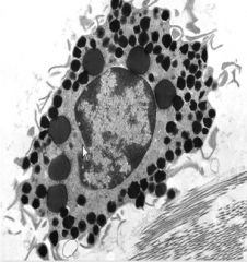
Bi-lobed nucleus, distinct eosinophillic granules which are lysosomes.
A. Eosinophil B. Fibroblasts C. Lymphocytes D. Macrophages E. Mast Cells F. Monocytes G. Neutrophils H. Plasma Cells |
A. Eosinophil
|
|
|
|
An increase in this type of cell can indicate presence of a parasitic infection.
A. Eosinophil B. Fibroblasts C. Lymphocytes D. Macrophages E. Mast Cells F. Monocytes G. Neutrophils H. Plasma Cells |
A. Eosinophil
|
|
|
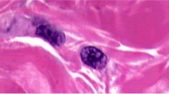
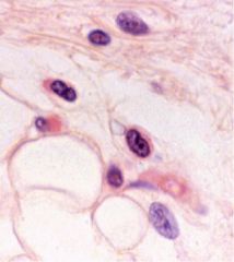
Identify the CT cell type.
|
Active fibroblast
- Nucleus is oval or spindle-shaped, very euchromatic, nucleoli - Cytoplasm not discernable at LM level - EM reveals abundant RER, Golgi |
|
|
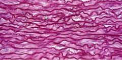
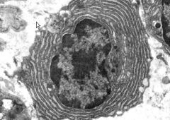
Identify the CT cell type.
|
Elastic fiber
- squigglies |
|
|
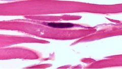
Identify the CT cell type.
|
Inactive fibroblast
- Nucleus very spindle-shaped, very heterochromatic (thus very dark) - Cytoplasm not apparent with LM; very thin EM - Low metabolic rate |
|
|
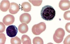
Identify the CT cell type.
|
Monocyte
o Large, round cell, 12-20 micrometers in diameter, distinct horseshoe-shaped nucleus o Precursors of macrophages o Are found in connective tissue and peripheral blood |
|
|
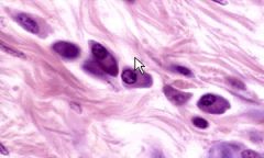
Identify the CT cell type.
|
Macrophage
o Very phagocytic o Indented nucleus, cytoplasm visible o Lysososmes and phagosomes in their cytoplasm |
|
|
Identify the cell type.
|
Lymphocyte
o Large, heterochromatic nucleus with thin thin ring of cytoplasm around it o Small, 6 to 7 micrometers in diameter o T and B lymphocytes exists o B lymphocytes become plasma cells |
|
|
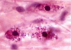
Identify this cell type.
|
Plasma Cell
o Oval shape, eccentric nucleus o Basophilic cytoplasm with prominent “negative” golgi o Round nucleus, clumped chromatin “clock-face” o Produce antibodies, “protein factories” |
|
|
Identify this cell.
|
Plasma Cell
o Oval shape, eccentric nucleus o Basophilic cytoplasm with prominent “negative” golgi o Round nucleus, clumped chromatin “clock-face” o Produce antibodies, “protein factories” |
|
|
Identify this cell type.
|
Mast Cell
o Small, centrally located neucleus, granules contain GAGs, heparin and histamine o Becomes activated in allergic reactions |
|
|
Identify this cell type.
|
Mast Cell
o Small, centrally located neucleus, granules contain GAGs, heparin and histamine o Becomes activated in allergic reactions |
|
|
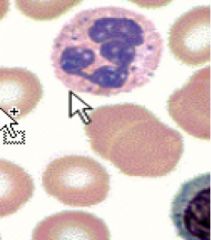
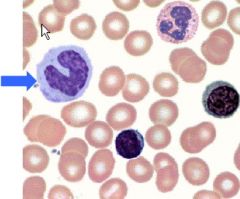
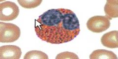
Identify this type of CT cell.
|
Neutrophils
o Most prevelant WBC o Multi-lobed nucleus with 3-5 lobes, 12 -15 micrometers o Two types of granules (one with enzymes and one with non-enzymes) o Highly phagocytic (responsible for cardinal signs of inflammation, or dolor (pain), calor (warmth), tumor (swelling), rubor (redness) o After intracellular digestion, remnants are stored or eocytosed o During this process most neutrophils die: the accumulation of dead bacteria and dead neutrophils can form pus |
|
|
Identify this type of cell.
|
Eosinophil
o Bi-lobed nucleus, about 12-14 micrometers o Distinct eosinophillic granules whichar e lysosomes o Phagocytic and increase in number with parasitic infections |
|
|
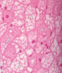
What type of CT cell is this?
|
Brown Adipose
o multiple droplets of lipids (multilocular) o central nucleus o used in thermoregulation o babies and hibernating animals have a lotta this |
|
|
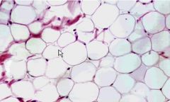
Identify the CT cell type.
|
Yellow or White adipose
o Cells synthesize and store lipid for nutrition and insulation o Nucleus and cytoplasm are displaced by large lipid droplet o May appear as small clusters of cells or large accumulations |
|
|
|
Which statement is FALSE?
A. Neutrophils are responsible for the cardinal signs of infection. B. Monocytes differentiate Macrophages C. Mast cells found in CT only, not blood D. T lymphocytes differentiate into Plasma cells E. Platelets are formed by megakaryocytes in bone marrow |
D. T lymphocytes differentiate into Plasma cells
Only B lymphocytes differentiate into plasma cells |
|
|
|
What type of cell fits the following description:
• Biconcave, 7.5 micrometer in diameter • Filled with hemoglobin • Actin-containing cytoskeleton • No cellular organelles A. Erythrocytes B. Leukocytes C. Platelets |
A. Erythrocytes
|
|
|
|
List the 5 different leukocyte cells presented in lecture. Which is most abundant? Which is least abundant?
|
• Neutrophils: 40-75% of WBc’s
• Lymphocytes: 20-50% of WBCs • Eosinophils: 5% of WBCs (unless we have parasitic infection, in which case this will be higher) • Monocytes: 1-5% of WBCs • Basophils: 0.5% of WBCs |
|
|
|
Basophils, the least common of WBC, is similar to __________ because of its nucleus and similar to __________ because of its granules.
|
Basophils have a bi-lobed nucleus, similar to eosinophils ; similar to mast cells because of granules that contain heparin and histamine.
|
|
|
|
What does this describe:
• non-nucleated, disk like cell FRAGMENT • promotes blood clotting • do have organelles in them • formed by megakaryocytes in bone marrow • responsible for clot formation |
Platelets!
|
|
|
|
Where and by what cells are platelets formed?
|
Platelets are formed by megakaryoctes in bone marrow.
|
|
|
|
Regarding hematopoiesis, what do the mesoblastic, hepatic and myeloid periods refer to, respectively?
|
Early early blood cells stem from mesenchymal cells in yolk sac and body stalk (from hypoblast). These are called mesoblastic. Eventually then blood cells are made in the liver for a while (hepatic) and then finally when bone marrow is present, it is made in bone marrow (myeloid).
|
|
|
|
Pluripotential hematopoietic stem cells arise from a single type of stem cell and proliferate to form.... (hint: 2)
|
Lymphoid multipotential cells (migrate to lymphoid organs, eventually produce B and T lymphocytes).
Myeloid multipotential cells (remain in bone marrow but give rise to the rest of WBC precursors - neutrophilic granulocyte, eosinophilic granulocyte, basophilic granulocyte, monocytes - as well as magakryocytes and erythrocytes. |
|
|
|
___________ have fibers that measure less than 7 micrometers in diameter.
A. Loose CT B. Dense Irregular CT C. Dense Regular CT D. Elastic CT E. Reticular CT |
A. Loose CT
|
|
|
|
____________ is a passageway for nerves and blood vessels.
A. Loose CT B. Dense Irregular CT C. Dense Regular CT D. Elastic CT E. Reticular CT |
A. Loose CT
|
|
|
|
________ have fiber bundles that measure at least 7 micrometers in diameter and dermis of skin is a good example.
A. Loose CT B. Dense Irregular CT C. Dense Regular CT D. Elastic CT E. Reticular CT |
B. Dense Irregular CT
|
|
|
|
Have collagen bundles in parallel rows. Tendons and ligaments are examples.
A. Loose CT B. Dense Irregular CT C. Dense Regular CT D. Elastic CT E. Reticular CT |
C. Dense Regular CT
|
|
|
|
Which type of CT is composed of mainly inactive fibroblasts?
A. Loose CT B. Dense Irregular CT C. Dense Regular CT D. Elastic CT E. Reticular CT |
C. Dense regular CT
|
|