Reading...
![]()
Play button
![]()
Play button
![]()
Use LEFT and RIGHT arrow keys to navigate between flashcards;
Use UP and DOWN arrow keys to flip the card;
H to show hint;
A reads text to speech;
48 Cards in this Set
- Front
- Back
|
Which of the following is FALSE about the cytoskeleton in general:
A. exhibits highly dynamic structure B. is responsible for cell movement C. provides mechanism for intracellular movement D. is present in both eukaryotic and prokaryotic cells |
D. Cytoskeleton is actually only found in eukaryotic cells
|
|
|
What are the functions of the cytoskeleton?
|
- Cell structure
- "Toughness" - Movement of materials within cells - Actual movement of the cell itslef |
|
|
What three protein structures is the cytoskeleton composed of and what are those components in turn composed of?
|
- Actin microfilaments (composed of actin)
- Microtubules (composed of tubulin) - Intermediate filaments (composed of fibrous proteins such as vimentin, keratin and lamin) |
|
|
Name the three protein structures that compose the cytoskeleton and where they can generally be found or what they generally attach to.
|
- Actin filaments are usually found concentrated at cortex just beneath the plasma membrane.
- Microtubules are attached on one end to the organizing center called a centrosome. - Intermediate filaments can form a nuclear lamina and can be found extending across the cytoplasm from one cell-cell junction to another. |
|
|
The plasma membrane is a critical component of the cytoskeleton and has a proteinaceous skeleton sometimes referred to as the _______. Human RBCs have a complex of membrane proteins in this called _____________ to maintain the RBC's structural integrity.
|
Cortex; spectrin dimers
|
|
|
What are spectrin dimers?
(hint: what type of cell are they associated with?) |
They are a complex of membrane proteins int eh cortex that allow RBC's to maintain structural integrity yet still be flexible. Spectrin dimers associate "head to head" to form tetramers that are linked together by short actin filaments and other associated proteins.
|
|
|
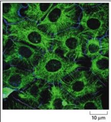
The _______________ provide a tough, flexible network that maintains the cell's integrity by resisting stresses and strains.
A. Actin filaments B. Intermediate Filaments C.Microtubules |
B. Intermediate Filaments
|
|
|
What component of the intermediate filament is mutated in the condition of Epidermolysis bulosa complex.
|
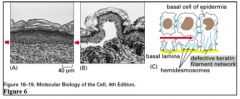
Keratin.
The mutation allows easy blistering of the epidermis as cells rupture in the basal layer. In severe cases, as the baby passes through the birth canal, the infant is born with most of the skin removed! |
|
|
The nuclear lamina, associated with both the _______ and the __________, is constructed from a special class of intermediate filament proteins called _________.
|
Nuclear lamina is associated with both chromatin and the inner nuclear membrane. Nuclear lamina is made of intermediate filaments proteins called lamins.
|
|
|
True or False:
The nulcear lamina is associated with both the outer and inner lumenal subunits of nuclear pores. |
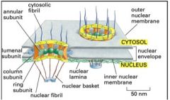
False. The nuclear lamina is associated with both the chromatin and the inner nuclear membrane.
|
|
|
Intermediate filaments are also found in concentric layers around the outside on the nuclear membrane and radiate outward from the nucleus to the ___________________ where they may attach to a desmosome or hemidesmosome.
|
plasma membrane
|
|
|
True or False:
In general, intermediate filaments are present in greater amounts in cells that endure mechanical stress such as epithelial cells. |
True
|
|
|
Which of the following statements about intermediate filaments is FALSE?
A. mutation in the lamin causes Epidermolysis bulosa simplex B. they are called intermediate because of their size (8-10 nm in diameter) which is between the size of actin (5-9nm in diam) and microtubules (25 nm in diam). C. There are four sub types of Intermediate Filmanets D. the ultra structure is made of 8 tetramers packed in a rope-like fashion |
A. Epidermolysis bulosa is caused by another intermediate filament mutation of the KERATIN, not the lamin.
|
|
|
Name the types of Intermediate Filament Proteins in vertebrate cells and where they can be found. (Hint there are four)
|
1. Nuclear (lamins A, B & C), in Nuclear Lamina
2. Vimentin-like (Vimentin, Desmin, Glial fibrillary, Peripherin), found in many cells, muscle, glial cells, some neurons 3. epithelial (keratins), found in epithelial-derived cells, hairs and nails 4. Axonal (Neurofilament proteins NF-L, NF-M, NF-H, found in neurons |
|
|
Describe the intermediate filament ultra structure.
|
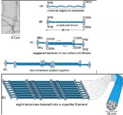
Start with an IF monomer (A) then pair it with an identical monomer to make a dimer (B) which coils. Two of these then line up side by side to form anti-parallel tetramer (C), Two tetramers are then staggered next to each other (D) then eight of the tetramers are twisted into a ropelike filament (E)
|
|
|
The ___________ networks of adjacent cells are indirectly connected to one another through desmosomes and to the basal lamina through hemi-desmosomes.
|
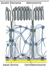
keratin filaments
|
|
|
Microtubules are long, stiff polymers made of the protein _____ which consists of two subunits, ___________. Microtubules extend throughout the cytoplasm and control the movement and location of ___________ and other cell components.
|
made of Tubulin, with subunits alpha and beta. Control movement and location of membrane-bounded organelles and other cell components.
|
|
|
_______ are approx. 25 nm in diameter, and is a hollow tube constructed of 13 side-by-side "rods".
A. Actin B. Intermediate Filaments C. Microtubules |
C. Microtubules
|
|
|
The two ends of a microtubule are different chemically and have polarity. What characteristics do the two different ends possess?
|
The PLUS end grows more rapidly during the assembly process (polymerization). The MINUS end is bound to a nucleation center called the nucleator which can be a centrosome, basal body, or spindle pole.
|
|
|
How does colchicine, used to treat __________, affect microtubules and what does that ultimately do to treat the condition?
|
Colchicine is used to treat gout. Because microtubules have a role in cell motility, inhibition of microtubules by Colchicine blocks the ability of inflammatory cells to migrate into inflamed joints. Additionally, inhibited microtubules reduces the phagocytosis of the accumulated urate crystals and the release of inflammatory mediators. Colchicine binds tightly to tubulin, preventing polymerization.
|
|
|
A drug similar to colchicine (for the treatment of ____________) but is reversible, is called _________.
|
gout; colecemid
|
|
|
List the five drugs mentioned in lecture that interfere with microtubule polymerization.
|
Colchicine, Colecemid (treatment of gout) Vinblastine and vincristine (treatment for various cancers b/c cause tubulin dimer aggregation thus interfering with microtubule polymerization. Taxol (cancers) bind directlyto the microtubule to prevent depolymerization, arresting them.
|
|
|
The minus end of the microtubule are bound to ________, of which there are three types. Name them.
|
minus end binds to nucleator, of which can be centrosome, basal body, or spindle pole.
|
|
|
What is an axoneme and what is it made of?
|
Axoneme is a structure that produces the movement of a cilium or flagellum. Composed of entirely microtubules and their associated proteins, this helps function in cell motility.
|
|
|
A microtubule-associated protein called _____________ interacts with adjacent microtubules to generate a sliding force to move the complex. This has a "motor domain" that hydrolyzes ATP to move along the microtubule towards its minus end.
|
Ciliary Dynein
|
|
|
Microtubules also play a critical function during cell division. THE MTOC, the centrosome, houses two cylindrical structures called _______ which are positioned at right angles to each other forming an L-shaped configuration.
|
centrioles
|
|
|
Microtubules are associated with vesicle transport via _________, which moves vesicles outwards towards the plus ends of the microtubule (towards the cytoplasm) whereas _________ move vesicles towards the negative ends (toward the nucleus).
|
Kinesins move towards plus end (towards cytoplasm) ; dyneins move vesicles towards the negative ends (towards the nucleus).
|
|
|
Kineisns move vesicles outwards towards the [ + / - ] end of the microtubule, towards the [ cytoplasm / nucleus ]. Dyneins move vesicles outwards towards the [ + / - ] end of the microtubule, towards the [ cytoplasm / nucleus ].
|
Kineisns, + , cytoplasm.
Dyneins, - , nucleus. |
|
|
Normally the minus ends of microtubules are stabilized by the __________ and the plus ends by __________.
A. Capping Structure involving GTP ; Nucleating center (Centrosome) B. Nucleating center (centrosome) ; capping structure involving GTP |
B. Nucleating center (centrosome) ; capping structure involving GTP
|
|
|
Actin is present as either a soluble monomer (G Actin) or in filaments (F Actin). Like tubulin, it has a nucleotide-binding site and polymerizes as ______________.
A. Actin-ATP B. Actin-GTP |
Actin-ATP
|
|
|
True or False:
A slow-acting GTPase activity regulates the stability of the actin polymer. |
FALSE. Not, GTP but ATP!
|
|
|
The actin filament has a plus end where growth is [ rapid / slow ]; and a minus end where growth is [ rapid /slow].
|
The actin filament's PLUS end grows RAPIDLY; the minus end grows slowly. (Unlike MTs, BOTH ends of actin grow, albeit at different speeds.)
|
|
|
Which one of the following is NOT a basic actin function?
A. participates in dynamic activities in the leading edge of crawling cells B. participates in the structural role in the cortex of some epithelial cells C. carries out a functional role in skeletal muscle cells D. radiates outward from nucleus from plasma membrane attaching to desmosomes and hemidesmsomes |
D. radiates outward from nucleus from plasma membrane attaching to desmosomes and hemidesmsomes
This actually describes an intermediate filament function! see p. 756 of syllabus |
|
|
Which cytoskeleton component uses microspikes and filopodia to move?
|
Actin, when crawling!
|
|
|
What actin-associated protein maintains the actin bundles when crawling?
|
Fimbrin.
|
|
|
Behind cell cortex protrusions are actin filaments organized as a gel with filaments crossing each other at approximately right angles. What is the actin-associated protein that supports these crossover points?
A. Alpha-Actin B. Filamin C. Fimbrin |
Filamin
|
|
|
The dynamic behavior of actin also requires filament-severing proteins represented by ________ that can bind to actin monomers and regulates their ability to enter filaments.
A. Filamin and Fimbrin B. Gelsolin and Thymosin C. Spectrin and Villin |
B. Gelsolin and Thymosin
|
|
|
The skeletons of microvilli are composed of what type of cytoskeletal component?
|
Microvilli skeletons are composed of parallel ACTIN filaments held together by Villin and Fimbrin. The plus end of the filaments in the bundles are attached to a dense amorphous material at the tip of the microvilli while the negative ends extend into the cortex and bind to the plasma membrane by a complex of Myosin-I and calcium binding protein Calmodulin.
|
|
|
Actin is associated with [ Myosin-I / Myosin-II ] in muscle.
|
Myosin II
|
|
Describe how this works.
|
Myosin II has a long rod-like segment attached to the "motor molecule" portion. These rods associate to form the thick filament. As the myosin utilizes ATP to creep along the actin filaments, the sarcomere shortens and muscle contraction occurs.
|
|
|
Which of the following is the correct pairings?
A. kineisin moves towards the + end to the cytoplasm ; dyneins move towards the - end to the nucleus B. kineisin moves towards the - end to the cytoplasm ; dyneins move towards the + end to the nucleus C. dyneins moves towards the + end to the cytoplasm ; kineisin move towards the - end to the nucleus |
A. kineisin moves towards the + end to the cytoplasm ; dyneins move towards the - end to the nucleus
|
|
|
Name the three classifications of cell junctions.
|
Occluding Junctions, Anchoring Junctions, Communicating Junctions.
Occluding: causes directional movement towards specialized cell receptors (tight junctions) Anchors: anchors cells to one another and matrix (adherens and desmosomes attachment sites) Communicating: Gap Junctions and Chemical Synapses |
|
|
True or False:
Cell junctions are essential for tissue function. |
duh. yepp
|
|
|
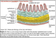
What type of junction is exhibited by the example of lumen of the gut and blood supply?
|
For example,molecules in the lumen of the gut (glucose for example) cannot pass through the intercellular space and therefore must be transported through the cell (in the direction necessary to bring the glucose to the blood supply in our example). Without these tight junctions, there would be a non-selective passage of
molecules from one end to the other side of the cell. |
|
|
What type of junction connects the cytoskeleton of a cell to those of its neighbor or to extracellular matrix?
|
Anchoring junction.
|
|
|
This type of anchoring junction is made of actin filaments. Specifically this type of anchoring involves adhesion belt and differentiation of cells.
|
Cadherins
|
|
|
Intermediate filament cell-cell attachment are called ______________ whereas cell-matrix attachment is called ________________.
|
Intermediate filament cell-cell attachment is called desmosome ; cell-matrix attachment is called hemidesmosome.
|
|
|
Name the two types of communicating junctions and the diff between the two / or examples.
|
Gap junctions. Direct connections such that they cells are electrically and metabolically linked. Ex. heart cells.
Chemical synapse: cells communicate even though they are not in physical contact. Ex. nueron, neurotransmitter, synaptic cleft. |