Reading...
![]()
Play button
![]()
Play button
![]()
Use LEFT and RIGHT arrow keys to navigate between flashcards;
Use UP and DOWN arrow keys to flip the card;
H to show hint;
A reads text to speech;
13 Cards in this Set
- Front
- Back
|
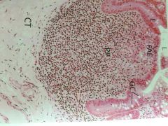
lymph nodule
(part of Peters patch) lumen lined by simp!e columnar epithelium with goblet cells Fae = follicle associated epithelium = no goblet and shorter, lymphocytes has no germinal center ct sm |
|
|
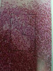
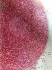
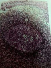
lymph nodule high magnification
lighter staining germinal center corona = darker staining corona =limiter amount cytoplasm = dense nucleus = small lymphocytes |
|
|
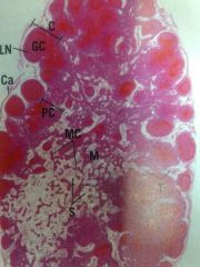
lymph node
kidney shaped convex and concave surface surrounded by capsule trabeculae projectct from capsule into substance cortex = peripheral part medulla = center = lighter staining paracortex = between medulla and cortex lymphatic nodules with germinal centers sinusoid's = gaps in medulla with medullary cords |
|
|
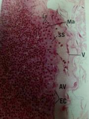
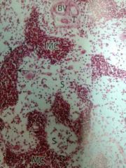
lymph node up close
afferent lymph vessels =vessels subscapular sinus = lymphocytes and macrophages sinus line by endothelial cells |
|
|
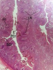
lymph node cortex
cortex = lymphatic nodules lymph node surrounded by adipose capsule trabeculae germinal center = light corona = dark staining |
|
|
lymph node medulla
sinusoids medullary cords = macrophages lymphocytes plasma trabeculae =ct blood vessels |
|
|
tonsil =Palatine
aggregate of lymphatic nodules germinal centers covered by stratified squamous nonkeratinised epithelium lining deep crypts ct capsule |
|
|
pharyngeal tonsil
aggregate lymphatic nodules germinal centers epithelial lining = pseudo stratified ciliated columnar lose collagenated ct |
|
|
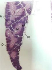
thymus
capsule = incompletely divides into lobules by ct septa cortex darker staining medulla lighter staining |
|
|
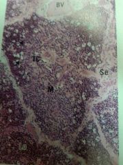
lobule thymus
medulla pale staining cortex dark staining = light patches are epithelial reticular cells and macrophages medulla of lobules continuous with each other blood vessels in septa |
|
|
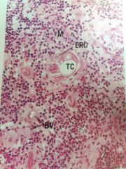
medulla of thymus
tymic corpuscles = epithelial reticular cells blood vessels |
|
|
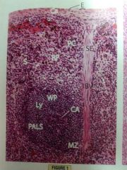
spleen
capsule = thick collagenous ct =surrounded by simple squamous epithelium septa =enter spleen from capsule =convey blood vessels white pull = dark staining around bv =pals configuration red pulp = sinusoids and pulp cords |
|
|
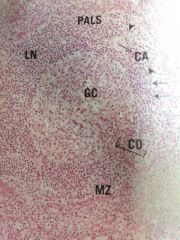
pals of spleen
germinal center central artery corona dark staining marginal zone |