![]()
![]()
![]()
Use LEFT and RIGHT arrow keys to navigate between flashcards;
Use UP and DOWN arrow keys to flip the card;
H to show hint;
A reads text to speech;
65 Cards in this Set
- Front
- Back
|
There are also two bony ridges connecting the two trochanters; the intertrochanteric line posteriorly and the trochanteric crest anteriorly. T/F |

False intertrochanteric line Anteriorly and the trochanteric crest posteriorly. |
|
|
ligamentum teres attaches to the fovea. T/F |
True |
|
|
The fovea is a small depression present on the head of femur bone. T/F |
True |
|
|
The neck of femur bone is cylindrical, projecting in a superior and lateral direction. T/F |
False Superior and medial |
|
|
The neck of femur bone is set at an angle of approximately 120° degrees to the shaft. T/F |
False 135° This angle of projection allows for an increased range of movement at the hip joint. |
|
|
Greater trochanter – the most medial palpable projection of bone that originates from the posterior aspect, just lateral to the neck. |
False Lateral Anterior aspect, just lateral to the neck |
|
|
What are the muscles that originate from the greater tronchanter : |
Gluteus medius, Gluteus minimum. Piriformis Vastus lateralis |
|
|
An avulsion fracture of the greater trochanter can occur as a result of forceful contraction of the gluteus minimus. T/F |
False Gluteus medius |
|
|
Lesser tronchanter projects from the posteromedial side of the femur, just inferior to the neck-shaft junction. T/F |

True |
|
|
________ is the site of attachment for iliopsoas (forceful contraction of which can cause an avulsion fracture of the lesser trochanter). |
Lesser tronchanter |
|
|
Intertronchanteric crest is located on the posterior surface of the femur. T/F |
True |
|
|
There is a rounded tubercle on its superior half called the quadrate tubercle; T/F |

True |
|
|
What muscle attaches to the quadrate tubercle located on the superior half of intertronchanteric crest |

Quadratus femoris |
|
|
The shaft of the femur descends in a slight lateral direction. T/F |
Medial |
|
|
A cross section of the shaft in the middle is circular but flattened posteriorly at the proximal aspect only. T/F |
and distal aspects. |
|
|
On the posterior surface of the femoral shaft, there are roughened ridges of bone, called the linea aspera (Latin for rough line. T/F |
True |
|
|
Linea aspera splits proximally to form the medial and lateral supracondylar lines. The f lat popliteal surface lies between them. |
False
Distally |
|
|
Proximally, the lateral border of the linea aspera becomes the pectineal line |
False Medial |
|
|
The lateral border of the linea aspera becomes the gluteal tuberosity, where the gluteus maximus attaches. T/F |

True |
|
|
Distally, the linea aspera widens and forms the floor of the popliteal fossa |

True |
|
|
The medial and lateral borders of the distal linea aspera forms the medial and lateral supracondylar lines. T/F |
True |
|
|
The medial supracondylar line ends at the adductor tubercle, where the adductor magnus attaches. T/F |

True |
|
|
The patella (kneecap) is located at the front of the knee joint, within the patellofemoral groove of the femur.T/F |
True |
|
|
Patella Inferior aspect is attached to the quadriceps tendon and superior aspect to the patellar ligament. |
False Superior - quadriceps tendon Inferior - patellar ligament |
|
|
Patella classified as a sesamoid type bone due to its position within the quadriceps tendon. T/F |
True |
|
|
__________ is the largest sesamoid bone in the body. |
Patella |
|
|
Patella Enhances the leverage that the quadriceps tendon can exert on the femur, increasing the efficiency of the muscle. T/F |
True |
|
|
The tibia is the main bone of the lower leg, forming what is more commonly known as the shin. T/F |
True |
|
|
The tibia is the second largest bone in the body and it is a key weight-bearing structure. T/F |
True |
|
|
The proximal tibia is widened by the medial and lateral condyles, which aid in weight-bearing. T/F |
True |
|
|
The condyles form a flat surface, known as the tibial plateau. This structure articulates with the femoral condyles to form the key articulation of the knee joint. |
True |
|
|
The shaft of the tibia is prism-shaped. T/F |
True |
|
|
The proximal aspect of the anterior border is marked by the tibial tuberosity; the attachment site for the patella ligament. T/f |

True |
|
|
The soleal line is the site of origin for part of the soleus muscle, and extends inferomedially, eventually blending with the medial border of the tibia. T/F |

True |
|
|
There is usually a nutrient artery distal end to the soleal line. |
False proximal |
|
|
Lateral border of the tibia bone also known as the interosseous border. It gives attachment to the interosseous membrane that binds the tibia and the fibula together. T/F |

True |
|
|
The medial malleolus is a bony projection continuing inferiorly on the medial aspect of the tibia. T/F |
True |
|
|
On the posterior surface of the tibia, there is a groove through which the tendon of tibialis posterior passes. T/f |

True |
|
|
Laterally is the fibular notch, where the fibula is bound to the tibia – forming the distal tibiofibular joint. T/f |
True |
|
|
The main function of fibula is to act as an attachment for muscles, and not as a weight-bearer. T/F |
True |
|
|
It has three main articulations mention them |
Proximal tibiofibular Distal tibiofibular Ankle joint |
|
|
Proximal tibiofibular joint – articulates with the medial condyle of the tibia. T/f |

False |
|
|
Distal tibiofibular joint – articulates with the f ibular notch of the tibia. T/f |

True |
|
|
Ankle joint – articulates with the talus bone of the foot. T/f |
True |
|
|
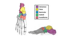
Bones of the foot can be divided into three groups: mention them |
•Tarsal •Metatarsal •Phalanges |
|
|
Tarsals – a set of seven regularly shaped bones. They are situated proximally in the foot in the ankle area. |

False Irregularly shaped bones |
|
|
Metatarsal connects the phalanges to the tarsals. There are f ive in number – one for each digit. T/f |
True |
|
|
The foot can also be divided up into three regions: mention them |
(i) Hindfoot – talus and calcaneus; (ii) Midfoot – navicular, cuboid, and cuneiforms; (iii) Forefoot – metatarsals and phalanges. |
|
|
The talus is the most superior of the tarsal bones. T/f |

True |
|
|
Talus has three articulations: mention them |
Superiorly – ankle joint – between the talus and the bones of the leg (the tibia and fibula). Inferiorly – subtalar joint – between the talus and calcaneus. Anteriorly – talonavicular joint – between the talus and the navicular. |
|
|
The main function of the talus is to transmit forces from the tibia to the heel bone (known as the calcaneus). T/f |

True |
|
|
Talus is wider anteriorly compared to posteriorly which provides additional stability to the ankle. T/f |
True |
|
|
Whilst numerous ligaments attach to the talus, no muscles originate from or insert onto it. T/f |
True This means there is a high risk of avascular necrosis as the vascular supply is dependent on fascial structures. |
|
|
The calcaneus is the largest tarsal bone and lies underneath the talus where it constitutes the heel. T/f |

True |
|
|
Calcaneus has three articulations. T/F |

False. It has two articulations Superiorly – subtalar (talocalcaneal) joint – between the calcaneus and the talus. Anteriorly – calcaneocuboid joint – between the calcaneus and the cuboid |
|
|
The posterior aspect of the calcaneus is marked by calcaneal tuberosity, to which the Achilles tendon attaches. T/F |
True |
|
|
The intermediate row of tarsal bones contains one bone, the navicular (given its name because it is shaped like a boat). T/f |

True |
|
|
Positioned medially, Navicular bone articulates with the talus posteriorly, all three cuneiform bones anteriorly, and the cuboid bone laterally. T/F |

True |
|
|
On the plantar surface of the navicular, there is a tuberosity for the attachment of part of the tibialis posterior tendon. T/f |
True |
|
|
The distal row, there are four tarsal bones – the cuboid and the three cuneiforms. These bones articulate with the metatarsals of the foot. T/f |

True |
|
|
The cuboid is furthest lateral, lying anterior to the calcaneus and behind the fourth and fifth metatarsals. As its name suggests, it is cuboidal in shape. T/f |

True |
|
|
The inferior (plantar) surface of the cuboid is marked by a groove for the tendon of fibularis longus. T/f |
True |
|
|
The three cuneiforms (lateral, intermediate (or middle) and medial) are wedge shaped bones. T/f |
True |
|
|
The shape of the bones helps form a transverse arch across the foot. They are also the attachment point for several muscles. T/f |

True |
|
|
Medial cuneiform – tibialis anterior, (part of) tibialis posterior and fibularis Kong's Lateral cuneiform – flexor hallucis brevis |
True |