Reading...
![]()
Play button
![]()
Play button
![]()
Use LEFT and RIGHT arrow keys to navigate between flashcards;
Use UP and DOWN arrow keys to flip the card;
H to show hint;
A reads text to speech;
97 Cards in this Set
- Front
- Back
|
What week do limbs begin to develop embryonically?
|
Initiation of limb development starts at week three
|
|
|
The lower extremity limb buds start at what vertebral levels?
|
L3-L5
|
|
|
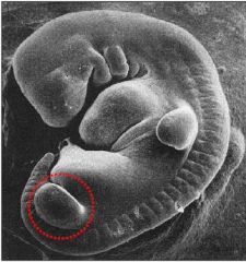
At what week does development of the LE limb buds begin?
|
Day 28 (week 4)
|
|
|
LE follows UE development and catches up at what week?
|
8
|
|
|
The core of the limb bud is derived from what tissue?
|
mesenchyme
|
|
|
The limb bud is surrounded by what?
|
An epithelial lining called "Ectodermal cap"
|
|
|
What is the signal source at the distal part of the limb bud?
|
AER (like the seam at the end of a hot pocket"
"growth along Proximal to distal axis. |
|
|
LE morphogenesis occurs from between what weeks?
|
From week 4-8
|
|
|
Describe the state of morphogenesis at Week 4
|
Elevation of tissue at the level L3-L5
|
|
|
Describe the state of morphogenesis at Week 4+
|
Rounded proximal portions of the LE limb buds can be distinguished
from the more tapered distal parts |
|
|
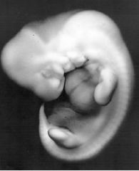
Describe the state of morphogenesis at week 5+
|
All three LE sections are present (Thighs Legs Feet are distinct), [Foot plates] are present.
|
|
|
Describe morphogenesis of LE at week 6+
|
[toe rays] are visible in the foot plates, but the rims of the foot plates remain smooth (cartilage models are forming)
|
|
|
Describe morphogeneis at the end of week 6!!! What day?
|
Day 47, medial rotation begins, toe rays are MORE prominent but rims of foot plates are still smooth. NOTE: LE's begin to flex toward the [parasagital] plane
|
|
|
Describe morphogenisis at week 7
|
LEs longer and feet have begun to approach eachother at midline (medial rotation continues)
NOTCHES in rims of footplates |
|
|
Describe Morphogenesis at week 8
|
LE’s longer and all regions are defined including the toes.
(Programmed cell death has occurred in the tissues between the toe rays –individual toes are seen.) Medial rotation of LE is complete. |
|
|
Amelia
|
Absence of one or more limbs
|
|
|
Meromelia
|
Absence of part of a limb
|
|
|
Phocomelia
|
Phoco= seal
absence of proximal limb structures (flipper like distal portions are present) |
|
|
Polydactyly
|
presence of extra digits or parts of digits
|
|
|
Syndactyly
|
Fusion of digits
|
|
|
Lateral Plate Mesoderm
SOMATIC part gives rise to what? |
Body wall and limb
|
|
|
What week are the tiniest buds present indicating limb development?
|
Week3
|
|
|
LPM SOMATIC mesoderm gives rise to what parts of the UE/LE?
|
Bones, Joints, Cartilage, Ligaments, Tendons, Fascia, Bood/lymphatic vessels
(General CT, BONES AND BLOOD) |
|
|
Cartilage models of bones form how, when?
|
Cartilage models of LE bones
mesenchymal core at the initially develop as condensations within the beginning of week 6. |
|
|
When do primary ossificatino centers begin to appear in the long bones?
|
Primary centers of ossification begin to appear in the long bones of the LE in the
8th to 12th weeks. |
|
|
LPM is divided into what two sections?
|
Loop on neural tube side: Somatic
Loop on notochord side: Visceral: Guts |
|
|
How is nural crest relevant to LE?
|
Gives rise to peripheral Posterior Root ganglia and nervous system, as well as schwann cells and some melanocytes
|
|
|
How is notochord relevant to LE?
|
Notochord becomes Nucleus pulposus which can herniate into the spinal cord
|
|
|
PAM (NOT LPM) divides into what 3 "tomes?"
|
Dermatome: Dermis
Myotome: Sk MM of LE Sclerotome: Vertebral Column |
|
|
What is chondrification? when does it begin?
|
Appearance of hyaline cartilage models of long bones, begins in week 6 in LE.
Begins as condensations of mesenchymal core |
|
|
When does endochondral ossification begin?
|
8th -12th week after primary centers of ossification appear.
|
|
|
in what part of the long bones do primary centers of ossification appear? When?
|
Diaphyses of LE long bones 8-12 weeks
|
|
|
Secondary centers of ossification in LE are called what? When do they begin to ossify?
|
Epiphyses, a few secondary might start around birth but the majority start after.
|
|

Week?
|

4
|
|
Week?
|

4
|
|

Week?
|

4+
|
|
Week?
|

5
|
|

week?
|

5
|
|

Week?
|

6
|
|

What week?
|

6
|
|

Week?
|

8
|
|

Whas dis? think developing LE
|

Mesenchymal condensations giving rise to hyaline cartilage outlines of the bones of LE
|
|

Whats this? From whence did it come?
|

mesenchyme from Somatic LPM
|
|

Was dis? what week is this?
|

AER, 4+
|
|

Name for condition
|

amelia
|
|

This is the process of what?
|
Endochondral ossification
|
|

name of condition
|
Phocomelia
|
|

diagnosis
|
Syndactyly
|
|
|
Where does paraxial mesoderm begin to develop?
|
along the sides of the neural tube
|
|
|
Somites are derived from what?
|
Paraxial Mesoderm
|
|
|
Somites give rise to what in the LE?
|
Muscles
|
|
|
Muscles of the LE are derived from what cells?
|
Somitic mesoderm (from somites) NOT MESENCHYME FROM LPM SOMATIC CELLS, DONT MISS THIS ON THE EXAM!!!!!!!
|
|
|
How do MM cells get into the already packed mesenchymal areas making bones, ct, and vessels?
|
Myogenic cells from the myotome (part of the somite) will migrate into the limb bud and
fuse to form the skeletal muscle of the LE. The connectives tissues present within the limb bud (derived from somAtic mesoderm) Guide and form the scaffolding for this muscle formation process. |
|
|
What are the derivations of the Dorsal, middle, and ventral parts of the Somite?
|
Dermatome (dorsal) dermis (not epidermis)
Myotome (middle) MM and some NN Sclerotome (stomach side) gives rise to Spinal Vertebrae (I tried to make that easy to remember D, M, S |
|
|
Somite migration cells travel towards the neural tube and towards the belly, going towards the belly they have to cross a point of no return, called:
|
Lateral SOMITIC fronteir
It's like the rio grande, on mexico side they're all Mexicans, once they cross they have a grip of names that they can become. My favorite is friend, Than's favorite is Wife, Hayden's favorite is chingaso. |
|
|
What is the primaxial domain?
|
Myotime that migrates to the Primary axis of the body, the spinal region.
Associated with Neural tube and notochord, |
|
|
What do myotomes and dermatome form in the primaxial domain?
|
Myotome: True back muscles
Dermatome: Dermis of the back |
|
|
What is the Abaxial domain?
|
Ab= away from the body's main axis (when we were fish we didn't have these somites, when we began to walk on land they were formed, Abaxial- away from the axis) to body wall and limbs.
YOu know what Myotome and dermatomes do. |
|
|
Describe the traditional way of labeling Primaxial and abaxial.
|
Myotome was divided into Epimere (like Primaxial- true back) and Hypomere (like abaxial- body wall and limb)
but the MM are organized as a dorsal and ventral MM mass guided by the bones. This initially was extremely segmented, but muscles twist and fuse and segementation is lost. |
|
|
What is the MM/Somite rule?
|
Muscles are derived from the somites of the same area that innervates them
LE is area L2,3,4 |
|
|
Define Dorsal and Ventral muscle masses
|
Initially the generic limb buds can be described as having: : Dorsal surfaces (or sides): continuous with back
Ventral surfaces (or sides): continuous with “belly” |
|
|
Define the association of movement and Dorsal/ventral muscle masses
|
Musculature associated with these surfaces or sides: Dorsal (surface) muscle mass: primarily extensors (& abductors)
Ventral (surface) muscle mass: primarily flexors (& adductors |
|
|
What in the freak is the Limb bud axis? What areas are they?
|
imaginary line drawn through the femur to the 2nd digit inferiorly.
Preaxial portion/border: tibia and hallux (medial) Postaxial portion/border: Fibula and the 5th |
|
|
Pre/Post axial MM are associated with what actions?
|
Preaxial: from ventral MM Mass; Flexors/adductors)
Postaxial: from dorsal MM (extensors, abductors) PreVee FAd (only need to memorize one) |
|
|
What happens to pre/postaxial regions with the medial rotation of the femur?
|
Preaxial moves to medial poserior
Postaxial moves to lateral/anterior |
|
|
Medial rotation of the femur happens on what pt of the femur?
|
Occurs embryonically from the distal half of the femur (hip NOT affected by the rotation)
|
|
|
Preaxial muscles are from what MM mass, become what MM?
|
Ventral MM mass,
Iliopsoas, Pectineus, Adductor longus, magnus, other flexors and adductors |
|
|
Postaxial muscles are from what MM mass, become what MM?
|
Dorsal MM mass, Gluteal MM, extensors and abductors
|
|
|
What pt of the femur is affected by rotation?
|
distal 1/2, so the muscles that act on everything distal to the hip, like at the knee, STJ, and ankle, are affected by rotation.
|
|
|
Knee: preaxial MM, from what mass?
|
Ventral Muscle mass
Become hamstrings (biceps femoris, semitendinosus, membranosus) and knee flexors |
|
|
The medial and posterior compartments of the thigh are derived from what "axial Muscles" and therefore what MM mass?
|
Preaxial MM, ventral MM Mass
|
|
|
Knee: postaxial MM, from what mass?
|
Dorsal MM mass, becomes quadriceps femoris, knee extensors
|
|
|
Preaxial MM, from what MM mass become what cmopartments of the leg?
|
Ventral MM mass, posterior leg, plantar foot
|
|
|
Preaxial MM, from what MM Mass, have what action at ankle, STJ, toes?
|
Plantarflexion
Inversion Flexion |
|
|
Postaxial MM, from what MM mass, become what compartment of the leg and foot?
|
Dorsal MM mass become anterior and lateral leg compartments/ dorsum of foot (extensors and abductors)
|
|
|
Postaxial MM, from what MM mass, have what action at ankle, STJ, toes?
|
Dorsiflexion
Inversion or eversion Extension |
|
|
INTERMISSION
|
Take a sip, take a deuce, say a prayer, tie a noose. Now back for another round.
|
|
|
what do posterior primary rami innervate?
|
True back MM and dermis of the back.
|
|
|
During development of LE the Neural tube contributes what?
|
Neural tube- Spinal cord- alpha motor neurons- Anterior Roots (exiting to LE)
|
|
|
Describe the formation of initial spinal N during LE development
|
Migrating axonal processes from motor neurons in the developing anterior horns of the spinal cord and the peripheral processes of sensory neurons located in the developing posterior root ganglia will join to form the initial spinal nerves.
|
|
|
During dvelopment of LE the Neural Crest contributes what?
|
(Not neural tube) PRG cells (sensory aspect) to form posterior horns, Schwann cells that myelinate PNS, Satellite cells
|
|
|
What happens after inital spinal N are formed in development of the LE?
|
Spinal nerves split to form posterior and anterior primary rami.
|
|
|
When do NN inervate the LE?
|
As soon as the limb bud forms, anterior primary rami will penetrate into the limb bud mesenchyme where they will innervate the LE muscles and dermis.
|
|
|
What rami innervates the LE?
|
Anterior, Posterior goes to true back mm and overlying dermis
|
|
|
What happens to each anterior primary rmaus as it enters the base of the LE limb bud?
|
It splits into an anterior and posteiror dividsion
|
|
|
The Anterior division from the anterior primary ramus innervate what?
|
Anterior divisions innervate preaxial MM,
Flexor Compartments and adductors and the dermis and skin overlying the preaxial Muscles So.. to add to the other mnemonic PreVee FAd Ant-PreVee Fad Anterior Division to Preaxial (Ventral MM mass) that does Flexion/adduction |
|
|
The posterior division from the anterior primary ramus innervate what?
|
Posterior division to postaxial muscles that will form the extensor compartments and abductors and the dermis of the skin overlying postaxial Muscles
|
|
|
What domain are the MM of the LE? therefore what rami innervate them?
|
Abaxial domain (hypomere)
Anterior primary rami |
|
|
What happens as a result of rotation of LE to the myotome arrangement that was so orderly?
|
An adult M is formed from more than one myotome and its inervation comes from more than one anterior primary ramus
|
|
|
what naming difference is there from the brachial plexus to LE?
|
No trunks.
|
|
|
Explain the Mnemonic again:
|
Ant-PreVe FAd
Anterior division innervates the Preaxial domain (ventral muscle mass) that are the Flexors/Adductors All you need to know is one. |
|
|
Explain the naming of lumbar and sacral plexuses
|
Anterior primary rami from I2-S3 that then divide into ant/posterior divisions, that then come together to named nerve (example sciatic is parts of anterior/posterior divisions of certain anterior rami)
|
|
|
Nerves formed from posterior divisions of the anterior primary rami innervate what? What are the names?
|
Femoral, common fib
Extensors and abductors and lateral rotators (postaxial) and dermatomes on extensor surface |
|
|
Nerves formed from anterior divisions of anterior primary rami innervate what? what are the names?
|
Ant-PreVe FAd
innervate flexors/adductors and medial rotators and dermatomes on flexor surfaces Obturator N Tibial N (medial and lateral plantar brs) |
|

What is the Pink,
Purple? Blue" |
Primaxial
Abaxial Lateral somitic frontier "Manifest destiny" |
|

What are these three items in the limb bud? Top to bottom
|
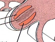
Dorsal MM mass
Chondrification (hyaline from mesenchymal) Ventral MM Mass Ant-PreVe FAd |
|
|
Explain Limb bud axes
|

|