![]()
![]()
![]()
Use LEFT and RIGHT arrow keys to navigate between flashcards;
Use UP and DOWN arrow keys to flip the card;
H to show hint;
A reads text to speech;
30 Cards in this Set
- Front
- Back
|
Differentiate between Leukaemia and Lymphoma |
Leukaemia is malignancy of WBCs in the Blood and Bone marrow ('emia' - blood) Lymphoma is malignancy of WBCs in Lymph nodes and other solid tissues |
|
|
Classification of Leukaemia |
There are 4 classes ● Acute Lymphoblastic Leukaemia/ALL (usually affect the Common Lymphoid Progenitor cell) ● Acute Myeloid Leukaemia/ AML (usually affects Common Myeloid Progenitor) ● Chronic Lymphocytic Leukaemia/CLL (Majorly affects B cells) ● Chronic Myeloid Leukaemia/CLL (affects all mature myeloid leukocytes; neutrophils, basophils, eosinophils, monocytes) |
|
|
The most common type of leukaemia is |
ALL |
|
|
Acute leukaemia have rapid onset and are rapidly fatal. T/F |
True |
|
|
Which organ destroys old or non-viable WBCs |
Spleen (destroys RBCs and WBCs) |
|
|
Gum swelling is seen in which leukaemia |
AML (monocytic type). |
|
|
Organs infiltrated in ALL are |
Lymph node, Thymus, Liver, Spleen |
|
|
Organs infiltrated in AML are |
CNS, Testes, soft tissues |
|
|
Disseminated Intravascular Coagulation (DIC) occurs in which acute leukaemia |
Acute Promyelocytic Leukaemia (morphological subtype of AML) |
|
|
Acute leukaemia is diagnosed when the number of blasts within marrow is what % |
Over 20% |
|
|
What are the electrolyte findings in Acute leukaemia |
● Increased phosphate levels ● Increased LDH ● Increased K+ ● Decreased Ca2+ All these are due to destruction/lysis of the incompetent blast cells. |
|
|
Difference between ALL and AML on light microscopy |
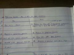
|
|
|
Does ALL occur in both children and adults |
Yes it does, but 80% of it is in children |
|
|
What genetic disorders predispose to ALL |
D.A.N 1) Down Syndrome 2) Neurofibromatosis Type 1 3) Ataxia
|
|
|
According to immunological classification of ALL, the common ALL (cALL) is |
Early Pre B cells (CD10+). It is called so because it is the most usual in children. |
|
|
Which ALL occurs mainly in males |
T cell ALL |
|
|
Based on genetic consideration, ALL in adults is usually related to |
Philadelphia Chromosome. |
|
|
Mention the 2 types of AML |
1) De-novo AML 2) Secondary AML (that is secondary to a disease) |
|
|
Erythroid leukaemia of AML is also called |
DiGuglielmo disease D.E like direct entry (DiGuglielmo disease - Erythroleukaemia) |
|
|
The only leukaemia that presents with Thrombocytosis is |
CML. Others present with thrombocytopenia |
|
|
B cell surface receptors in CLL |
CD19, CD23 (B cell) CD5 (T cell)
|
|
|
CLL is monoclonal because |
It expresses just one form of light chain (either lambda or kappa) |
|
|
Which lymphocyte is majorly affected in CLL |
B cell (95% of times) |
|
|
Lymphadenopathy in CLL is usually found in what regions of the body |
Neck, axillae and inguinal region |
|
|
If CD5 (usually on T cell) is expressed on B cell, it is called |
Lineage infedelity |
|
|
Differential diagnosis of CLL |
1) B-PLL 2) T-PLL/CLL 3) Hairy cell leukaemia 4) Non-Hodgkin lymphoma with circulating lymphoma cells 5) Mononucleosis and viral infections 6) ALL |
|
|
Mention the 2 staging known for CLL |
1) Rai staging (L+L+S+A+P)2) Binet staging 1) Rai staging (L+L+S+A+P)2) Binet staging 1) Rai staging (L+L+S+A+P)2) Binet staging |
|
|
What is Binet staging |
Classifies CLL based on number of lymph nodes affected (lymphadenopathy), presence of anaemia and thrombocytopenia. There are 3 stages: ● Stage A : < 3 lymph nodes affected, no anaemia, no thrombocytopenia ● Stage B : > 3 lymph nodes affected, no anaemia, no thrombocytopenia ● Stage C : > 3 lymph nodes affected, with anaemia and thrombocytopenia. |
|
|
Richter syndrome is a complication of |
CLL (Chronic Lymphoblastic Leukaemia) |
|
|
Chemotherapy for CML is with |
Hydroxyurea |