Reading...
![]()
Play button
![]()
Play button
![]()
Use LEFT and RIGHT arrow keys to navigate between flashcards;
Use UP and DOWN arrow keys to flip the card;
H to show hint;
A reads text to speech;
40 Cards in this Set
- Front
- Back
|
How is skeletal muscle involved in metabolic function?
|
- body temperature
- interacting with thyroid hormone - involvement in glucose metabolism |
|
|
How is it best to understand the functional attributes of muscles?
|
Include the gross architecture of the entire muscle, the microscopic features of the cells that make up the muscle, and the biochemical makeup of the muscle fibers.
|
|
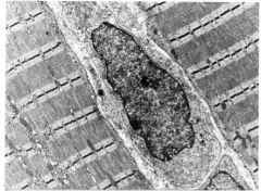
In this picture locate the Z and M lines and the A, I and H bands, mitochondria and granular glycogen in the sarcoplasm.
|
From Valentine's notes:
In this ultrastructural photograph you can see portions of two skeletal muscle fibers with a fibroblast in between. The fibroblasts are the cells that produce the interstitial collagen. In this photograph you should be able to identify the Z and M lines and the A, I, and H bands. Mitochondria and granular glycogen can also be seen within the sarcoplasm. Be sure that you can identify these structures. |
|
|
What would happen to a muscle cell with dysfunctional mitochondria or with no glycogen?
|
Both come down to oxidative phosphorylation - no mitos, no oxidative phosphorylation; no glycogen, no glucose to feed Kreb's cycle to feed oxidative phosphorylation.
|
|
|
What are terminal cisternae extensions of?
|

The sarcoplasmic reticulum.
|
|
|
What are the T-tubules an extension of?
|
The sarcolemma, but they are linked to the SR through a slow release Ca channel activating the ryanodine receptor at the triad.
|
|
|
Where is Ca stored in skeletal muscle?
|

The terminal cisternae (extensions of the SR)
|
|
|
What does the SR regulate?
|

Ionized Ca release and uptake.
|
|
|
Where is Ca stored in skeletal muscle?
|

The terminal cisternae
|
|
|
T/F the slow Ca channel in skeletal muscle is involved in generation of the action potential?
|
F. It is involved in activating the ryanodine receptor through a channel conformational change. This change causes release of Ca from SR terminal cisternae into the sarcoplasm and contraction of the myofibrils.
|
|
|
What would abnormalities in the ryanodine receptor of skeletal muscle cause?
|
Malignant hyperthermia, as the activation of excitation-contraction coupling would be unregulated. This leads to excessive muscle contraction and heat generation.
|
|

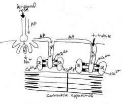
Be sure that you understand the series of electrical and molecular events that are involved in muscle contraction and relaxation. Identify the myosin and actin filaments, the A and I bands, and the Z line in the diagram.
|

Diagrammatic representation of excitation-contraction coupling in normal skeletal muscle. Action potentials (AP) generated by nerves are propagated along the sarcolemma to the t-tubule and travel down the t-tubule; depolarization of the t-tubules activates the slow calcium channels at the triads that interact, through a protein-protein interaction mechanism, with the ryanodine receptors within the membrane of the terminal cisternae of the sarcoplasmic reticulum (SR). This causes release of calcium from the SR into the sarcoplasm and results in contraction of the myofibrils.
|
|
|
Are the Ca ions that enter the cell through the protein-protein interaction involved in the action potential activity?
|
No, blocking the calcium channel activity of these structures does not interfere with excitation contraction coupling. It is the protein-protein interaction with the ryanodine receptor that is important.
|
|
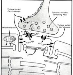
The numbers in this diagram are in sequence. Name the steps
|

(1) opening of presynaptic voltage-gated calcium channels as a result of a decrease in membrane potential, (2) calcium activated fusion of synaptic vesicles to the nerve terminal membrane and release of acetylcholine (ACh), (3) binding of ACh to ACh receptors causing influx of sodium ions into the muscle cell and generation of a muscle membrane action potential, (4) muscle membrane action potential activated opening of muscle membrane voltage gated sodium channels resulting in propogation of the action potential, and (5) opening of voltage gated calcium channels in the transverse tubules leading to ryanodine receptor activation on the membrane of the terminal cisternae of the sarcoplasmic reticulum, and release of stored calcium into the sarcoplasm to result in actin/myosin interaction and contraction.
|
|
|
How would hypocalcemia effect skeletal muscle?
|
The ability of the T tubules and sarcoplasmic reticulum at the triads to regulate the cycling of calcium ions into and out of the sarcoplasmic reticulum is critical for the muscle to exhibit sustained or repeated contraction and to allow relaxation. Calcium plays a vital role in this process, and alterations of available calcium ions can result in severe skeletal muscle dysfunction. A common example of this occurs in dairy cattle with “milk fever” in which hypocalcemia (lowered blood calcium) results in severe muscle weakness and the animal becomes recumbant. Therapy with intravenous calcium-containing solutions can result in rapid recovery.
|
|
|
How do gradations in the force of muscle contractions occur?
|
AP's spread from the neuromuscular junction to initiate all or nothing contractions. To vary the force of the muscle contractions, you vary the number of recruited fibers.
|
|
|
Which type of muscle are high in mitochondria and fat content and low in glycogen content.
|
Type 1, slow-twitch
pOstural, slow (One, pOstural, slOw, Oxidative, mitOchondrial plethora) |
|
|
Which muscle type is oxidative, fatigue resistant and aerobic?
|
Type 1, slow-twitch
pOstural, slow (One, pOstural, slOw, Oxidative, mitOchondrial plethora) |
|
|
Describe type 2A muscles
|
Fast twitch, oxidative and glycolytic, fatigue resistant.
|
|
|
Which muscle type has intermediate numbers of mitochondria, fat and glycogen content?
|
Type 2A Fast-Twitch
|
|
|
T/F Muscle type 2A can be described as fast-twitch, glycolytic, fatigue sensitive, and anaerobic.
|
False, 2B Fast-twitch is the anaerobic muscle. It has low mitochondrial numbers and fat content. It also has high glycogen content.
|
|
|
What do denervated muscle cells undergo?
|
Atrophy.
|
|
|
Will exercise result in increased fiber numbers?
|
No, but it will result in increased fiber diameter.
|
|
|
What is the mosaic pattern? What do alterations to the pattern indicate?
|
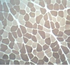
the intermingling of the type 1 (light staining) and type 2 (dark staining) fibers. Alterations of this normal mosaic pattern are often due to pathologic conditions that affect the muscle’s innervation.
|
|
|
What is a motor unit?
|
The group of muscle fibers innervated by a single nerve is called the motor unit.
|
|
|
What is the signifigance of the size of motor units?
|
The size of motor units also influences the type of movement the muscle is capable of performing. For example, extraocular muscles have many nerves, each innervating a small number of fibers. This allows for fine movements of the globe. In contrast, the quadriceps muscle has fewer nerves innervating a larger number of fibers, which allows for maximum strength of contraction without as much fine motor control.
|
|

These are all possible arrangements of skeletal muscle fibers within muscles. Name them.
|
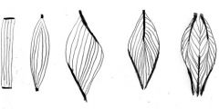
In order from left to right:
Parallel, Fusiform, Unipennate, Bipennate, Multipennate |
|
|
How does the contraction of cardiac muscles differ from the contraction of skeletal muscle?
|
Cardiac myocytes contain T tubules and sarcoplasmic reticulum similar to skeletal muscle fibers, but the sarcoplasmic reticulum is less well-developed.
They have intercalated disks for the transmissal of the contractile impulses. They have inherent rhythmicity. |
|
|
What are the two purposes of intercalated disks?
|
1. Firmly bind adjacent cells together to prevent separation in contraction.
2. Provide areas for communication of the impulses for contraction. |
|
|
What are the Z-bands of smooth muscle?
|
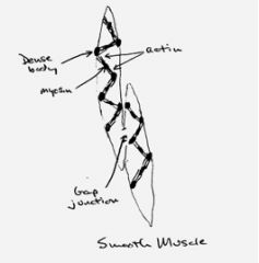
The intermediate, actin and myosin filament structures interacting.
It has been suggested that the contractile force developed by the sliding contractile filaments is transmitted to the intermediate filament harness causing shortening of the long axis of the fiber. |
|
|
How do smooth muscle depolarizations initiate?
|
Spontaneous membrane depolarization, circulating hormones, local environments (like pH), quick muscle stretch (like when you swallow too much and can feel it getting squeezed down).
|
|
|
How do smooth muscle stimulations propagate?
|
Cell to cell through gap junctions.
|
|
|
T/F Smooth muscle cells have pacemakers.
|
True, though not like in the heart. Areas of high neurotransmittor content may act as “pacemakers” to control the force and direction of peristalsis.
|
|
|
Is there sarcoplasmic reticulum in smooth muscle?
|
It is poorly developed.
|
|
|
Are there T-tubules in smooth muscle?
|
No.
|
|
|
Compare and contrast the biochemistry of smooth muscle contraction to skeletal muscle contraction. Start with Ca.
|
Smooth muscle contraction also involves calcium, but the biochemical events are different than those occurring in skeletal muscle contraction. Smooth muscle does not contain the molecule troponin that is so important in initiating contraction of skeletal muscle. Instead, calmodulin and inositol triphosphate are thought to play major roles in excitation-contraction coupling in smooth muscle.
|
|
|
What type of muscle (smooth, cardiac, or skeletal) has the longest contraction?
|
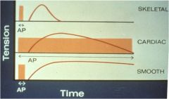
Smooth. The cycling time for the formation and release of cross bridges between actin and myosin and subsequent release is much longer in smooth muscle than in skeletal muscle
|
|
|
What type of muscle (smooth, cardiac, or skeletal) contraction requires the least amount of ATP?
|
Smooth.
|
|
|
What type of muscle (smooth, cardiac, or skeletal) is capable of a graded contraction, rather than all or none?
|
Smooth
|
|
|
Did you know that there are undifferentiated cells in skeletal muscle that are available to be made into myocytes when repair is necessary?
|
It's true. They are called satellite cells and they can re-establish skeletal muscle.
Smooth muscle can divide and repair on its own. Cardiac muscle can't. |