![]()
![]()
![]()
Use LEFT and RIGHT arrow keys to navigate between flashcards;
Use UP and DOWN arrow keys to flip the card;
H to show hint;
A reads text to speech;
26 Cards in this Set
- Front
- Back
|
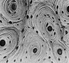
Cartilage |
Semi-rigid connective tissue; articular surfaces of bones within joints; younger people have more cartilage because it ossifies over time |
|
|
Bone |
Hard form of connective tissues Functions: support/protection for vital internal structures; mechanical basis for movement; storage of Ca/Ph; supply of new blood cells (marrow) |
|
|
Periosteum |
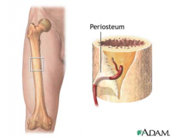
Fibrous connective tissue surrounding the bone Functions: provide nourishment (blood supply); nociceptive nerve endings (sensing pain); can lay down more cartilage or bone (fracture healing); provide interface for attachment of tendons and ligaments |
|
|
Perichondrium |
Fibrous connective tissue surrounding cartilage in developing bone Functions: provide nourishment (blood supply); nociceptive nerve endings (sensing pain); can lay down more cartilage or bone (fracture healing); provide interface for attachment of tendons and ligaments |
|
|
Long bones |
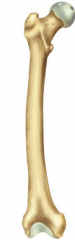
Tubular with shaft and two ends Examples: femur, tibia, humerus |
|
|
Short bones |
Cuboidal in ankle and wrist; only in carpus and tarsus Examples: scaphoid, cuneiforms |
|
|
Flat bones |
Usually serve protective functions Examples: sternum, scapula |
|
|
Sesamoid bones |
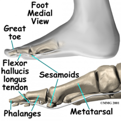
Embedded within a tendon; add mechanical advantage and protect tendons from wear Examples: medial/lateral sesamoids of great toe (FHB), patella |
|
|
Accessory bones |
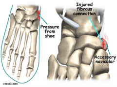
Form additional bone forming center that hasn't joined with main bone Examples: accessory navicular in foot |
|
|
Cortical (compact) |
An outer layer of bone surrounding a central medullary canal of cancellous bone; rigid support; slow turnover; very dense and strong (thick white part of bones on X-ray); thickest in the diaphysis (shaft) of long bones |
|
|
Cancellous (spongy) |
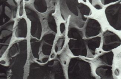
Less dense than cortical; increased turnover; more elastic; medullary canal filled with bone marrow Two types: red (hematopoiesis) or yellow (inert & fatty) |
|
|
Types of cells |
Osteoblasts; osteocytes; osteoclasts |
|
|
Osteoblasts |
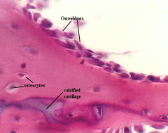
Derived from mesenchymal stem cells; forms bone organic matrix and its mineralization (osteoid); also known as immature bone cells |
|
|
Osteocytes |
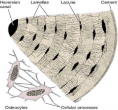
90% of cells in mature skeleton; within mineralized matrix in lacunae; connected to cell processes via cannuliculi; nutrient transport, Ca/phos regulation; also known as osteoblasts that have become encased in bone matrix during bone tissue production and may serve as sensors of mechanical stimuli within bone tissue |
|
|
Osteoclasts |
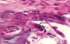
Arise from hematopoietic stem cells; if activated, bone breakdown (fracture remodeling, calcium mobilization); responsible for remodeling of bone to reduce its volume |
|
|
Intramembranous bone formation |
Mechanism by which a long bone grows in width; osteoblasts differentiate directly from preosteoblasts and lay down seams of osteoid; does NOT involve cartilage anlage |
|
|
Endochondral bone formation |
Mechanism by which a long bone grows in length; osteoblasts line a cartilage precursor |
|
|
Cutting cones |
Primarily a mechanism to remodel bone; osteoclasts at the front of the cutting cone remove bone and trailing osteoblasts lay down new bone |
|
|
Fracture healing |
1. Inflammation: healing cells to fracture sight 2. Repair: periosteal callus forms along the periphery (intramembranous ossification initiated by preosteoblasts) 3. Remodeling: intramedullary callus forms in the center of the fracture (endochondral ossification) |
|
|
Sources of blood supply to long bones |
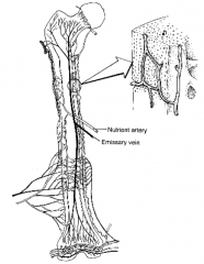
1. Nutrient artery system: run longitudinal within medullary cancal after piercing bone cortex (pierce the cortical bone at nutrient foramina) 2. Metaphyseal-epophyseal system: supply the ends of hones 3. Periosteal system: multiple brances at periphery of bone (majority of cortical or compact bone receivers) |
|
|
Hip |
Proximal femur, pelvic bones |
|
|
Thigh |
Femur |
|
|
Leg |
Tibia and fibula |
|
|
Foot |
Tarsus |
|
|
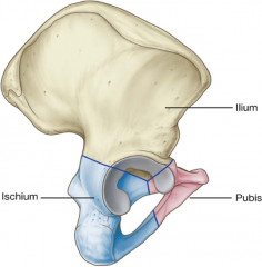
Pelvis |
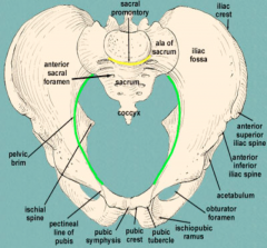
Sacrum + (innominate bones) ilium, ischium, pubis |
|
|
Innominate bones (pelvis) |
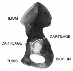
Ilium, ischium, pubis joined at triradiate cartilage to form acetabulum |