![]()
![]()
![]()
Use LEFT and RIGHT arrow keys to navigate between flashcards;
Use UP and DOWN arrow keys to flip the card;
H to show hint;
A reads text to speech;
42 Cards in this Set
- Front
- Back
|
Motor recruitment |
Progressively activating more and more motor units |
|
|
Small motor units contain? |
Few muscle fibers and are type 1 (slow,Oxidative, red) |
|
|
Large motor units contain? |
Large number of muscle fibers, type 2, (fast, glycolic, white) |
|
|
Muscle tone |
At any time some fibers in a muscle are contracted while others are relaxed. Essential for posture and balance. |
|
|
Muscle twitch |
Response of a whole muscle to a single stimulus |
|
|
Latent period |
Few milliseconds the muscle does not contract. Covers time of the action potential, calcium release from sarcoplasmic reticulum and molecular events before power stroke of myosin |
|
|
Treppe |
Muscle stimulated repeatedly, allowing sufficient time for relaxation between stimuli, responds with a series of separate twitches that progressively increase up to a max value after a few seonds. Caused by calcium buildup in sarcoplasm |
|
|
Tetanus |
Skeletal muscle stimulates so frequently it cannot relax after previous twitch. Contractions merge not a sustained contraction. Characterizes most of contractions made by muscles during voluntary activity. |
|
|
Isotonic contraction |
Muscle shortens and tension remains constant |
|
|
Isometric contraction |
Muscle length does not change but tension developed increases sharply |
|
|
Smooth muscle |
Cells are considerably shorter than skeletal muscle fibers. Each fiber has 1 nucleus. Contains actin and myosin. Lack striations,not well organized. Lack T-Tubules, troponin and tropomyosin. Contacts slower than skeletal muscle but greater degree of shortening and large movement can be produced. |
|
|
Types of smooth muscle |
Multi unit smooth muscle and visceral smooth muscle. |
|
|
Multi unit smooth muscle |
Cells are not well organized. Occurs as separate fiber rather than sheets. Separate fibers may contract independently. Contractions initiated by signals from nerves in autonomic nervous system. Iris of eye, walls of blood vessels, large passage ways of lungs, arrector pili muscle |
|
|
Visceral smooth muscle |
Single unit. Cells arranged in wrap around sheets. Found in walls of hollow organs such as stomach, intestine urinary tract, reproductive tract and small blood vessels. Cells are in close contact and can stimulate each other via gap junctions. |
|
|
Peristaltic movements |
Wave like movements in visceral smooth muscle. Can force the contents of hollow organs in particular direction. |
|
|
Pacemaker cells |
Self initiates Repeated waves of excitation. Creates rhythmicity in visceral smooth muscle |
|
|
Calmodulin |
Mediates excitation contraction coupling in smooth muscle. Calcium binding protien |
|
|
Controls smooth muscle contraction |
Nervous input from autonomic nervous system Hormones and related substances in circulation Substances produced withing a metabolizing tissue cam cause smooth muscle to relax. Stretching |
|
|
Intercalated disc |
Connect cardiac muscles end to end in branching networks. Contains gap junctions to spread electrical excitation. |
|
|
Muscle Orgin |
The end of the muscle which does not move |
|
|
Muscle instertion |
The end the muscle attached to the moving skeletal unit. |
|
|
Kinesiology |
Study of different types of muscles, lever systems and their movements |
|
|
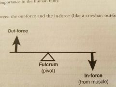
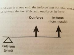
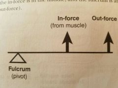
Out force |
Generated by the lever. Move hand lift weight |
|
|
In force |
Force the muscle applies to lever to accomplish movement |
|
|
First class lever |
|
|
First class lever examples |
Lifting the head Straightening arm at the elbow Movement of foot around fulcrum of ankle |
|
|
Second class lever i.e wheelbarrow |
|
|
Third class lever |
|
|
Third class lever examples |
Most common in body Flexing forearm at elbow |
|
|
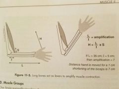
Formula for muscle amplification |
|
|
|
Flexor |
Decrease angle of joint |
|
|
Extensor |
Increase angle of joint |
|
|
Abductor |
Moves bone away from midline |
|
|
Adductor |
Moves bone closer to midlie |
|
|
Supinator |
Turn palm upward or anteriorly |
|
|
Pronator |
Turns palm downward or posteriorly |
|
|
Satellite cells |
Stem cells that repair damaged muscle fibers. Quiescent myoblast. |
|
|
Muscular hyperplasia |
Increase in number of muscle fibers in a muscle. Humans skeletal muscles have very limited ability to form new fibers |
|
|
Muscular hypertrophy |
Increase in size of muscle fiber. Due to production of myofibrils, mitochondria and sarcoplasmic reticulum |
|
|
Muscular atrophy |
Wasting away of muscle fibers |
|
|
Myasthenia gravis |
Autoimmune disease where patient develops antibodies against acetylcholine receptors in neuromuscular junction |
|
|
Duchenne muscular distrophy |
Causes muscular atrophy. Hereditary disease. Mutation in general for dystrophin. Localized in sarcolemma. Integral for anchoring certain integral membrane glycoproteins which may control calcium flow into muscle |