![]()
![]()
![]()
Use LEFT and RIGHT arrow keys to navigate between flashcards;
Use UP and DOWN arrow keys to flip the card;
H to show hint;
A reads text to speech;
30 Cards in this Set
- Front
- Back
|
Hippocampus |
- Part of the limbic system - Interacts with endocrine and autonomic systems - Memory - Navigation |
|
|
Left brain |
- Linear reasoning - Mathematics - Present vs. past - Primarily the dominant structure |
|
|
Cerebellum |
- Visual signals - Proprioceptive input - Language - Simple problem solving - Balance and coordination |
|
|
The prosencephalon develops into |
- Telencephalon: cerebrum, hippocampus, amygdala, olfactory bulbs - Diencephalon: eyes, pituitary gland, hypothalamus, thalamus, epiphysis |
|
|
Evolutionary trends (Fish to human brain) |
- Human Hindbrain: relatively smaller (to fish brain) - Midbrain: relatively smaller - Forebrain: relatively larger |
|
|
Functions of the blood brain barrier |
- Reduces the immune systems access to the brain - Comprised of the cells that make up the smallest blood vessels of the brain - Physiological transport systems - Accounts for some drug actions: Morphine vs. heroin - Glucose and other important brain substrates are transported through active transporters |
|
|
Crista ampullaris |
Sensory receptor organ for rotational acceleration and deceleration housed within the ampulla of each semicircular canal of the inner ear |
|
|
Causes of Huntington's Disease |
- Extra CAG repeat on the end of chromosome 4 (insertion) - Degeneration of neuronal cells in the frontal lobe, basal ganglia, and caudate nucleus |
|
|
Factors of the mammalian diving reflex |
- Bradycardia: heart rate drops 20% - Peripheral vasoconstriction: decreased blood flow to extremities - Blood shift: plasma into thoracic cavity (diaphragm and neck); helps lungs from collapsing - The trigeminal nerve (V) innervates the vagus nerve (X) |
|
|
Quadriplegia |
- Transection of the cervical region - Results in paralysis of the four limbs |
|
|
Ruffini's corpuscles |
- Simple receptors: encapsulated - Mechanoreceptors (deep pressure and stretch) - Responds to deep continuous pressure - Deep in dermis, hypodermis, and joint capsules |
|
|
Six types of taste sensations |
1) Sweet 2) Salt 3) Sour 4) Bitter 5) Umami 6) Fat |
|
|
Function of extrinsic eye muscles |
Enable the eye to follow moving objects and maintain the shape of the eyeball |
|
|
Composition of the fibrous tunic |
- Forms the outermost coat of the eye and is composed of the sclera and cornea - The sclera protects the eye and anchors extrinsic muscles - The cornea lets light enter the eye |
|
|
Retinal |
- A light-absorbing molecule - It combines with opsins to form visual pigments - It is similar to and is synthesized from vitamin A - It contains two isomers: 11-cis and all-trans |
|
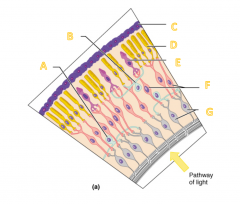
Label the diagram for letters A - G. |
A) Amacrine cell B) Horizontal cell C) Pigmented layer of retina D) Rod E) Cone F) Bipolar cells G) Ganglion cells |
|
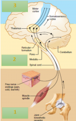
Label the reflex arc for letters A - F. |
A) Spinal cord B) Motor neuron C) Sensory neuron D) Muscle E) Muscle spindle F) Golgi tendon of organ |
|
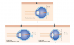
Label the refraction designation for each of the schematic drawings of the eye. |
A) Emmetropic eye B) Myopic eye C) Hyperopic eye |
|
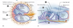
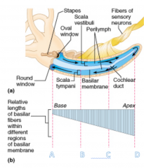
Label the parts of the cochlea. |
A) Outer hair cells B) Tectorial membrane C) Basilar membrane D) Cochlear duct E) Spiral organ (organ of Corti) |
|
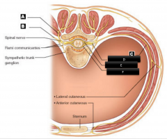
Label the spinal nerve branches. |
A) Dorsal ramus B) Ventral ramus C) Intercostal nerve D) Dorsal root ganglion E) Dorsal root F) Ventral root |
|
Three main levels of neural integration operate in the somatosensory (or any sensory) system: |
1) Receptor level: sensory receptors 2) Circuit level: processing in ascending pathways 3) Perceptual level: processing in cortical sensory areas |
|
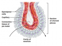
Label the function of the choroid plexus (arrow). |
- Inward arrow: Wastes and unnecessary solutes absorbed - Outward arrow: CSF forms as a filtrate containing glucose, oxygen, vitamins and ions (Na, Cl, Mg, etc.) |
|
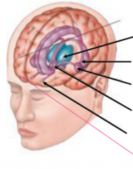
Label the structures of the limbic system and define their functions. |
Limbic system: deep structures in the cerebrum involved in emotion and memory (From top to bottom) 1) Thalamus: relays sensory information to cortex 2) Hippocampus: memory, navigation 3) Amygdala: reward, fear, mating urges 4) Hypothalamus: heart rate, blood pressure 5) Olfactory bulbs 6) Prefrontal cortex |
|
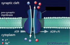
Name the active transporter and its function. |
- Na+/K+ ATPase - It maintains resting membrane potential |
|
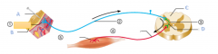
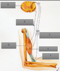
Label the components of a reflex arc. |
A) Stimulus B) Skin C) Spinal cord D) Interneuron 1) Receptor 2) Sensory neuron 3) Integration center 4) Motor neuron 5) Effector |
|
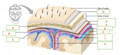
Label the meninges of the brain. |
A) Superior sagittal sinus B) Subdural space C) Subarachnoid space D) Periosteum E) Periosteal F) Meningeal G) Arachnoid mater H) Pia mater I) Arachnoid villus J) Blood vessel K) Falx cerebri |
|
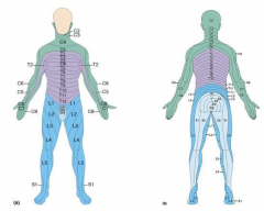
Name the structure and function. |
- Dermatome: an area of skin supplied by peripheral nerve fibers originating from a single dorsal root ganglion - If a nerve is cut, one loses sensation from that dermatome |
|
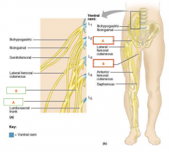
Name the nerves of the lumbar plexus. |
A) Femoral B) Obturator |
|
Label the frequencies of sound waves as they travel through the auditory canal. |
A) 20,000 Hz (High notes) B) 1500 Hz C) 500 Hz D) 20 Hz (Low notes) |
|
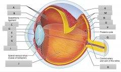
Label the parts of the eyeball for letters A - R. |
A) Ora serrata retinae B) Ciliary body C) Cornea D) Iris E) Pupil F) Anterior pole G) Anterior segment I) Lens J) Posterior segment (contains vitreous humor) K) Sclera M) Choroid N) Retina O) Macula lutea P) Fovea centralis Q) Optic nerve R) Optic disc (blind spot) |