![]()
![]()
![]()
Use LEFT and RIGHT arrow keys to navigate between flashcards;
Use UP and DOWN arrow keys to flip the card;
H to show hint;
A reads text to speech;
343 Cards in this Set
- Front
- Back
|
1. The term varus refers to A turned outward B turned inward C rotated medially D rotated laterallt |
B. Turned inward |
|
|
2. Demonstration of posterior fat pad on the lateral projection of the adult elbow can be caused by 1 trauma or other pathology 2 > 90 degree flexion 3 < 90 degree flexion A 1 only B 3 only C 1 and 2 D 1 and 3 |
D 1 and 3; trauma or other pathology, |
|
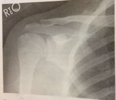
3. Select the most correct statement in figure 2-1 A chin has been depressed too much B chin needs to be extended more C head is tilted D shoulders are not depressed enough |
D shoulders are not depressed enough |
|
|
4. The coronoid process should be visualized in profile in which of the following positions A scap Y B AP scapula C medial oblique elbow D lateral oblique elbow |
C medial oblique elbow |
|
|
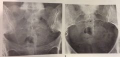
5. The male bony pelvis differs from the female pelvis in which of the following ways 1 male pelvis has a larger pelvic inlet 2 female pubic arch is greater than 90 degrees 3 male ilium is more vertical A 1 only B 1 and 2 C 2 and 3 D all of the above |
C. 2 and 3; female pubic arch is greater than 90 degrees and male ilium is more vertical |
|
|
6. Which of the following techniques would provide a PA projection of the gastroduodenal surfaces of a barium filled high and transverse stomach A place pt in a 35 to 40 degree RAO position B place pt in a lateral position C angle CR 35 to 45 degree cephalad D angle CR 35 to 45 degrees caudad |
C angle the CR 35 to 45 degrees cephalad |
|
7. With the pt and X-ray tube positioned in fig 2-2, the following will be visualized 1 intercondyloid fossa 2 patellofemoral articulation 3 tangential patella A 1 only B 1 and 2 C 2 and 3 D all of the above |
C 2 and 3; patellofemoral articulation, tangential patella |
|
|
8. All of the following statements regarding respiratory structures are true except A right lung has 3 lobes B inferior portion of the lung is the apex C each lung is enclosed in serous membrane D main stem bronchi enter the lung hilus |
B the inferior portion of the lungs is the apex |
|
|
9. All of the following statements regarding an exact PA projection of the skull are true except A OML is perp to the IR B petrous pyramids fill orbits C MSP is parallel to IR D CR is perp to IR and exits nasion |
C MSP is parallel to the IR |
|
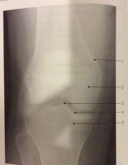
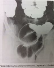
10. What could be done to improve the mediolateral projection of the knee in fig 2-3 A rotate the pelvis slightly forward B rotate the pelvis slightly backward C angle tube 5 degrees cephalad D angle tube 5 degrees caudad |
B rotate the pelvis slightly backward |
|
|
11. Pacemaker electrodes can be introduced thru a vein in the chest or upper extremity from where they are advanced to the A left atrium B right atrium C left ventricle D right ventricle |
D right ventricle |
|
|
12. Widening of the intercostal spaces is characteristic of which condition A pneumothorax B emphysema C pleural effusion D pneumonia |
Emphysema |
|
|
13. Which of the following structures is/are located in the right upper quadrant 1 hepatic flexure 2 gallbladder 3 ileocecal valve A 1 only B 1 and 2 C 2 and 3 D all of the above |
B 1 and 2; hepatic flexure and gallbladder |
|
14. Which of the following statements regarding the image in fig 2-4 is correct? A left kidney is more parallel to the IR B image was made in the left posterior oblique position C left ureter is better visualized D image was made post void |
A left kidney is more parallel to the IR |
|
|
15. During an upper GI exam, stomach of avg shape demonstrates a barium filled fundus and double contrast of the pyloris and duodenal bulb. The position used is most likely A AP erect B PA C RAO D LPO |
D. LPO |
|
|
16. Which of the following articulations participates in formation of the ankle mortise? 1 talotibial 2 talocalcaneal 3 talofibular A 1 only B 1 and 3 only C 2 and 3 only D 3 only |
B. 1 and 3 only; talotibial and talofibular |
|
|
17. Which projections of the foot will best demonstrate the longitudinal arch A mediolateral B lateromedial C lateral weight bearing D 30 degree medial oblique |
C lateral weight bearing |
|
|
18. Graves' disease is associated with A thyroid underactivity B thyroid overactivity C adrenal underactivity D adrenal overactivity |
B thyroid overactivity |
|
|
19. To best visualize the lower ribs, the exposure should be made A on normal inspiration B on inspiration, second breath C on expiration D during shallow breathing |
C on expiration |
|
|
20. In the AP axial projection (Towne) of skull, with the CR directed 30 degrees caudal to the OML and passing midway btwn the EAM, which of the following is best demonstrated A. Occipital bone B. Frontal bone C. Facial bones D. Basal foramina |
A. Occipital bone |
|
|
21. The right posterior oblique position (Judet) of the right acetabulum will demonstrate the 1. Anterior rim of the right acetabulum 2. Right iliac wing 3. Right anterior iliopubic column A. 1 only B. 1 and 2 C. 2 and 3 D. All three |
B. 1 and 2; anterior rim of right acetabulum and right iliac wing |
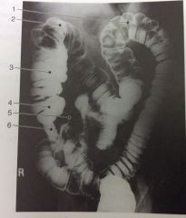
|
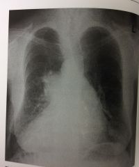
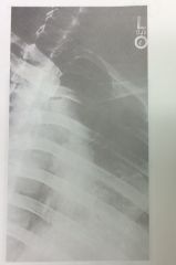
22. Figure 2-5 demonstrates which of the following conditions A. Right upper lobe atelectasis B. Left upper lobe atelectasis C. Pneumothorax D. Dextrocardia |
D.dextrocardia |
|
|
23. A frontal view of the sternum is best accomplished in which of the following positions A. AP B. PA C. RAO D. LAO |
C. RAO |
|
|
24. What is the name of the condition that results in the forward slipping of one vertebra on the one below it A. Spondylitis B. Spondylolysis C. Spondylolisthesis D. Spondylitis |
C. Spondylolisthesis |
|
|
25. During atrial systole, blood flows into the 1. Right ventricle via the mitral valve 2. Left ventricle via the bicuspid valve 3. Right ventricle via the tricuspid valve A. 1 only B. 1 and 2 C. 2 and 3 D. All three |
C. 2 and 3; left ventricle via the bicuspid valve and right ventricle via the tricuspid valve |
|
|
26. How should a chest examination to rule out air fluid levels be obtained on a pt having traumatic injuries A. Perform the exam in the trendelenburg position B. Erect inspiration and expiration images should be obtained C. Include a lateral chest exam performed in dorsal decubitus position D. Perform the exam AP supine at 44" SID |
C. Include a lateral chest exam performed in dorsal decubitus position |
|
|
27. All the following statements regarding the use of iodinated contrast agents with patients taking metformin hydrochloride are true except A. Metformin is used to help lower blood sugar levels in type 2 diabetic patients B. Patients on metformin who have IV iodinated contrast agent administration are at risk for renal failure C. Metformin should be withheld for 48 hours before IV iodinated contrast studies D. Metformin should be withheld for 48 hours after IV iodinated contrast studies |
C. Metformin should be withheld for 48 hours before IV iodinated contrast studies |
|

28. Which of the following methods was used to obtain the image seen in Fig 2-6 A. Erect PA,chin extended, OML forming 37 degrees to IR B. Erect PA,OML and CR perp to IR C. Erect PA, chin extended, OML 15 degree from horizontal D. Erect PA, chin extended, OML 30 degrees from horizontal |
C. Erect PA, chin extended, OML 15 degrees from horizontal |
|
29. Which of the following statements regarding the radiograph in fig 2-6 is(are) true 1. The position is used to demonstrate the frontal and ethmoid sinuses 2. The ethmoid sinuses are seen near the medial aspect of the orbits 3. The perp plate is visualized in midline of the nasal cavity A. 1 only B. 1 and 2. C. 1 and 3 D. All three |
D. All three: . The position is used to demonstrate the frontal and ethmoid sinuses, The ethmoid sinuses are seen near the medial aspect of the orbits, The perp plate is visualized in midline of the nasal cavity |
|
|
30. Which of the following is an important consideration to avoid excessive metacarpophalangeal joint overlap in the oblique projection of the hand? A. Oblique the hand no more than 45 degrees B. Use a support sponge for the phalanges C. Clench the fist to bring the carpals closer to IR D. Use ulnar flexion |
A. Oblique the hand no more than 45 degrees |
|
|
31. All the following positions are used frequently to demonstrate the sternoclavicular articulations except A. Weight bearing B. RAO C. LAO D. PA |
A. Weight bearing |
|
|
32. Which of the following positions is most likely to place the right kidney parallel to the IR A. AP B. PA C. RPO D. LPO |
D. LPO |
|
|
33. When examining a pt whose elbow is in partial flexion, how should an AP projection be obtained 1. With humerus parallel to IR, CR perp 2. With forearm parallel to IR, CR perp 3. Thru the partially flexed elbow, resting on the olecranon process, CR perp A. 1 only B. 1 and 2 C. 2 and 3 D. All three |
B. 1 and 2, With humerus parallel to IR, CR perp And With forearm parallel to IR, CR perp |
|
|
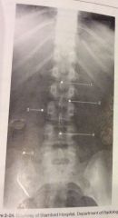
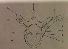
34. Which of the following positions is required to demonstrate small amounts of air in the peritoneal cavity A. Lateral decubitus, affected side up B. Lateral decubitus, affected side down C. AP trendelenburg D. AP supin |
A. Lateral decubitus,affected side up |
|
|
35. Which of the anatomical structures listed below is seen most anteriorly in a lateral projection of the chest A. Esophagus B. Trachea C. Cardiac apex D. Superimposed scapular borders |
C. Cardiac apex |
|
|
36. For an AP projection of the knee on a pt whose measurement from ASIS to tabletop is 21 cm, which CR direction will best demonstrate the knee joint A. 5 degrees caudad B. 10 degrees caudad C. 5 degrees cephalad D. 0 degrees (perpendicular) |
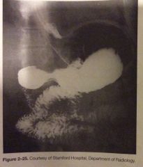
D. 0 degrees (perp) |
|
37. In which of the following projections was the image in fig 2-7 made A. AP B. Medial oblique C. Lateral oblique D. Acute flexion |
B. Medial oblique |
|

38. Which of the following anatomical structures is indicated by the number 2 in fig 2-7 A. Medial epicondyle B. Trochlea C. Capitulum D. Olecranon process |
D. Olecranon process |
|
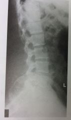
39. Which of the following is (are) well demonstrated in the lumbar spine in fig 2-8 1. Apophyseal articulations 2. Intervertebral foramina 3. Pedicles A. 1 only B. 1 and 2 C. 2 and 3 D. All three |
C. 2 and 3; intervertebral foramina and pedicles |
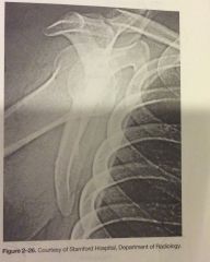
|
|
40. In which of the following tangential axial projections of the patella is complete relaxation of the quadriceps femoris required for an accurate diagnosis 1. Supine flexion 45 degrees (merchant) 2. Prone flexion 90 degrees (settegast) 3. Prone flexion 55 degrees (hughston) A. 1 only B. 1 and 2 C. 2 and 3 D. All three |
A. 1 only; supine flexion 45 degrees (merchant) |
|
|
41. Which of the following projections can be used to supplement the traditional "open mouth" projection when the upper portion of the odontoid process cannot be well demonstrated A. AP or PA thru the foramen magnum B. AP oblique with right and left head rotation C. Horizontal beam lateral D. AP axial |
A. AP or PA thru the foramen magnum |
|
|
42. The floor of the cranium includes all the following bones except A. Temporal bone B. Occipital bone C. Ethmoid bone D. Sphenoid bone |
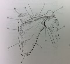
B. Occipital bone |
|
|
43. A lateral projection of the the hand in extension is often recommended to evaluate 1. Fracture 2. Foreign body 3. Soft tissue A. 1 only B. 2 only C. 2 and 3 D. 1 and 3 |
C. 2 and 3; foreign body and soft tissue |
|

44. In which of the following positions was the radiograph fig 2-9 taken A. RPO B. LPO C. AP axial D. Right lateral decubitus |
A. RPO |
|
45. The structure indicated as number 4 in fig 2-9 is the A. Terminal ilium B. Appendix C. Cecum D. Sigmoid |
C. Cecum |
|
|
46. The condition that results from a persistent fetal foramen ovale is A. An atrial septal defect B. A ventricular septal defect C. A patent ductus arteriosus D. Coarctation of the aorta |
A. Atrial septal defect |
|
|
47. Which of the following projections or positions will best demonstrate subacromial or subcoracoid dislocation A. Tangential B. AP axial C. Trains thoracic lateral D. PA oblique scapular Y |
D. PA oblique scapular Y |
|
|
48. With the patient recumbent on the X-ray table with the head lower than the feet, the patient is said to be in the A. Trendelenburg position B. Fowler position C. Decubitus position D. Sims position |
A. Trendelenburg position |
|
|
49. Which of the following positions can be used to demonstrate the axiliary ribs of the right thorax 1. RAO 2. LAO 3. RPO A. 1 only B. 1 and 2 C. 2 and 3 D. All three |
C. 2 and 3; LAO and RPO |
|
|
50. In which projection of the foot are the interspaces between the first and second cuneiforms best demonstrated A. Lateral oblique foot B. Medial oblique foot C. Lateral foot D. Weight bearing foot |
A. Lateral oblique foot |
|
|
51. The sternal angle is at approx the same level as the A. T2-3 interspace fifth thoracic vertebra B. T9-10 interspace C. T5 D. Costal margin |
C. T5 |
|
|
52. Which of the following structures is/are located in the right upper quadrant (RUQ) 1. Spleen 2. Gallbladder 3. Hepatic flexure A. 1 only B. 1 and 2 only C. 2 and 3 only D. All three |
C 2 and 3 only; gallbladder and hepatic flexure |
|
|
53. To demonstrate esophageal varices, the patient must be examined in A. The recumbent position B. The erect position C. The anatomical position D. The fowler position |
A. The recumbent position |
|

54. Which of the following statements regarding fig 2-10 is/are true 1. Correct degree of rotation is present 2. Midphalanges are foreshortened 3. Fingers are parallel to the IR A. 1 only B. 1 and 2 C. 2 and 3 D. All three |
B. 1 and 2; correct degree of rotation is present and Midphalanges are foreshortened |
|
|
55. The tissue that occupies the central cavity within the shaft of a long bone in an adult is A. Red marrow B. Yellow marrow C. Cortical tissue D. Cancellous tissue |
B. Yellow marrow |
|
|
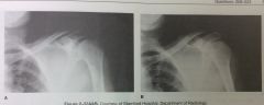
56. All of the following structures are associated with the posterior femur except A. Popliteal surface B. Intercondyloid fossa C. Intertrochanteric line D. Linea aspera |
C. Intertrochanteric line |
|
|
57. Which of the following projections of the ankle would best demonstrate the mortise A. Medial oblique 15 to 20 degrees B. Lateral oblique 15 to 20 degrees C. Medial oblique 45 degrees D. Lateral oblique 45 degrees |
A. Medial oblique 15 to 20 degrees |
|
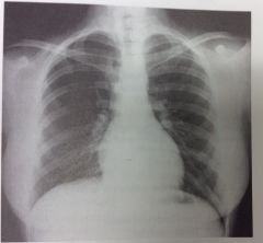
58. Which of the following statments with respect to the PA chest seen in fig 2-11 is/are correct 1. Adequate inspiration is demonstrated 2. Shoulders are rolled forward adequately 3. Rotation is demonstrated A. 1 only B. 1 and 2 only C. 2 and 3 only D. All three |
D. All three; adequate inspiration, shoulders rolled forward adequately and rotation is demonstrated |
|
|
59. Which of the following bony landmarks is in the same transverse plane as the symphysis pubis A. Ischial tuberosity B. Prominence of the greater trochanter C. Anterosuperior iliac spine D. Anteroinferior iliac spine |
B. Prominence of the greater trochanter |
|
|
60. A radiolucent sponge can be placed under the patient's waist for a lateral projection of the lumbosacral spine to 1. Make the vertebral column parallel with the IR 2. Place the intervertebral disk spaces perp to the IR 3. Decrease the amount of SR reaching the IR A 1 only B 1 and 2 C 2 and 3 D all three |
B 1 and 2; make the vertebral column parallel with the IR, decrease the amount of SR reaching the IR |
|
|
70. The junction of the sagittal and coronal sutures is the A diploe B lambda C bregma D pterion |
C bregma |
|
|
61. To reduce the amt of scattered radiation reaching the IR in CR/DR of the lumbosacral region, which of the following is/are recommended 1. Close collimation 2. Lead mat on table posterior to the pt 3. Decreased SID A 1 only B 1 and 2 C 2 and 3 D all three |
B 1 and 2; close collimation, lead mat on table posterior to the pt |
|
|
62. Which of the following is/are distal to the tibial plateau 1. Intercondyloid fossa 2. Tibial condyles 3. Tibial tuberosity A 1 only B 1 and 2 C 2 and 3 D all three |
C 2 and 3; tibial condyles, tibial tuberosity |
|
|
63. Evaluation criteria for a lateral projection of the humerus include 1. Epicondyles parallel to the IR 2. Lesser tubercle in profile 3. Superimposed epicondyles A 1 only B 1 and 3 C 2 and 3 D all three |
C 2 and 3; lesser tubercle in profile l, superimposed epicondyles |
|
|
64. Which position of the shoulder demonstrates the lesser tubercle in profile medically A AP B External rotation C internal rotation D neutral position |
C internal rotation |
|
|
65. With the pt in the PA position, which of the following tube angle and direction combinations is correct for an axial projection of the clavicle A 5 to 15 degrees caudad B 5 to 15 degrees cephalad C 15 to 30 degrees cephalad D 15 to 30 degrees caudad |
D 15 to 30 degrees caudad |
|
|
66. Which of the following fx classifications describes a small bony fragment pulled from a bony process A avulsion fx B torus fx C comminuted fx D compound fx |
A avulsion fx |
|
|
67. What portion of the humerus articulates with the ulna to help form the elbow joint A semilunar/trochlear notch B radial head C capitulum D trochlea |
D trochlea |
|
|
68. Movement of a part toward the midline of the body is termed A eversion B inversion C abduction D adduction |
D adduction |
|
|
69. During myelography, contrast medium is introduced into the A subdural space B subarachnoid space C epidural space D epidermal soace |
B subarachnoid space |
|
71. Which of the following statements is/are true regarding the radiograph in fig 2-12 1. Pt placed in RAO position 2. MCP is about 60 degrees to the IR 3. Acromion process is free of superimposition A.1 only B. 1 and 2 only C. 2 and 3 D. All three |
D. All three; pt placed in RAO position with MCP about 60 degrees to the IR and the acromion is free of superimposition |
|
|
72. Examples of synovial pivot articulations include the
1. Atlantoaxial joint 2. radioulnar joint 3. Temporomandibular joint A. 1 only B. 1 and 2 C. 2 and 3 D. All three |
B 1 and 2, atlantoaxial and radioulnar joints |
|
|
73. The lumbar transverse process is represented by what part of the "Scotty dog" seen in a correctly positioned oblique lumbar spine A. Eye B. Nose C. Body D. Ear |
B. Nose |
|
|
74. An injury to a structure located on the side opposite that of the primary injury is referred to as A. Blowout B. Le Fort C. Contracture D. Contrecoup |
D.contrecoup |
|
|
75. In which of the following positions can the sesamoid bones of the foot be demonstrated to be free of superimposition with the metatarsals or phalanges A. Dorsoplantar metatarsals/toes B. Tangential metatarsals/toes C. 30 degree medial oblique foot D. 30 degree lateral oblique foot |
B. Tangential metatarsals/toes |
|
|
76. Which of the following conditions is limited specifically to the tibial tuberosity A. Ewing sarcoma B. Osgood-Schlatter disease C. gout D. Exostosis |
B. Osgood-schlatter disease |
|
|
77. AP stress studies of the ankle may be performed 1. To demonstrate fxs of the distal tib/fib 2. Following inversion or eversion injuries 3. To demonstrate a ligament tear A. 1 only B. 1 and 2 only C. 2 and 3 only D. All three |
C. 2 and 3; following inversion or eversion injuries and demonstrate a ligament tear |
|
|
78. Which of the following is/are part of the bony thorax 1. Manubrium 2. Clavicles 3. 24 ribs A. 1 only B. 1 and 2 C. 1 and 3 D. All three |
C. 1 and 3; Manubrium and 24 ribs |
|
|
79. Aspirated foreign bodies in older children and adults are most likely to lodge in the A. Right main stem bronchus B. Left main stem bronchus C. Esophagus D. Proximal stomach |
A. Right main stem bronchus |
|
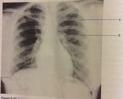
80. The PA chest radiograph shown in fig 2-13 demonstrates 1. Rotation 2. Scapulae removed from lung fields 3. Adequate inspiration A. 1only B. 1 and 2 C. 2 and 3 D. All three |
B. 1 and 2; rotation and scapulae removed from lung fields |
|

81. The letter B in fig 2-13 indicates A. Left anterior rib B. Right posterior rib C. Left posterior rib D. Right anterior rib |
A. Left anterior rib |
|
|
82. With the pt seated at the end of the X-ray table, elbow flexed 80 degrees and the CR directed 45 degrees laterally from the shoulder to the elbow joint, which of the following structures will be demonstrated best A. Radial head B. Ulnar head C. Coronoid process D. Olecranon process |
C. Coronoid process |
|
|
83. The structures forming the brain stem include 1. Pons 2. Medulla oblongata 3. Midbrain A. 1 and 2 B. 1 and 3 C. 2 and 3 D. All three |
All three; pons, medulla oblongata and midbrain |
|
|
84. The CR will parallel the intervertebral foramina in which of the following projections 1. Lateral cervical spine 2. Lateral thoracic spine 3. Lateral lumbar spine A.1 only B. 1 and 2 C. 2 and 3 D. All three |
C. 2 and 3; lateral thoracic and lateral lumbar |
|
|
85. What structure can be located midway btwn the ASIS and pubic symphysis A. Dome of the acetabulum B. Femoral neck C. Greater trochanter D. Iliac crest |
A. Dome of the acetabulum |
|

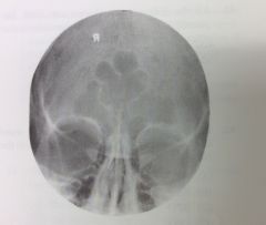
86. The structure labeled 3 in fig 2-14 is the A. Maxillary sinus B. Sphenoidal sinus C. Ethmoidal sinus D. Frontal sinus |
B. Sphenoidal sinus |
|
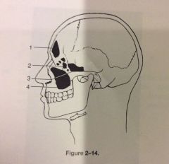
87. Which of the following would best evaluate the structure labeled 4 in fig 2-14 A. PA axial Caldwell method B. Waters method C. Lateral projection D. SMV Projection |
B. Waters method |
|
|
88. Which of the following positions demonstrates the sphenoid sinuses 1. Modified waters (mouth open) 2. Lateral 3. PA axial A 1 only B 1 and 2 C. 2 and 3 D. All three |
B. 1 and 2; modified waters (mouth open) and lateral |
|
89. The radiograph shown in fig 2-15 demonstrates the articulation btwn the 1. Talus and calcaneus 2. Calcaneus and cuboid 3. Talus and navicular A. 1 only B. 1 and 2 C. 2 and 3 D. All three |
C. 2 and 3; calcaneus and cuboid, talus and navicular |
|
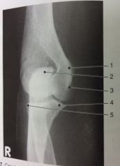
90. Identify the structure labeled 1 on AP projection in fig 2-16 A. lateral condyles B. Lateral epicondyle C. Medial condyle D. Medial epicondyle |
D. Medial epicondyle |
|
|
91. The articular facets of L5/S1 are best demonstrated in a(n) A. AP projection B. 30 degree oblique C. 45 degree oblique D. AP axial |
B. 30 degree oblique |
|
|
92. The pt's chin should be elevated during chest radiography to A. Permit the diaphragm to move to its lowest position B. Avoid superimposition on the apices C. Assist in maintaining an upright position D. Keep the MSP parallel |
B. Avoid superimposition on the apices |
|
|
93. The secondary center of ossification in long bones is the A. Diaphysis B. Epiphysis C. Metaphysis D. Apoptosis |
B. Epiphysis |
|
|
94. Medial displacement of a tibial fx would be best demonstrated in the A. AP B. Lateral C. Medial oblique D. Lateral oblique |
A. AP |
|
|
95. The lumbar lamina is represented by what part of the "Scotty dog" seen in a correctly positioned oblique lumbar spine view A. Eye B. Nose C. Body D. Neck |
C. Body |
|
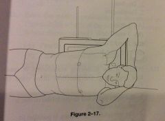
96. All the following statements regarding the position shown in fig 2-17 are true except A. Left pleural effusion could be demonstrated B. Right pneumothorax could be demonstrated C. Left lateral decubitus position is illustrated D. CR directed vertically to the level of T7 |
D. CR directed vertically to the level of T7 |
|
|
97. Which of the following positions would best demonstrate the proximal tibia fibulae articulation A. AP B. 90 degrees mediolateral C. 45 degree internal rotation D. 45 degree external rotation |
C. 45 degree internal rotation |
|
|
98. At what level do the carotid arteries bifurcate A. Foramen magnum B. Trachea C. Pharynx D. C4 |
D. C4 |
|
|
99. During a double contrast BE, which of the following positions would afford the best double contrast visualization of the lateral wall of the descending colon and the medial wall of the ascending colon A. AP or PA erect B. Right lateral decubitus C. Left lateral decubitus D. Ventral decubitus |
B. Right lateral decubitus |
|
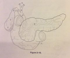
100. What is the structure indicated by the number 8 in fig 2-18 A. Common hepatic duct B. Common bile duct C. Cystic duct D. Pancreatic duct |
B. Common bile duct |
|

101. What is the structure indicated by the number 7 in figure 218 A. Common hepatic duct B. Common bile duct C. Cystic duct D. Pancreatic duct |
C. Cystic duct |
|
|
102. Which of the following conditions is often the result of urethral obstruction or stricture A. Pyelonephritis B. Nephritis C. Hydronephrosis D. Cystourethritis |
C. Hydronephrosis |
|
|
103. Which of the following examinations involves the introduction of a radiopaque contrast medium thru a uterine cannula A. Retrograde pyelogram B. Voiding cystourethrogram C. Hysterosalpingogram D. Myelogram |
C. Hysterosalpingogram |
|
|
104. All the following statements regarding large bowel radiograph are true except A. Large bowel must be completely empty prior to exam B. Retained fecal material can obscure pathology C. Single-contrast studies help to demonstrate intraluminal lesions D. Double contrast studies help to demonstrate mucosal lesion |
C. Single contrast studies help to demonstrate intraluminal lesions |
|
|
105. In a lateral projection of the normal knee, the 1. Fibulae head should be somewhat superimposed on the proximal tibia 2. Patellofemoral joint should be visualized 3. Femoral condyle should be superimposed A. 1 only B. 2 only C. 1 and 3 only D. All three |
D. All three:1. Fibulae head should be somewhat easy superimposed on the proximal tibia2. Patellofemoral joint should be visualized3. Femoral condyle said should be superimposed |
|
|
106. All elbow fat pads are best demonstrated in which position A. AP B. Lateral C. Acute flexion D. AP partial flexion |
B. Lateral |
|
|
107. The term used to describe expectoration of blood from the bronchi is A. Hemoptysis B. Hematemesis C. Chronic obstructive pulmonary disease D. Bronchitis |
A. Hemoptysis |
|
|
108. Double contrast exams of the stomach or large bowel are performed to better visualized the A. Position of the organ B. Size and shape of the organ C. Diverticula D. Gastric or bowel mucosa |
D. Gastric or bowel mucosa |
|
|
109. Which of the following are mediastinal structures 1. Heart 2. Trachea 3. Esophagus A. 1 only B. 1 and 2 C. 2 and 3 D. All three |
D. All three, heart, trachea and esophagus |
|
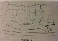
110. Which of the following statements is/are true regarding the position illustrated in fig 2-19 1. Right ureter is parallel to IR (adjacent to table) 2. Left kidney is parallel to IR (elevated) 3. Degree of obliquity should be about 30 deg A. 1 only B. 1 and 2 only C. 2 and 3 only D. All three |
D. All three;. Right ureter is parallel to IR (adjacent to table)2. Left kidney is parallel to IR (elevated)3. Degree of obliquity should be about 30 degrees |
|
|
111. In which position of the shoulder is the greater tubercle seen superimposed on the humeral head A. AP B. External rotation C. Internal rotation D. Neutral position |
C. Internal rotation |
|
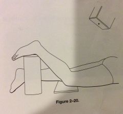
112. With the patient positioned as illustrated in fig 2-20, which of the following structures is best demonstrated A. Patella B. Patellofemoral articulation C. Intercondyloid fossa D. Tibial tuberosity |
C. Intercondyloid fossa |
|
113. Which of the following structures is illustrated by the number 2 in fig 2-21 A. Maxillary sinus B. Coronoid process C. Zygomatic arch D. Coracoid process |
C. Zygomatic arch |
|
|
114. Which of the following articulations may be described as diarthrotic 1. Knee 2. Intervertebral joints 3. TMJ A. 1 only B. 2 only C. 1 and 3 only D. All three |
C. 1 and 3; knee and TMJ |
|
|
115. Ulnar flexion/deviation will best demonstrate which carpals 1. Medial 2. Lateral 3. Scaphoid A. 1 only B. 1 and 2 C. 2 and 3 D. All three |
C. 2 and 3; lateral carpals and scaphoid |
|
116. What should be done to better demonstrate the coracoid process shown in fig 2-22 A. Use a perp CR B. Angle the CR about 30 degrees cephalad C. Angle the CR about 30 degrees caudad D. Angle the MSP 15 degrees toward the affected side |
B. Angle the CR about 30 degrees cephalad |
|
|
117. Structures comprising the neural or vertebral, arch include 1. Pedicles 2. Laminae 3. Body A. 1 only B. 1 and 2 C. 2 and 3 D. All three |
B. 1 and 2, pedicles and laminae |
|
|
118. In which type of fx are the splintered ends of bone forced thru the skin A. Closed B. Compound C. Compression D. Depressed |
B. Compound |
|
|
119. The thoracic zygapophyseal/apophyseal joints are demonstrated with the A. Coronal plane 90 degrees to the IR B. Midsagittal plane 90 degrees to the IR C. Coronal plane 20 degrees to the IR D. Midsagittal plane 20 degrees to the IR |
D. Midsagittal plane 20 degrees to the IR |
|
|
120. Which of the following may be used to evaluate the glenohumeral joint 1. Scap Y 2. Inferosuperior axial 3. Transthoracic lateral A. 1 only B. 1 and 2 C. 2 and 3 D. All three |
D. All three, scap Y, inferosuperior axial and transthoracic lateral |
|
|
121, The long flat structures that project posteromedially from the pedicles are the A. Transverse process B. Vertebral arches C. Laminae D. Pedicles |
C. Laminae |
|
|
122. The type of ileum characterized by cessation of peristalsis is termed A. Mechanical B. Paralytic C. Asymptomatic D. Sterile |
B. Paralytic |
|
|
123. Radiography of which of the following structures in the AP or PA position will inherently result in an image demonstrating shape distortion of the anatomical part 1. Kidney 2. Scaphoid 3. Sigmoid A. 1only B. 1 and 2 C. 2 and 3 D. All three |
D. All three, kidney, scaphoid and sigmoid |
|
|
124. Which of the following procedures will best demonstrate the cephalic, basilic and subclavian veins A. Aortofemoral arteriogram B. Upper limb venogram C. Lower limb venogram D. Renal venogram |
B. Upper limb venogram |
|
125. Which of the following statements is/are correct with respect to the images shown in fig 2-23 1. Image A was made with cephalad angle 2. Image B was made with cephalad angle 3. Images A & B were made with CR directed 15 degrees cephalad A. 1 only B. 2 only C. 2 and 3 D. All three |
A. 1 only Image A was made with cephalad angle |
|
|
126. The body habitus characterized by a long and narrow thoracic cavity and low midline stomach and gallbladder is the A. Asthenic B. Hyposthenic C. Sthenic D. Hypersthenic |
A. Asthenic |
|
|
127. Which of the following should be performed to rule out subluxation or fx of the cervical spine A. Oblique cervical spine, seated B. AP cervical spine, recumbent C. Horizontal beam lateral D. Laterals in flexion and extension |
C. Horizontal beam lateral |
|
|
128. Which of the following is proximal to the carpal bones A. Distal interphalangeal joints B. Proximal interphalangeal joints C. Metacarpals D. Radial styloid process |
D. Radial styloid process |
|
|
129. Which of the following statements regarding the scap Y projection of the shoulder is/are true 1. MSP should be about 60 degrees to IR 2. Scapular borders should be superimposed on the humeral shaft 3. Oblique projection of the shoulder is obtained A. 1only B. 1 and 2 C. 2 and 3 D. All three |
C. 2 and 3; Scapular borders should be superimposed on the numeral shaft, Oblique projection of the shoulder is obtained |
|
|
130. Which of the following are characteristics of the hypersthenic body type 1. Short wide transverse heart 2. High and peripheral large bowel 3. Diaphragm positioned low A. 1 and 2 B. 1 and 3 C. 2 and 3 D. All three |
A. 1 and 2; short wide transverse heart, high and peripheral large bowel |
|
|
131. Glossitis refers to inflammation of the A. Epiglottis B. Salivary glands C. Tongue D. Ossicles |
C. Tongue |
|
|
132. With the patients head In a PA position and the CR directed 20 degrees cephalad, which part of the mandible will be best visualized A. Symphysis B. Rami C. Body D. Angle |
B. Rami |
|
|
133. During IV urography, the prone position generally is recommended to demonstrate 1. The filling of the ureters 2. The renal pelvis 3. The superior calyces A. 1 only B. 1 and 2 C. 1 and 3 D. All three |
B. 1 and 2; filling of the ureters, renal pelvis |
|
|
134. The plane that passes vertically thru the body, dividing it into anterior and posterior halves, is termed the A. MSP B. MCP C. Sagittal plane D. Transverse plane |
B. MCP mid coronal plane |
|
|
135. To demonstrate a profile view of the glenoid fossa, the patient is AP recumbent and obliques 45 degrees A. Toward the affected side B. Away from the affected side C. With the arm at the side in the anatomic position D. With the arm in external rotation |
A. Toward the affected side |
|
136. Which of the following anatomic structures is indicated by the number 1 in fig 2-24 A. Body of L3 B. Body of L4 C. Spinous process D. Transverse process |
C. Spinous process |
|
|
137. During an air-contrast BE, in what part of the colon is air most likely to be visualized with the body in the AP recumbent position A. Transverse colon B. Descending colon C. Ascending colon D. Left and right colic flexures |
A. Transverse colon |
|
|
138. Central Ray angulation may be required for 1. Magnification of anatomic structures 2. Foreshortening or self superimposition 3. Superimposition of overlying structures A. 1 only B. 1 and 2 C. 2 and 3 D. All three |
C. 2 and 3; foreshortening or self imposition, superimposition of overlying structures |
|
|
139. Which of the following is recommended to better demonstrate the tarsometatarsal joints in a dorsoplantar projection of the foot A. Invert the foot B. Evert the foot C. Angle the CR 10 degrees posteriorly D. Angle the CR 10 degrees anteriorly |
C. Angle the CR 10 degrees posteriorly |
|
|
140. Valid evaluation criteria for a lateral projection of the forearm requires that 1. The epicondyle be parallel to the IR 2. Radius and ulna be superimposed distally 3. Radial tuberosity should face anteriorly A. 1 only B. 1 and 2 C. 2 and 3 D. All three |
C. 2 and 3; radius and ulna be superimposed distally, radial tuberosity should face anteriorly |
|
|
141. Which of the following positions will provide an AP projection of the L5/S1 interspace A. Pt AP with 30 to 35 degree angle cephalad B. Pt AP with 30 to 35 degree angle caudad C. Pt AP with 0 degree angle D. Pt lateral, coned to L5 |
A. Pt AP with 30 to 35 degree angle cephalad |
|
|
142. Subject/object unsharpness can result from all of the following except when A. Object shape does not coincide with the shape of the X-ray beam B. Object plane is not parallel with X-ray tube and/or IR C. Anatomic objects of interest is/are in the path of CR D. Anatomic objects of interest is/are at a distance from the IR |
C. Anatomic objects of interest is/are in the path of CR |
|
|
143. Patients are instructed to remove all jewelry, hair clips, metal prostheses, coins and credit cards before entering the room for an evaluation in A. Sonography B. CT C. MRI D. Nuc Med |
C. MRI |
|
|
144. The true lateral position of the skull uses which of the following principles 1. Inter pupillary line perp to the IR 2. MSP perp to IR 3. IOML parallel to the transverse axis of IR A. 1 only B. 1 and 2 C. 1 and 3 D. All three |
C. 1 and 3; Interpupillary line perp to IR; IOML parallel to the transverse axis of IR |
|
145. In which of the following positions was the radiograph shown in fig 2-25 probably made A. Supine recumbent B. Prone recumbent C. PA upright D. Supine trendelenburg |
B. Prone recumbent |
|
|
146. A kyphotic curve is formed by which of the following 1. Sacral vertebrae 2. Thoracic vertebrae 3. Lumbar vertebrae A. 1 only B. 1 and 2 C. 3 only D. 1 and 3 only |
B. 1 and 2; sacral and thoracic vertebrae |
|
|
147. Which of the following is/are required for a lateral projection of the skull 1. IOML is parallel to the IR 2. MSP is parallel to the IR 3. CR enters 3/4 in superior and anterior to the EAM A. 1 only B. 1 and 2 C. 2 and 3 D. All three |
B. 1 and 2; IOML and MSP are parallel to the IR |
|
|
148. That ossified portion of a long bone where cartilage has been replaced by bone is known as the A. Diaphysis B. Epiphysis C. Metaphysis D. Apophysis |
C. Metaphysis |
|
|
149. Which of the following positions will most effectively move the gallbladder away from the vertebrae in an asthenic pt A. LAO B. RAO C. LPO D. Erect |
A. LAO |
|
|
150. The ileocecal valve normally is located in which of the following body regions A. Right iliac B. Left iliac C. Righ lumbar D. Hypogastric |
A. Right iliac |
|
|
151. Which of the following is/are true regarding radiograph of examinations of the acromioclavicular joints 1. Procedure is performed in the erect position 2. Use of weights can improve demonstration of the joints 3. Procedure should be avoided if dislocation or seperation is suspected A. 1 only B. 1 and 2 C. 1 and 3 D. 2 and 3 |
B. 1 and 2; procedure is performed in erect position, use of weights can improve demonstration of the joints, |
|
|
152. A type of cancerous bone tumor occurring in children and young adults and arising from bone marrow is A. Ewing sarcoma B. Multiple myeloma C. Enchondroma D. Osteochondroma |
A. Ewing sarcoma |
|
|
153. Arteries and veins enter and exit the medial aspect of each lung at the A. Root B. Hilus C. Carina D. Epiglottis |
B. Hilus |
|
|
154. Which of the following skull positions will demonstrate the cranial base, sphenoidal sinuses, atlas and Odontoid process A. AP axial B. Lateral C. Parietocanthial D. SMV |
D. SMV |
|
155. Which of the following statements is/are true with respect to the radiograph shown in fig 2-26 1. Acromion process is seen partially superimposed on the 3rd rib 2. Projection is performed to evaluate the scapula 3. Projection is performed to evaluate the acromioclavicular articulation A. 1 only B. 2 only C. 1 and 2 D. 2 and 3 |
B. 2 only; projection is performed to evaluate the scapula |
|
|
156. Which of the following is/are located on the anterior aspect of the femur 1. Patellar surface 2. Intertrochanteric crest 3. Linea aspera A. 1 only B. 1 and 2 C. 2 and 3 D. All three |
A. 1 only; patellar surface |
|
|
157. An intrathecal injection is associated with which of the following exams A. Intravenous urogram B. Retrograde pyelogram C. Myelogram D. Cystogram |
C. Myelogram |
|
158. In fig 2-27, the structure indicated as # 7 is which of the following A. Neck of rib B. Tubercle of rib C. Transverse process D. Head of rib |
D. Head of rib |
|
|
159. Which of the following statements is/are correct with respect to evaluation criteria for a PA projection of the chest for lungs 1. Sternal extemities of clavicles are equidistant from a vertebral borders 2. 10 posterior ribs are demonstrated above the diaphragm 3. Esophagus is visible in the midline A. 1 only B. 1 and 2 C. 2 and 3 D. All three |
B. 1 and 2; sternal extremities of clavicles are equidistant from a vertebral borders, 10 posterior ribs are demonstrated above the diaphragm |
|
|
160. In which of the following positions/projections will the talocalcaneal joint be visualized A. Dorsoplantar projection of foot B. Plantodorsal projection of os calcis C. Medial oblique position of foot D. Lateral foot |
B. Plantodorsal projection of os calcis |
|
|
161. In the lateral projection of the ankle the 1.talotibial joint is visualized 2. Talofibular joint is visualized 3. Tibia and fibula are superimposed A. 1 only B. 1 and 2 C. 1 and 3 D. All three |
C. 1 and 3; talotibial joint is visualized, tibia and fibula are superimposed |
|
162. The position illustrated in fig 2-28 may be obtained with the pt 1. Supine and CR angled 30 degrees caudad 2. Supine and CR angled 30 degrees cephalad 3. Prone and CR angled 30 degrees cephalad A. 1 only B. 2 only C. 1 and 3 D. 2 and 3 |
B. 2 only; supine and CR angled 30 degrees cephalad |
|
|
163. All the following positions are likely to be employed for both single and double contrast exams of the large bowel except A. Lateral rectum B. AP axial rectosigmoid C. Right and left lateral decubitus abdomen D. RAO and LAO abdomen |
C. Right and left lateral decubitus abdomen |
|
|
164. Which of the following statements regarding the Norgaard method, "ball catcher's position" is/are correct 1. Bilateral AP oblique hands are obtained 2. It is used for early detection of rheumatoid arthritis 3. Hands are obliques about 45 degrees, palm up A. 1 only B. 1 and 2 C. 2 and 3 D. All three |
D. All three; Bilateral AP oblique hands are obtained, It is used for early detection of rheumatoid arthritis, Hands are obliques about 45 degrees, palm up |
|
|
165. Which of the following can be used to demonstrate the intercondyloid fossa 1. Prone, knee flexed 40 degrees, CR directed caudad, 40 degrees to the popliteal fossa 2. Supine, IR under flexed knee, CR directed cephalad to knee, perp to tibia 3. Prone, patella parallel to IR, heel rotated 5 to 10 degrees lateral, CR perp to knee joint A. 1 only B. 1 and 2 C. 2 and 3 D. All three |
B. 1 and 2, Prone, knee flexed 40 degrees, CR directed caudad, 40 degrees to the popliteal fossa, Supine, IR under flexed knee, CR directed cephalad to knee, perp to tibia |
|
166. The scapula shown in fig 2-29 demonstrates 1. Posterior aspect 2. Costal surface 3. Sternal articular surface A. 1 only B. 1 and 2 C. 1 and 3 D. All three |
A. 1 only; posterior aspect |
|
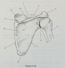
167. In fig 2-29 which of the following is represented by the number 3 A. Acromion process B. Scapular spine C. Coracoid process D. Acromioclavicular joint |
A. Acromion process |
|

168. In fig 2-29 which of the following is represented by the number 7 A. Medial border B. Lateral border C. Inferior angle D. Superior angle |
B. Lateral border |
|
|
169. With the patient in the PA position and the OML and CR perp to IR, the resulting radiograph will demonstrate the petrous pyramids A. Below the orbits B. In the lower third of the orbits C. Completely within the orbits D. Above the orbits |
C. Completely within the orbits |
|
|
170. When evaluating a PA axial projection of the skull with a 15 degree caudal angle, the radiographer should see 1. Petrous pyramids in the lower third of the orbits 2. Equal distance from the lateral border of the skull to the lateral rim of the orbit bilaterally 3. Symmetrical petrous pyramids A. 1 and 2 B. 1 and 3 C. 2 and 3 D. All three |
D. All three; Petrous pyramids in the lower third of the orbits, Equal distance from the lateral border of the skull to the lateral rim of the orbit bilaterally, Symmetrical petrous pyramids |
|
|
171. Which of the following barium-filled anatomic structures is best demonstrated in the LPO position A. Hepatic flexure B. Splenic flexure C. Sigmoid colon D. Ileocecal valve |
A. Hepatic flexure |
|
|
172. The uppermost portion of the iliac crest is at approx the same level as the A. Costal margin B. Umbilicus C. Xiphoid tip D. Fourth lumbar vertebrae |
D. Fourth lumbar vertebrae |
|
|
173. What is the position of the stomach in a hypersthenic pt A. High and vertical B. High and horizontal C. Low and vertical D. Low and horizontal |
B. High and horizontal |
|
|
174. In the anterior oblique position of the cervical spine, the structures best seen are the A. Intervertebral foramina nearest the IR B. Intervertebral foramina furthest from IR C. Interacts ulnar joints D. Intervertebral joints |
A. Intervertebral foramina nearest the IR |
|
|
175. During chest radiography, the act of inspiration 1. Elevates the diaphragm 2. Raises the ribs 3. Depresses the abdominal viscera A. 1 only B. 1 and 2 C. 2 and 3 D. All three |
C. 2 and 3; raises the ribs and depresses the abdominal viscera |
|
|
176. In the lateral projection of the scapula, the 1. Vertebral and auxiliary borders are superimposed 2. Acromion and coracoid processes are superimposed 3. Inferior angle is superimposed on the ribs A. 1 only B. 1 and 2 C. 1 and 3 D. All three |
A. 1 only, vertebral and axillary borders are superimposed |
|
177. Which of the following statements is/are true regarding fig 2-30 1. The image was made in the LAO position 2. CR should enter more inferiority 3. Sternum is projected onto the left side of the thorax A. 1 only B. 2 only C. 2 and 3 D. All three |
C. 2 and 3; CR should enter more inferiority, Sternum is projected onto the left side of the thorax |
|
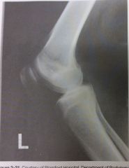
178. To better visualize the knee joint space in the radiograph in fig 2-31, the radiographer should A. Flex the knee more acutely B. Flex the knee less acutely C. Angle the CR 5 to 7 degrees cephalad D. Angle the CR 5 to 7 degrees caudad |
C. Angle the CR 5 to 7 degrees cephalad |
|
|
179. Which of the following is/are demonstrated in an AP projection of the cervical spine 1. Intervertebral disk spaces 2. C3-7 cervical bodies 3. Apophyseal joints A. 1 only B. 1 and 2 C. 2 and 3 D. All three |
B. 1 and 2; intervertebral disk spaces and C3-7 cervical bodies |
|
|
180. With which of the following does the trapezium articulate A. Fifth metacarpal B. First metacarpal C. Distal radius D. Distal ulna |
B. First metacarpal |
|
|
181. Which of the following statements is/are true regarding a PA axial projection of the paranasal sinuses 1. OML is elevated 15 degrees from the horizontal 2. Petrous pyramids completely fill the orbits 3. Frontal and ethmoidal sinuses are visualized A. 1 only B. 1 and 2 only C. 1 and 3 only D. All three |
C. 1 and 3; OML is elevated 15 degrees from the horizontal, frontal & ethmoidal sinuses are visualized |
|
|
182. Tracheotomy is an effective technique used to restore breathing when there is A. Respiratory pathway obstruction above the larynx B . Crushed tracheal rings owing to trauma C. Respiratory pathway closure owing to inflammation and swelling C. All of the above |
A. Respiratory pathway obstruction above the larynx |
|
|
183. To demonstrate the first two cervical vertebrae in the AP projection, the pt is positioned so that A. Glabllomeatal line is vertical B. Acanthiomeatal line is vertical C. Line btwn the mentum and the mastoid tip is vertical D. Line btwn the maxillary occlusal plane and the mastoid tip is vertical |
D. Line btwn the maxillary occlusal plane and the mastoid tip is vertical |
|
|
184. For which of the following conditions is operative cholangiography a useful tool 1. Patency of the biliary ducts 2. Biliary tract calculi 3. Gallbladder calculi A. 1 only B. 1 and 2 C. 2 and 3 D. All three |
B. 1 and 2; binary tract calculi and patency of biliary ducts |
|
|
185. For the avg pt, the CR for a lateral projection of a barium filled stomach should enter A. Midway btwn the MCP and the anterior abdominal surface B. Midway btwn the vertebral column and the lateral border of the abdomen C. At the MCP at the level of the iliac crest D. Perp to the level of L2 |
A. Midway btwn the MCP and the anterior abdominal surface |
|
|
186. Which of the following positions is obtained with the patient lying supine on the radiographer table with the CR directed horizontally to the iliac crest A. Left lateral decub position B. Right lateral decub position C. Ventral decub position D. Dorsal decub position |
D. Dorsal decub position |
|
|
187. Which of the following is/are appropriate techniques for imaging a pt with a possible traumatic spine injury 1. Instruct the pt to turn slowly and stop if anything hurts 2. Maneuver the X-ray tube instead of moving the pt 3. Call for help and use the log rolling method to turn the patient A. 1 only B. 1 and 3 C. 2 and 3 D. All three |
C. 2 and 3; Maneuver the X-ray tube instead of moving the pt, Call for help and use the log rolling method to turn the patient |
|
|
188. Which of the following positions is used to demonstrate vertical patellar fxs and the patellofemoral articulation A. AP knee B. Lateral knee C. Tangential patella D. Tunnel view |
C. Tangential patella |
|

189. The structure labeled #5 in fig 2-32 is the A. Body of C1 B. Body of C2 C. Odontoid process D. Anterior arch of C1 |
D. Anterior arch of C1 |
|
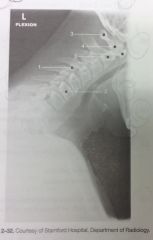
190. The structure labeled #4 in fig 2-32 is A. Body of C1 B. Body of C2 C. Odontoid process D. Anterior arch of C1 |
C. Odontoid process |
|
|
191. Which of the following exams is used to demonstrate vesicoureteral reflux A. Retrograde urogram B. Intravenous urogram C. Voiding cystourethrogram D. Retrograde cystogram |
C. Voiding cystourethrogram |
|
|
192. Which of the following should be demonstrated in a true AP projection of the clavicle 1. Clavicular body 2. Acromioclavicular joint 3. Sternocostal joint A. 1 only B. 1 and 2 C. 2 and 3 D. All three |
B. 1 and 2; clavicular body and acromioclavicular joint |
|
|
193. In which of the following projections is the talofibular joint best demonstrated A. AP B. Lateral oblique C. Medial oblique D. Lateral |
C. Medial oblique |
|
|
194. Free air in the abdominal cavity is best demonstrated in which of the following positions A. AP projection, left lateral decub B. AP projection, right lateral decub C. PA recumbent position D. AP recumbent position |
A. AP projection, left lateral decub |
|
|
195. Which of the following sequences correctly describes the path of blood flow as it leaves the left ventricle A. Arteries, arterioles, capillaries, veins and venules B. Arterioles, arteries, capillaries, veins and venules C. Veins, venules, capillaries, arteries, arterioles D. Venules, veins, capillaries, arterioles, arteries |
A. Arteries, arterioles, capillaries, veins and venules |
|
|
196. Which of the following projections of the elbow should demonstrate the radial head free of ulnar superimposition A. AP B. Lateral C. Medial oblique D. Lateral oblique |
D. Lateral oblique |
|
|
197. Which of the following projections will best demonstrate acromioclavicular separation A. AP recumbent, affected shoulder B. AP recumbent, both shoulders C. AP erect, affected shoulder D. AP erect, both shoulders |
D. AP erect, both shoulders |
|
|
198. Which of the following statements regarding myelography is/are correct 1. Spinal puncture may be performed in the prone or flexed lateral position 2. Contrast medium distribution is regulated thru X-ray tube angulation 3. Pt's neck must be in extension during trendelenburg positions A. 1 only B. 1 and 2 C. 1 and 3 D. All three |
C. 1 and 3; spinal puncture may be performed in the prone or flexed lateral position; pt's neck must be in extension during trendelenburg positions |
|
|
199. The term that refers to parts away from the source or beginning is A. Cephalad B. Proximal C. Distal D. Lateral |
C. Distal |
|
|
200. With the patient PA, the MSP centered to the grid, OML forming a 37 degree angle with the IR and the CR perp and exiting the acantion, which of the following is best demonstrated A. Occipital bone B. Frontal bone C. Facial bones D. Basal foramina |
C. Facial bones |
|
|
201. The inhalation of liquid or solid particles into the nose, throat or lungs is referred to as A. Asphyxia B. Aspiration C. Atelectasis D. Asystole |
B. Aspiration |
|
|
202. Endoscopic retrograde cholangiopancreatography usually involves 1. Cannulation of the hepatopancreatic ampulla 2. Introduction of contrast into the common bile duct 3. Introduction of barium directly into the duodenum A. 1 only B. 1 and 2 C. 1 and 3 D. All three |
B. 1 and 2; cannulation of the hepatopancreatic ampulla, introduction of contrast into the common bile duct |
|
|
203. Which of the following is/are associated with a Colles fx 1. Transverse fx of the radial head 2. Chip fx of the ulnar styloid 3. Posterior or backward displacement A. 1 only B. 1 and 3 C. 2 and 3 D. All three |
C. 2 and 3; chip fx of the ulnar styloid, posterior or backward displacement |
|
|
204. The axiolateral or horizontal beam, projection of the hip requires the IR to be placed 1. Parallel to the CR 2. Parallel to the long axis of the femoral neck 3. In contact with the lateral surface of the body A. 1 only B. 1 and 2 C. 2 and 3 D. All three |
C. 2 and 3; parallel to the long axis of the femoral neck, in contact with the lateral surface of the body |
|
|
205. Which of the following guidelines should be followed when performing exam on pedi pts A. Use restraint only when necessary B. Always use physical or mechanical restraint C. Use physical restraint only D. Use mechanical restraint only |
A. Use restraint only when necessary |
|
|
206. Which of the following interventional procedures can be used to increase the diameter of a stenosed vessel 1. Percutaneous transluminal angioplasty PTA 2. Stent placement 3. Peripherally inserted central catheter PICC A. 1 only B. 1 and 2 C. 1 and 3 D. All three |
B. 1 and 2; PTA and stent placement |
|
|
207. Important considerations for radiographic exam of traumatic injuries to the upper extemity include 1. Joint closest to the injured site should be supported during movement of limb 2. Both joints must be included in long bone studies 3. Two views, at 90 degrees to each other's are required A. 1 only B. 1 and 2 C. 2 and 3 D. All three |
C. 2 and 3; Both joints must be included in long bone studies, Two views, at 90 degrees to each other's are required |
|
|
208. Correct prep for a pt scheduled for an UGI series is most likely to be A. Iodinated contrast administration evening before exam; water only in the morning B. NPO after midnight C. Cathartics and cleansing enemas D. NPO after midnight, cleansing enemas and empty bladder before scout image |
B. NPO after midnight |
|
|
209. The contraction and expansion of arterial walls in accordance with forceful contraction and relaxation of the heart are called A. Hypertension B. Elasticity C. Pulse D. Pressure |
C. Pulse |
|
|
210. The AP trendelenburg position is often used during an upper GI exam to demonstrate A. Duodenal loop B. Filling of the duodenal bulb C. Hiatal hernia D. Hypertrophic pyloric stenosis |
C. Hiatal hernia |
|
|
211. Which of the following positions would be the best choice for a right shoulder exam to rule out fx A. Internal and external rotation B. AP and tangential C. AP and AP axial D. AP and scap Y |
D. AP and scap Y |
|
|
212. Which of the following projections will best demonstrate the tarsal navicular free of superimposition A. AP oblique, medial rotation B. AP oblique, lateral rotation C. Mediolateral D. Lateral weight bearing |
A. AP oblique, medial rotation |
|
|
213. Which of the following bones participates in the formation of the obturator foramen 1. Ilium 2. Ischium 3. Pubis A. 1 and 2 B. 1 and 3 C. 2 and 3 D. All three |
C. 2 and 3; ischium and pubis |
|
|
214. Which of the following radiologic procedures requires that a contrast medium be injected into the renal pelvis via a catheter placed within the ureter A. Nephrotomography B. Retrograde urography C. Cystourethrography D. IVU |
B. Retrograde urography |
|
|
215. The AP projection of the coccyx requires that the CR be directed 1. 15 degrees cephalad 2. 2" superior to the pubic symphysis 3. To a level midway btwn the ASIS and pubic symphysis A. 1 only B. 2 only C. 1 and 2 only D. 1 and 3 only |
B. 2 only, 2" superior to pubic symphysis |
|
|
216. Which of the following views would best demonstrate arthritic changes is the knee A. AP recumbent B. Lateral recumbent C. AP erect D. Medial oblique |
C. AP erect |
|
|
217. Which of the following positions will demonstrate the lumbosacral apophyseal articulation A. AP B. Lateral C. 30 degree RPO D. 30 degree LPO |
C. 30 degree RPO |
|
218. Which of the following statements is/are true regarding the images shown in fig 2-33 1. Image A is positioned in internal rotation 2. Image B is positioned in internal rotation 3. The greater tubercle is better demonstrated in image A A. 1 only B. 2 only C. 1 and 3 D. 2 and 3 |
D. 2 and 3;Image B is positioned in internal rotation, The greater tubercle is better demonstrated in image A |
|
|
219. Which of the following will best demonstrate the size and shape of liver and kidneys A. Lateral abdomen B. AP abdomen C. Dorsal decubitus abdomen D. Ventral decubitus abdomen |
B. AP abdomen |
|
|
220. Correct preparation for a pt scheduled for a lower GI series is most likely to be A. Iodinated contrast evening before exam; water only in the morning B. NPO after midnight C. Cathartics and cleansing enemas D. NPO after midnight, cleansing enemas, and empty bladder before scout image |
C. Cathartics and cleansing enemas |
|
|
221. The AP axial projection, or frog leg position of the femoral neck places the pt in a supine position with the affected thigh A. Addicted 25 degrees from the horizontal B. Abducted 25 degrees form vertical C. Adducted 40 degrees from horizontal D. Abducted 40 degrees from vertical |
D. Abducted 40 degrees from vertical |
|
|
222. Which of the following precautions should be observed when radiographing a pt who has sustained a traumatic injury to the hip 1. When a fx is suspected,manipulation of the affected extremity should be performed by a physician 2. The AP axis lateral projection should be avoided 3. To evaluate the entire region, the pelvis typically is included in the initial exam A. 1 only B. 1 and 3 C. 2 and 3 D. All three |
B. 1 and 3; when a fx is suspected, manipulation of the affected extremity should be performed by a physician; to evaluate the entire region, the pelvis typically is included in the initial examination |
|
|
223. Which of the following projections requires that the humeral epicondyle be perp to the IR 1. AP humerus 2. Lateral forearm 3. Internal rotation shoulder A. 1 only B. 1 and 2 only C. 2 and 3 D. All three |
C. 2 and 3; lateral forearm and internal rotation shoulder |
|
|
224. Prior to the start of an IVU, which of the following procedures should be carried out 1. Have pt empty the bladder 2. Review the pt's allergy history 3. Check the pt's creatinine level A. 1 only B. 2 only C. 1 and 2 only D. All three |
D. All three,Have pt empty the bladder, Review the pt's allergy history, Check the pt's creatinine level |
|
|
225. To demonstrate the entire circumference of the radial head, exposures must be made with the 1. Epicondyles perp to the cassette 2. Hand pronated and supinated as much as possible 3. Hand lateral and in internal rotation A. 1 only B. 1 and 2 C. 1 and 3 D. All three |
D. All three, Epicondyle so perp to the cassette, Hand probated and supinate do as much as possible, Hand lateral and in internal rotation |
|
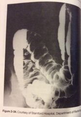
226. The image shown in fig 2-34 was made in what position A. AP or PA chest B. Dorsal decub C. Left lateral decub D. Right lateral decub |
D. Right lateral decub |
|
|
227. In myelography, the contrast medium generally is injected into the A. Cisterna magna B. Individual intervertebral disks C. Subarachnoid space btwn the first and second lumbar vertebrae D. Subarachnoid space btwn the third and fourth lumbar vertebrae |
D. Subarachnoid space btwn the third and fourth lumbar vertebrae |
|
|
228. To evaluate the interphalangeal joints in the oblique and lateral positions, the fingers A. Rest on the cassette for immobilization B. Must be supported parallel to IR C. Are radiographed in natural flexion D. Are radiographed in palmar flexion |
B. Must be supported parallel to IR |
|
|
229. Which of the following is/are effective in reducing exposure to sensitive tissue for frontal views during scoliosis exams 1. Use of PA 2. Use of breast shields 3. Use of compensating filtration A. 1 only B. 1 and 2 C. 2 and 3 D. All three |
D. All three; use of PA, use of breast shields, use of compensating filtration |
|
|
230. Which type of articulation is evaluated in arthrography A. synarthrodial B. Diarthrodial C. Amphiarthrodial D. Cartilaginous |
B. Diarthrodial |
|
|
231. The laryngeal prominence is formed by the A. Thyroid gland B. Thyroid cartilage C. Vocal cords D. Pharynx |
B. Thyroid cartilage |
|
|
232. In the AP projection of the ankle, the 1. Plantar surface of the foot is vertical 2. Fibula projects more distally than the tibia 3. Calcaneous is well visualized A. 1 only B. 1 and 2 C. 2 and 3 D. All three |
B. 1 and 2; fibula projects more distally than the tibia, calcaneous is well visualized |
|
|
233. Which of the following exams most likely would be performed to dx Wilm's tumor A. BE B. UGI C. IVU D. Bone survey |
C. IVU |
|
|
234. To visualize or open the right sacroiliac joint, the pt is positioned A. 30 -40 degrees LPO B. 30-40 degrees RPO C. 25-30 degrees LPO D. 25-30 degrees RPO |
C. 25-30 degrees LPO |
|
|
235. Deoxygenated blood from the head the thorax is returned to the heart by the A. Pulmonary artery B. Pulmonary veins C. Superior vena cava D. Thoracic aorta |
C. Superior vena cava |
|
|
236. Which of the following women is likely to have the most homogenous glandular breast tissue A. Postpubertal adolescent B. 20 yr old with one previous pregnancy C. Menopausal woman D. Post menopausal 65 yr old |
A. Postpubertal adolescent |
|
|
237. Standard radiographic protocols may be reduced to include two views, at right angles to each other, in which of the following situations A. BE B. Spine radiography C. Skull radiography D. Emergency and trauma radiography |
D. Emergency and trauma radiography |
|
|
238. Which of the following is a condition in which an occluded blood vessel stops blood flow to a portion of the lungs A. Pneumothorax B. Atelectasis C. Pulmonary embolism D. Hypoxia |
C. Pulmonary embolism |
|
|
239. Following the ingestion of a fatty meal, what hormone is secreted by the duodenal mucosa to stimulate contraction of the gallbladder A. Insulin B. Cholecystokinin C. Adrenocorticotropic hormone D. Gastrin |
B. Cholecystokinin |
|
|
240. Which of the following projections is most likely to demonstrate the carpal pisiform free of superimposition A. Radial flexion/deviation B. Ulnar flexion/deviation C. AP (medial) oblique D AP (lateral) oblique |
C. AP (medial) oblique |
|
|
241. Myelography is a dx exam used to demonstrate 1. Internal disk lesions 2. Post traumatic swelling of the spinal cord 3. Posterior disk herniation A. 1 only B. 2 only C. 2 and 3 D. All three |
C. 2 and 3; post traumatic swelling of the spinal cord, posterior disk herniation |
|
|
242. Which of the following blood chemistry levels must be the radiographer check prior to excretory urography 1. Creatinine 2. Blood urea nitrogen (BUN) 3. Red blood cells (RBCs) A. 1 only B. 1 and 2 C. 2 and 3 D. All three |
B. 1 and 2; creatinine and BUN |
|
|
243. Which of the following are components of a trimalleolar fx 1. Fx lateral malleolus 2. Fx medial malleolus 3. Fx posterior tibia A. 1 only B. 1 and 3 C. 2 and 3 D. All three |
D. All three, fx lateral malleolus, medial malleolus and posterior tibia |
|
|
244. The functions of which body system include mineral homeostasis, protection and triglyceride storage A. Endocrine B. Integumentary C. Skeletal D. Muscular |
C. Skeletal |
|
|
246.. Ingestion of barium sulfate is contraindicated in which of the following situations 1. Suspected perforation of a hollow viscous 2. Suspected large bowel obstruction 3. Pre operative pt's A. 1 only B. 1 and 3 C. 2 and 3 D. All three |
D. All three; . Suspected perforation of a hollow viscous, Suspected large bowel obstruction, Pre operative pt's |
|
|
245.
The four major arteries supplying the brain include the 1. Brachiocephalic artery 2. Common carotid arteries 3. Vertebral arteries A. 1 and 2 B. 1 and 3 C. 2 and 3 D. All three |
C. 2 and 3; common carotid arteries, vertebral arteries |
|
|
247. Which of the following is a major cause of bowel obstruction in children A. Appendicitis B. Intussusception C. Regional enteritis D. Ulcerative colitis |
B. Intussusception |
|

248.
Which of the following is/are well demonstrated in the lumbar spine shown in fig 2-35 1. Zygapophyseal/apophyseal articulations 2. Intervertebral foramina 3. Inferior articular processes A. 1 only B. 1 and 2 C. 1 and 3 D. All three |
C. 1 and 3; zygapophyseal/apophyseal joints and inferior articular processes |
|
|
249.
Which of the following statements is/are correct, with respect to a left lateral projection of the chest 1. MSP must be perfectly vertical and parallel to the IR 2. Right posterior ribs will be projected slightly posterior to the left posterior ribs 3. Arms must be raised high to prevent upper arm soft tissue superimposition on lung field A. 1 only B. 1 and 2 C. 1 and 3 D. All three |
D. All three; MSP must be perfectly vertical and parallel to the IR, Right posterior ribs will be projected slightly posterior to the left posterior ribs, Arms must be raised high to prevent upper arm soft tissue superimposition on lung fieldA. 1 only |
|
250.
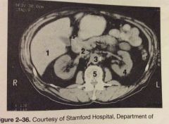
Which of the following is represented by the number 3 in fig 2-36 A. Inferior vena cava B. Aorta C. Gallbladder D. Psoas muscle |
B. Aorta |
|
|
251.
Which of the following bones particpates in the formation of the knee joint 1. Femur 2. Tibia 3. Patella A. 1 and 2 B. 1 and 3 C. 2 and 3 D. All three |
A. 1 and 2; femur and tibia |
|
|
252.
All the following are palpable bony landmarks used in radiography of the pelvis except A.. Femoral neck B. Pubic symphysis C. Greater trochanter D. Iliac crest |
A. Femoral neck |
|
|
253.
Lateral deviation of the nasal septum may be best demonstrated in the A. Lateral projection B. PA axial (Caldwell) projection C. Parietoacanthial (waters) projections D. AP axial (Towne) projection |
C. Parietoacanthail (waters) projection |
|
|
254.
Which of the following structures should be visualized thru the foramen magnum in an AP axial projection (towne) of the skull for occipital bone 1. Posterior clinoid processes 2. Dorsum sella 3. Posterior arch of C1 A. 1 only B. 2 only C. 1 and 2 D. 2 and 3 |
C. 1 and 2; posterior clinoid processes, dorsum sella |
|

255.
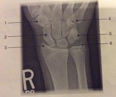
What structure labeled #5 in fig 2-37 A. Base of the 2nd metacarpal B. Pisiform C. Trapezium D. Trapezoid |
B. Pisiform |
|
256.
What is the structure labeled #3 in fig 2-37 A. Trapezium B. Scaphoid C. Ulnar styloid D. Radial styloid |
D. Radial styloid |
|
|
257.
In the anterior oblique position of the cervical spine, the CR should be directed A. Parallel to C4 B. Perp to C4 C. 15 degrees cephalad to C4 D. 15 degrees caudad to C4 |
D. 15 degrees caudad to C4 |
|
|
258.
Which of the following is a functional study used to demonstrate the degree of AP motion present in the cervical spine A. Open mouth projection B. Moving mandible AP C. Flexion and extension laterals D. Right and left bending AP |
C. Flexion and extension laterals |
|
|
259.
If a pt's zygomatic arch has been traumatically depressed or the pt has flat cheekbones, the arch may be demonstrated by modifying the SMV projection and rotating the pt's head A. 15 degrees toward the side being examined B. 15 degrees away from side being examined C. 30 degrees toward the side being examined D. 30 degrees away from side being examined |
A. 15 degrees toward the side being examined |
|
|
260.
Which of the following factors can contribute to hypertension 1. Obesity 2. Smoking 3. Stress A. 1 only B. 1 and 2 C. 2 and 3 D. All three |
D. All three; obesity, smoking and stress |
|
261.
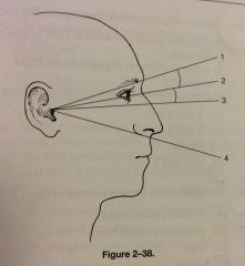
What is the degree of difference btwn the baselines numbered 2 and 3 in fig 2-38 and used for various projections of the skull A. 7 degrees B. 8 degrees C. 15 degrees D. 23 degrees |
A. 7 degrees |
|

262.
Referring to fig 2-38 which of the following positions requires that baseline number 3 be parallel to the IR A. Parietoacanthial B. PA axial (Caldwell) C. AP axial (Towne) D. SMV |
D. SMV |
|
|
263.
Orthoroentgenography or radiographic measurements of long bones of an upper or lower extremity, requires which of the following accessories 1. Bell-Thompson scale 2. Bucky tray 3. Cannula A. 1 only B. 1 and 2 C. 1 and 3 D. All three |
B. 1 and 2; bell Thompson scale and Bucky tray |
|
|
264.
Which of the following is/are demonstrated in a lateral projection of the cervical spine 1. Intervertebral foramina 2. Zygapophyseal/apophyseal joints 3. Intervertebral joints A. 1 only B. 1 and 2 C. 2 and 3 D. All three |
C. 2 and 3; Zygapophyseal/apophyseal joints, Intervertebral foramina |
|
|
265. In a lateral projection of the nasal bones, the CR is directed A. 1/2" posterior to the anterior nasal spine B. 3/4" posterior to the glabella C. 3/4" distal to the nasion D. 1/2" anterior to the EAM |
C. 3/4" distal to the nasion
|
|
|
266.
To make a pt as comfortable as possible during a single contrast BE, the radiographer should 1. Instruct pt to relax the abdominal muscles to prevent intra-abdominal pressure 2. Instruct pt to concentrate on breathing deeply to reduce colonic spasm 3. Prepare a warm barium suspension (98-105') to aid in retention A. 2 only B. 1 and 2 C. 2 and 3 D. All three |
B. 1 and 2: Instruct pt to relax the abdominal muscles to prevent intra-abdominal pressure, Instruct pt to concentrate on breathing deeply to reduce colonic spasm |
|
|
267. Which of the following positions will best demonstrate the right apophyseal articulations of the lumbar vertebrae A. PA B. Left lateral C. RPO D. LPO
|
C. RPO |
|
|
268.
Structures involved in blowout fx include the 1. Orbital floor 2. Inferior rectus muscle 3. Zygoma A.1 only B. 1 and 2 C. 2 and 3 D. All three |
B. 1 and 2; orbital floor, inferior rectus muscle |
|
|
269.
Inspiration and expiration projections of the chest are performed to demonstrate 1. Partial or complete collapse of pulmonary lobes 2. Air in the pleural cavity 3. Foreign body A. 1 only B. 1 and 2 C. 1 and 3 D. All three |
D. All three; Partial or complete collapse of pulmonary lobes, Air in the pleural cavity, Foreign body |
|
|
270.
Shoulder arthrography is performed to 1. Evaluate humeral luxation 2. Demonstrate complete or partial rotator cuff tear 3. Evaluate the glenoid labrum A. 1 only B. 1 and 2 C. 2 and 3 D. All three |
C. 2 and 3; Demonstrate complete or partial rotator cuff tear, Evaluate the glen oil labrum |
|
|
271.
Which of the following positions will separate the radial head ,neck and tuberosity from superimpostion on the ulna A. AP B. Lateral C. Medial oblique D. Lateral oblique |
D. Lateral oblique |
|
|
272.
The most significant risk factor for breast cancer is A. Age B. Gender C. Family history D. Personal history |
B. Gender |
|
|
273.
Which of the following structures is located at the level of the interspace btwn the 2nd and 3rd thoracic vertebrae A. Manubrium B. Jugular notch C. Sternal angle D. Xiphoid process |
B.jugular notch |
|
|
274.
For the AP projection of the scapula, the 1.pt's arm is abducted at right angles to the body 2. Pt's elbow is flexed with the hand supinated 3. Exposure is made during quiet breathing A. 1 and 2 B. 1 and 3 C. 3 only D. All three |
D. All three;pt's arm is abducted at right angles to the body, Pt's elbow is flexed with the hand supinated, Exposure is made during quiet breathing |
|
|
275.
The innominate bone is located in the A. Middle cranial fossa B. Posterior cranial fossa C. Foot D.pelvis |
D. Pelvis |
|
|
276.
The sternoclavicular joints are best demonstrated with the pt PA and A. In a slight oblique position, affected side adjacent to the IR B. In a slight oblique position,affected side away from the IR C. Erect and weight bearing D. Erect with and without weights |
A. In a slight oblique position, affected side adjacent to the IR |
|
|
277.
Which of the following sinus groups is demonstrated with the pt positioned as for a parietoacanthial projection (waters) with the CR directed thru the pt's open mouth A.frontal B. Ethmoidal C. Maxillary D. Sphenoidal |
D. Sphenoidal |
|
|
278.
Below diaphragm ribs are better demonstrated when A. Respiration is suspended at the end of full inspiration B. Exposed using shallow breathing technqiue C. Pt is in the recumbent position D. Pt is in the AP erect position |
C. Pt is in the recumbent position |
|
|
279.
Which of the following positions is essential in radiography of the paranasal sinuses A. Erect B. Recumbent C. Oblique D. Trendelenburg |
A.erect |
|
|
280.
What projection of the os calsis is obtained with the leg extended, the plantar surface of the foot vertical and perp to the IR, and the CR directed 40 degrees cephalad A. Axial plantodorsal projection B. Axial dorsoplantar projection C. Lateral projection D. Weight bearing lateral projection |
A. Axial plantodorsal projection |
|
|
281.
During GI radiography, the position of the stomach may vary depending on 1. Respiratory phase 2. Body habitus 3. Pt position A. 1 and 2 B. 1 and 3 C. 2 and 3 D. All three |
D. All three; respiratory phase, body habitus and pt position |
|
|
282.
With a pt in the PA position and the OML perp to the table, a 15 to 20 degree caudal angular ion would place the petrous ridges in the lower third of the orbit. To achieve the same result in a baby or small child, it is necessary for the radiographer to modify the angular ion to A. 10 to 15 degrees caudal B. 25 to 30 degrees caudal C. 15 to 20 degrees cephalic D. 3 to 5 degrees caudal |
A. 10 to 15 degrees caudal |
|

283.
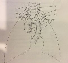
The structure labeled number 6 in fig 2-39 is the A. Left subclavian artery B. Brachiocephalic artery C. Right common carotid artery D. Left vertebral artery |
B. Brachiocephalic artery |
|
284.
The structure labeled number 3 in fig 2-39 is the A. Left subclavian artery B. Brachiocephalic artery C. Right common carotid artery D. Left vertebral artery |
D.left vertebral artery |
|
|
285.
In the lateral projection of the foot, the 1. Plantar surface should be perp to IR 2. Metatarsals are superimposed 3. Talofibular joint should be visualized A. 1 only B. 1 and 2 C. 2 and 3 D. All three |
B. 1 and 2; plantar surface should be perp to IR and metatarsals are superimposed |
|
|
286.
HSG may be performed for demonstration of 1. Uterine tube potency 2. Mass lesions in the uterine cavity 3. Uterine postion A. 1 and 2 B. 1 and 3 C. 2 and 3 D. All three |
D. All three; Uterine tube potency, Mass lesions in the uterine cavity, Uterine postion |
|
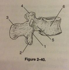
287.
The number 2 in fig 2-40 represents which of the following structures A. Body B. Pedicles C. Inferior articular process D. Superior articular process |
C. Inferior articular process |
|
|
288.
To demonstrate the mandibular body in the PA position, the A. CR is directed perp to the IR B. CR is directed cephalad to IR C. Skull is obliqued away from the affected side D. Skull is obliqued toward the affected side |
A. CR is directed perp to IR |
|
|
289.
Which of the following equipment is necessary for ERCP 1. Fluroscopic unit with imaging device and tilt table capabilities 2. Fiber optic endoscope 3. Polyethylene catheters A. 1 and 2 B. 1 and 3 C. 2 and 3 D. All three |
D. All three; Fluroscopic unit with imaging device and tilt table capabilities, Fiber optic endoscope, Polyethylene catheters |
|
|
290.
All of the following statements regarding pediatric positioning are true except A. For radiography of the kidneys, the CR should be directed midway btwn the diaphragm and the symphysis pubis B. If a pediatric pt is in respiratory distress, a chest radiograph should be obtained in the AP projection rather than in the standard PA proj C. Chest radiography on a neonate should be performed in the supine position D. Radiography of pediatric pt's with a myelomeningocele defect should be performed in the supine position |
D. Radiography of pediatric pt's with a myelomeningocele defect should be performed in the supine position |
|
|
291.
The act of expiration will cause the 1. Diaphragm to move inferiorly 2. Sternum and ribs to move inferiorly 3. Diaphragm to move superiorly A. 1 only B. 1 and 2 C. 2 and 3 D. All three |
C. 2 and 3; Sternum and ribs to move inferiorly3. Diaphragm to move superiorly |
|
|
292.
A pt unable to extend his or her arm is seated at the end of the X-ray table, elbow flexed 90 degrees. The CR is directed 45 degrees medially. Which of the following structures will be demonstrated best 1. Radial head 2. Capitulum 3. Coronoid process A. 1 only B. 1 and 2 C. 2 and 3 D. All three |
B. 1 and 2; radial head and capitulum |
|
|
293. Which of the following articulations with the bases of the metatarsals 1. Heads of first row of phalanges 2. Cuboid 3. Cuneiforms A. 1 only B. 1 and 2 C. 2 and 3 D. All three |
C. 2 and 3 only; cuboid and cuneiforms |
|
|
294.
Which of the following statements regarding knee X-ray arthrography is/are true 1. Ligament tears can be demonstrated 2. Sterile technique is observed 3. MRI can follow X-ray A. 1 and 2 B. 1 and 3 C. 2 and 3 D. All three |
D. All three; Ligament tears can be demonstrated, Sterile technique is observed, MRI can follow X-ray |
|

295.
What projection was used to obtain the image seen in fig 2-41 A. AP internal rotation B. AP external rotation C. AP neutral position D. AP axial |
B. AP external rotation |
|
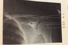
296.
The structure labeled #4 in fig 2-41 is the A. Acromion process B. Coracoid process C. Coronoid process D. Glenoid process |
B. Coracoid process |
|

297.
The structure labeled #5 in fig 2-41 is the A. Sternoclavicular joint B. Acromioclavicular joint C. Glenohumeral joint D. Acromion umbra joint |
C. Glenohumeral joint |
|
|
298.
Which of the following is demonstrated in a 25 degree RPO with the CR entering 1 inch medial to the elevated ASIS A. Left sacroiliac joint B. Right sacroiliac joint C. Left ilium D. Right ilium |
A. Left sacroiliac joint |
|
|
299.
The relationship btwn the ends of fx long bones is referred to as A. Angulation B. Apposition C. Luxation D. Sprain |
B. Apposition |
|
|
230.
A lateral projection of the larynx is occasionally required to rule out foreign body, polyps, or tumor. The CR should be directed A. Just below the EAM B. To the level of the mandibular angles C. To the level of the laryngeal prominence D. To the level of C7 |
C. To the level of the laryngeal prominence |
|
|
10/1.
The term valgus refers to A. Turned outward B. Turned inward C. Rotated medically D. Rotated laterally |
A. Turned outward |
|
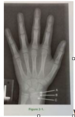
10/3. What is the portion of bone labeled C in pediatric PA hand image in fig 2-1 A. Diaphysis B. Epiphysis C. Metaphysis D. Apophyseal |
C. Metaphysis |
|
|
10/4.
The proximal radius & ulna are seen free of superimposition in the following projection A. Scap Y B. AP scapula C. Medial oblique elbow D. Lateral oblique elbow |
D. Lateral oblique elbow |
|
|
10/5.
The femal pelvis differs from the male pelvis in the following ways 1. Male greater/false pelvis is deep 2. Male acetabulum faces more laterally 3. Female coccyx is more vertical A. 1 only B. 1 and 2 C. 2 and 3 D. All three |
B. 1 and 2; male greater/false pelvis is deep, male acetabulum faces more laterally |
|
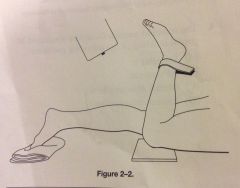
10/7.
The structures would be visualized when obtained as positioned in fig 2-2 could also be visualized when performed with the pt in the following position 1. Lateral recumbent 2. Seated 3. Erect AP A. 1 only B. 1 and 2 C. 2 and 3 D. All three |
B. 1 and 2; lateral recumbent, seated |
|
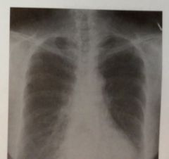
10/10.
An accurate critique of the PA projection of he chest seen in fig 2-3 would include the following 1. Pulmonary apices are demonstrated 2. Air filled trachea and carina are demonstrated 3. 10 posterior ribs seen above diaphragm A. 1 only B. 1 and 2 C. 2 and 3 D. All three |
D. All three; Pulmonary apices are demonstrated, Air filled trachea and carina are demonstrated, 10 posterior ribs seen above diaphragm |
|
|
10/12.
Flattening of the hemidiaphragm is characteristic of which of the following conditions A. Pneumothorax B. Pleural effusion C. Emphysema D. Pneumonia |
C. Emphysema |
|
|
10/13.
Structure located in the RLQ include the following 1. Cecum 2. Vermiform appendix 3. Sigmoid A. 1only B. 1 and 2 C. 2 and 3 D. All three |
B. 1 and 2; cecum and vermiform appendix |
|
|
10/17.
The upper surface of the foot may be described as 1. Plantar surface 2. Anterior surface 3. Dorsum A. 1 only B. 1 and 2 C. 2 and 3 D. All three |
C. 2 and 3; anterior surface and dorsum |
|
|
10/18.
The outermost wall of the digestive tract is the A. Mucosa B. Muscular C. Submucosa D. Serosa |
D. Serosa |
|
|
10/19.
Terms used to describe movement 1. Plantar flexion 2. Valgus 3. Oblique A. 1 only B. 1 and 2 C. 2 and 3 D. All three |
B. 1 and 2; plantar flexion and valgus |
|
|
10/20.
Sternoclavicular articulation are likely to be demonstrated in all except A. Weight bearing B. RAO C. LAO D. PA |
A. Weight bearing |
|

10/45.
The cecum is indicated by which number in fig 2-9 A. 3 B. 4 C. 5 D. 6 |
B. 4 |
|
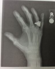
10/54.
Which of the following statements regarding fig 2-10 is/are true 1. Midphalanges are foreshortened 2. Degree of obliquity is too great 3. Interphalangeal joints are well demonstrated A. 1 only B. 1 and 2 C. 2 and 3 D. All true |
A. 1 only; Midphalanges are foreshortened |
|
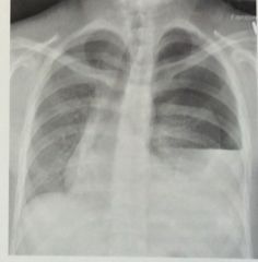
10/58.
What condition is are demonstrated in fig 2-11 1. Emphysema 2. Pneumothorax 3. Pleural effusion A. 1 only B. 1 and 2 C. 2 and 3 D. All three |
C. 2 and 3; pneumothorax and pleural effusion |
|
|
10/59
Which of the following bony landmarks is in the same transverse plane as L2 and L3 A. Inferior costal margin B. Greater trochanter C. Iliac crest D. ASIS |
A. Inferior costal margin |
|
|
10/62.
Which of the following is/are proximal to the tibial plateau 1. Intercondyloid fossa 2. Tibial condyles 3. Tibial tuberosity A. 1 only B. 1 and 2 C. 2 and 3 D. All three |
A. 1 only; intercondyloid fossa |
|
|
10/73. The lumbar vertebral lamina is represented by what part of the "Scotty dog" seen in a correctly positioned oblique lumbar spine A. Eye B. Nose C. Body D. Ear |
C. Body |
|
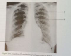
10/81.
The letter A in fig 2-13 indicates A. A left anterior rib B. A right posterior rib C. A left posterior rib D. A right anterior rib |
C. A left posterior rib |
|
|
10.83 The type of articulation lacking a joint cavity include 1. Fibrous 2. Cartilaginous 3. Synovial A. 1 only B. 1 and 2 C. 2 and 3 D. All three |
B 1 and 2; fibrous and cartilaginous |
|

10/86.
The structure labeled #2 is the A. Maxillary sinus B. Sphenoidal sinus C. Ethmoidal sinus D. Frontal sinus |
C. Ethmoidal sinus |
|
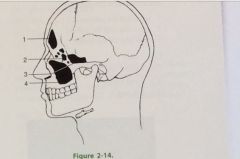
10/87.
Which of the following would best evaluate the structure labeled #3 in fig 2-14 A. PA axial projection (Caldwell) B. Parietoacanthial projection (waters) C. Lateral D. SMV |
D. SMV |
|
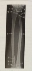
10/90.
Fig 2-16 identify the structure labeled #10 in AP projection of forearm A. Head of radius B. Head of ulna C. Radial styloid process D. Ulna styloid process |
B. Head of ulna |
|
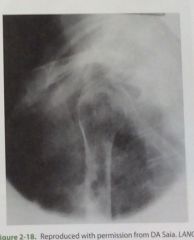
10/100.
Which of the following statements is/are true regarding the shoulder image 1. The unaffected area is adjacent to IR 2. Provides a lateral view 3. Is frequently performed in trauma situations A. 1 only B. 1 and 2 C. 2 and 3 D. All three |
C. 2 and 3; Provides a lateral view, Is frequently performed in trauma situations |
|
|
10/101.
Elements of correct positioning for PA projection of the chest include 1. Weight evenly distributed on feet 2. Elevation of chin 3. Shoulders elevated and rolled forward A. 1 only B. 1 and 2 C. 2 and 3 D. All three |
B. 1 and 2; Weight evenly distributed on feedback, Elevation of chin |
|
|
10/107.
The term used to describe the presence of blood in vomit is A. Hemoptysis B. Hematemesis C. COPD D. Bronchitis |
B. Hematemesis |
|
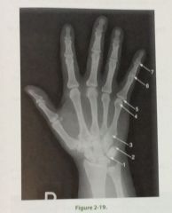
10/110.
In the PA projection of hand seen in fig 2-19 which # identifies proximal interphalangeal joint A. 4 B. 5 C. 6 D. 7 |
C. 6 |
|
|
10/114.
Which of the following articulations may be described as diarthrotic 1. Condyloid 2. Sella 3. Gomphosis A. 1 only B. 3 only C. 1 and 2 D. All three |
C. 1 and 2; condyloid and sella |
|
|
10/123.
The projection/method often used to detect carpal canal defect is A. PA proj wrist, radial deviation B. PA axial wrist, stecher method C. AP oblique hand, norgaard method D. Tangential wrist, gaynor-hart method |
D. Tangential wrist, gaynor-hart method |
|
|
10/124.
Narrowing of the upper airway as seen in pediatric croup, can be visualized A. AP B. Lateral C. Axial D. Lordotic |
A. AP |
|
|
10/126.
Body habitus having short and wide heart and lung area and a high transverse stomach is A. Asthenic B. Hyposthenic C. Sthenic D. Hypersthenic |
D. Hypersthenic |
|
|
10/131.
The most proximal portion of the pharynx A. Laryngopharynx B. Nasopharynx C. Epiglottis D. Oropharynx |
B. Nasopharynx |
|
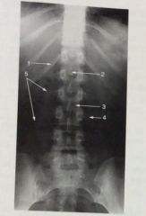
10/136.
In fig 2-24, the #2 indicates the following A. Body of L2 B. Spinous process of L1 C. Spinous process of L3 D. Transverse process of L3 |
B. Spinous process of L1 |
|

10/167.
Fig 2-29 what does number 2 represent A. Acromion process B. Scapular spine C. Coracoid process D. Acromioclavicular joint |
C. Coracoid process |
|

10/168.
What is represented by the number 9 in fig 2-29 A. Medial border B. Lateral border C. Inferior angle D. Superior angle |
A. Medial border |
|
|
10/179.
Which of the following is/are demonstrated in an AP axial projection of the c-spine 1. C3-7 cervical bodies 2. Intervertebral foramina 3. Zygapophyseal joints A. 1 only B. 1 and 2 C. 2 and 3 D. All three |
A. 1 only; C3-7 cervical bodies |
|
|
10/181.
Which of the following statements is/are true regarding a PA projection of the paranasal sinuses 1. OML is elevated 15 degrees from the horizontal 2. Petrous pyramids completely fill the orbits 3. Frontal and ethmoidal sinuses are visualized A. 1 only B. 1 and 2 C. 1 and 3 D. All three |
C. 1 and 3; OML is elevated 15 degrees from the horizontal, Frontal and ethmoidal sinuses are visualized |
|

10/189.
Structure labeled #3 in fig 2-32 is A. Spinous process of C2 B. Body of C2 C. Body of C1 D. Posterior arch of C1 |
D. Posterior arch of C1 |
|
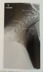
10/190.
Structure labeled #1 in fig 2-32 A. Zygapophyseal joint B. Intervertebral foramina C. Intervertebral disk space D. Vertebral body |
A. Zygapophyseal joint |
|
|
10/197.
An acromioclavicular separation will be best demonstrated in the following projection A. AP recumbent affected shoulder B. AP recumbent both shoulders C. AP erect affected shoulder D AP erect both shoulders |
D. AP erect both shoulders |
|
|
10/221.
In AP axial proj, or bilateral frog leg position of the following is most likely to place the long axis of the femoral necks parallel with plane of IR A. Addicted 25 degrees from horizontal B. Abducted 25 degrees from vertical C. Adducted 40 degrees from horizontal D. Abducted 40 degrees from vertical |
D. Abducted 40 degrees from vertical |
|
|
10/278.
Below diaphragm ribs are better demonstrated 1. Pt in AP erect position 2. Respiration is suspended at end of full exhalation 3. Pt is in recumbent position A. 1 only B. 1 and 2 C. 2 and 3 D. All three |
C. 2 and 3; Respiration is suspended at end of full exhalation, Pt is in recumbent position |
|
|
10/290.
Types of mechanical obstruction found in pediatric pts include 1. Volvulus 2. Intussusception 3. Paralytic ileum A. 1only B. 1 and 2 C. 2 and 3 D. All three |
B. 1 and 2; volvulus and intussusception |