![]()
![]()
![]()
Use LEFT and RIGHT arrow keys to navigate between flashcards;
Use UP and DOWN arrow keys to flip the card;
H to show hint;
A reads text to speech;
63 Cards in this Set
- Front
- Back
- 3rd side (hint)
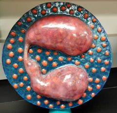
Name this WBC and its function |
Eosinophil- reduce inflammation |
Red dots in cytoplasm |
|
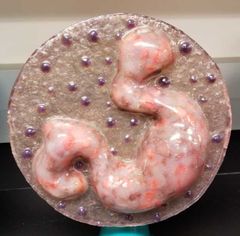
Name this WBC and its function |
Basophil- promotes inflammation releases histamine & heparin |
Blue/purple dots in cytoplasm |
|
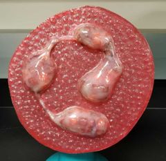
Name this WBC and its function |
Neutrophil- eat foreign objects |
3-5 lobed Nucleus |
|
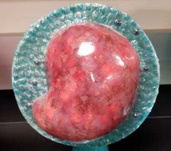
Name this WBC and its function |
Lymphocyte- helps to produce antibodies |
Smooth cytoplasm |
|
Name this WBC and its function |
Monocyte- breakdown antigens become macrophages |
Kidney shaped nucleus |
|
Name this WBC and its function |
Erythrocyte- (aka RBC) carries O2 |
|
|
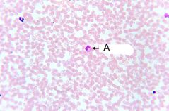
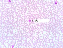
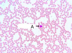
Label A and its function |
Eosinophil - reduces inflammation |
Bright pink/ red |
|
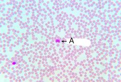
Label A and its function |
Monocyte- breakdown antigens |
Kidney shaped nucleus |
|
Label A and its function |
Neutrophil- eats foreign objects |
3-5 lobed nucleus |
|
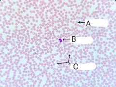
Label A-C |
A= erythrocyte B= leukocyte C= thrombocytes |
|
|
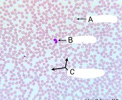
Label A-C |
A= leukocyte B= erythrocyte C= thrombocyte |
|
|
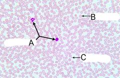
Label A-C |
A= erythrocyte B= leukocyte C= thrombocyte |
|
|
Label A and its function |
A= lymphocyte Helps produce antibodies |
|
|
|
Large granulation leukocytes are called __________. |
Granulocytes |
|
|
|
List the types of granulocytes |
Basophil Eosinophil Neutrophil |
|
|
|
Small granulation leukocytes are called ___________. |
Agranulocytes |
|
|
|
List the types of agranulocytes |
Monocytes Lymphocytes |
|
|
|
When does a monocyte become a macrophage? |
When it enters the tissue |
|
|
|
Name the leukocyte that is kidney shaped |
Monocyte |
|
|
|
Name the leukocyte that has a circular nucleus |
Lymphocyte |
|
|
|
Name the leukocyte that has a 3-5 lobed nucleus |
Neutrophil |
|
|
|
Name the leukocyte that has blue/ purple granulations in the cytoplasm |
Basophil |
|
|
|
Name the leukocyte that has reddish/pink granulations in the cytoplasm |
Eosinophil |
|
|
|
P wave represents _______ |
Atria depolarization (contraction) |
|
|
|
QRS complex represents ______ |
Ventricular depolarization (contraction) |
|
|
|
T wave represents ________ |
Ventricular repolarization (relax) |
|
|
|
What is the function of red blood cells? |
To carry respiratory gases |
|
|

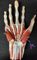
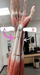
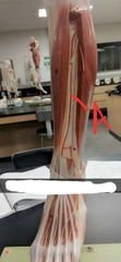
Label A |
Deep palmar arch |
|
|

Label A |
Superficial palmar arch |
|
|
Label A |
Superficial palmar arch |
|
|
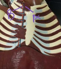
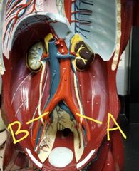
Label A and B on the anterior thorax |
A= internal thoracic vein B= internal thoracic artery |
|
|
|
What color are veins and arteries on the models |
Vein= blue Artery = red |
|
|
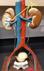
Label A |
Renal artery |
|
|
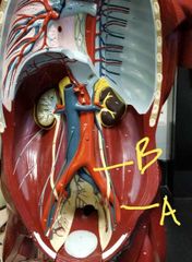
Label A and B |
A= left internal iliac artery B= left common iliac artery |
|
|
|
What arteries branch off the abdominal aorta? |
Separates into: Left common iliac artery and Right common iliac artery |
|
|
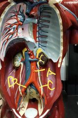
Label A-C |
A= abdominal aorta B= right common iliac artery C= left common iliac artery |
|
|
|
When does the external iliac artery become the femoral artery? |
Once its below the inguinal ligament (which looks white on the model) |
|
|
Label A and B |
A= right atrium B= left atrium |
|
|
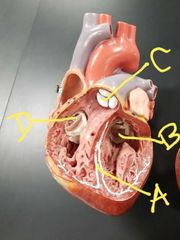
Label A-D |
A= interventricular septum B= bicuspid valve C= pulmonary semilunar valve D= tricuspid valve |
|
|
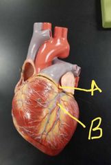
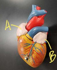
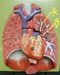
Label A and B |
A= left coronary artery B= anterior interventricular artery |
|
|
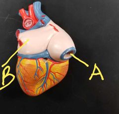
Label A and B |
A= inferior vena cava B= left atrium |
|
|
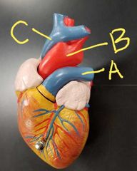
Label A- C |
A= pulmonary trunk B= aortic arch C= superior vena cava |
|
|
|
List the branches of the left coronary artery |
Anterior interventricular artery Circumflex artery Left marginal artery |
|
|
|
List the branches of the right coronary artery |
Right marginal artery Posterior interventricular artery |
|
|
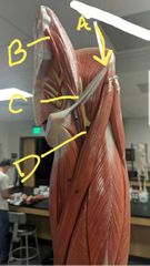
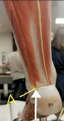
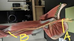
Label A-D on the leg |
A= inguinal ligament B= common iliac artery C= external iliac artery D= femoral artery |
|
|
Label A and B |
A= ulnar artery (pinky side) B= radial artery (thumb side) |
|
|
Label A |
Fibular artery (pinky toe side) |
|
|
Label A and B |
A= left common iliac artery B= right common iliac artery |
|
|
Label A and B |
A= left pulmonary artery B= left pulmonary vein |
|
|
Label A on the leg |
A= anterior tibial artery |
|
|
Label A and B on the arm |
A= subclavian artery B= brachial artery |
|
|
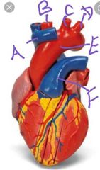
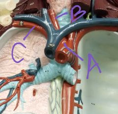
Label A-F |
A= superior vena cava B= Right brachiocephalic artery C= left common carotid artery D= left subclavian artery E= aortic arch F= pulmonary trunk |
|
|
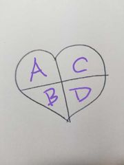
Label A-D (heart chambers) |
A= right atrium B= right ventricle C= left atrium D= left ventricle |
|
|
Blood from the right side flows out the _______. |
Pulmonary trunk to the lungs |
|
|
Blood from the left side flows out the _______. |
Aortic arch to the extremities |
|
|
|
Blood coming from the inferior vena cava and superior vena cava dumps into the _______. |
Right atrium |
|
|
|
Name the branches of the aortic arch |
Right brachiocephalic artery Left common carotid artery Left subclavian artery |
|
|
|
List the branches of the right brachiocephalic artery |
Right common carotid artery Right subclavian artery |
|
|
Label the structures |
A= vein (has valves, collapsible) B= arteries (no valves, circular shape) |
|
|
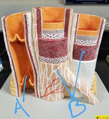
Label the layers A-C |
A= tunica adventitia (outer) B= tunica media (middle) C= tunica intima (inner) |
|
|
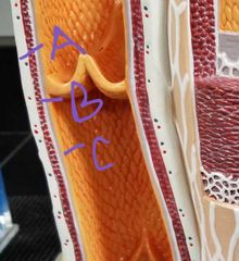
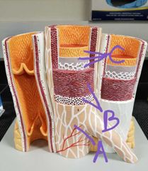
Label the layers A-C of the vein |
A= tunica externa (outer) B= tunica media (middle) C= tunica intima (inner) *Lumen houses the blood |
|
|
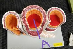
Label A |
The Lumen (houses the blood within an artery and vein) |
|
|
Label A-C |
A= right brachiocephalic artery B= right common carotid artery C= right subclavian artery |
|