![]()
![]()
![]()
Use LEFT and RIGHT arrow keys to navigate between flashcards;
Use UP and DOWN arrow keys to flip the card;
H to show hint;
A reads text to speech;
199 Cards in this Set
- Front
- Back
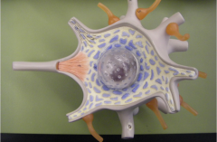
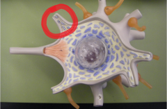
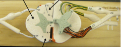
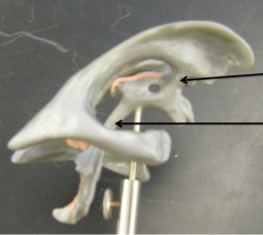
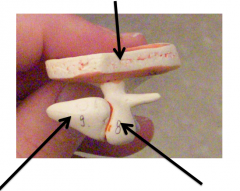
What are the orange nobs called?
|
synaptic knob
|
|

what are the blue specks?
|
Nissl body
|
|
|
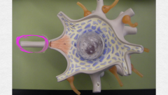
Axon
|
|
|
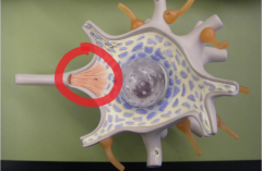
axon hillock
|
|
|
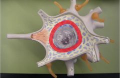
Nucleus
|
|
|
Dendrite
|
|
|
Nerve fiber
|
|
|
Nodes of Ranvier
|
|
|
Schwann cells
|
|
|
nucleus of Schwann cells
|
|
|
Axon of neuron
|
|
What is 1st bottom arrow? |
Endoneurium
|
|
|
myelin sheath
|
|

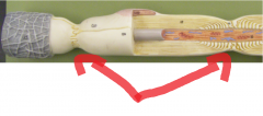
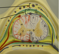
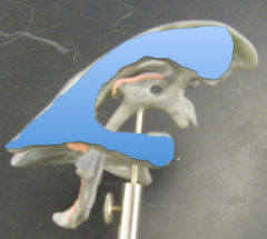
1st arrow
|
periosteum
|
|

2nd arrow
|
Dura Matter (white)
|
|
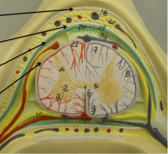
3rd arrow
|
Arachnoid Mater (green)
|
|

4th Arrow
|
Pia Matter (grey)
|
|
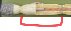
1st area
|
Epidural space
|
|

2nd area
|
subarachnoid space
|
|

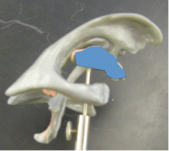
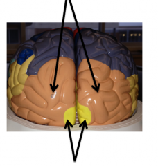
top arrow
|
Posterior median sulcus (small groove) |
|
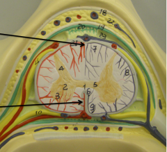
Bottom arrow
|
Anterior Median Fissure (large groove) |
|

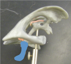
1st arrow
|
Posterior funiculus (white matter) |
|
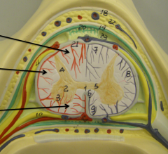
2nd arrow
|
Lateral funiculus (white matter) |
|

3rd arrow
|
Anterior Funiculus (white matter) |
|
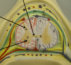
1st arrow
|
Gray commissure
|
|

2nd arrow
|
posterior horn (grey matter) |
|

3rd arrow
|
Anterior Horn (grey matter) |
|

top 1st
|
Dorsal root Ganglion
|
|

Top 2nd
|
Dorsal Root
|
|
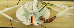
top 3rd
|
Grey matter
|
|

Bottom 1st
|
Ventral Root
|
|

Bottom 2nd
|
White matter
|
|
Top middle
|
Posterior median sulcus
|
|

bottom middle green arrow
|
Anterior median fissure
|
|
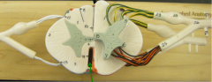
top 1st
|
Posterior horn (grey matter) |
|

top 2nd
|
grey commissure
|
|

bottom first
|
Lateral Horn (grey Matter) |
|

bottom second
|
Anterior Horn (grey Matter) |
|

top
|
Posterior Funiculus (white matter) |
|
middle left
|
Lateral Funiculus (white matter) |
|

bottom
|
Anterior Funiculus (white matter) |
|
shaded
|
Lateral ventricle
|
|
|
3rd ventricle
|
|
|
4th ventricle
|
|

1st arrow
|
interventricular foramen
|
|
2nd arrow
|
cerebral aqueduct
|
|

1st bottom
|
frontal lobe of cerebrum
|
|

2nd bottom
|
Temporal lobe of cerebrum
|
|
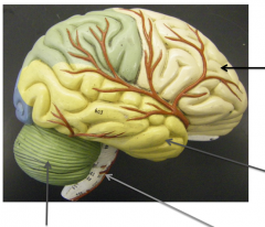
3rd right
|
Brainstem
|
|

4th top right
|
Cerebellum
|
|
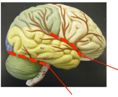
Bottom 1st
|
Transverse fissure
|
|

2nd middle
|
lateral sulcus
|
|
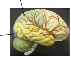
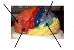
left
|
occipital lobe of cerebrum
|
|

top
|
parietal lob of cerebrum
|
|
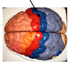
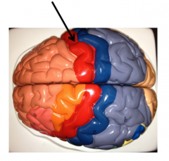
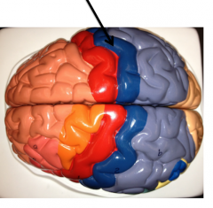
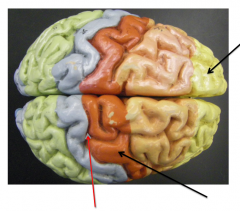
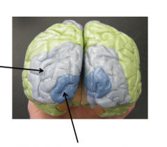
groove between red and blue gyri
|
central sulcus
|
|
|
precentral gyrus/ primary motor cortex (red)
|
|
|
postcentral gyrus/primary somatosensory cortex (blue) |
|
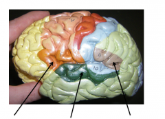
1st top
|
prefrontal cortex
|
|

2nd top
|
sensory association area
|
|

1st bottom
|
Broaca's area
|
|

2nd bottom
|
Wernicke's area
|
|

top |
Visual association areas
|
|
bottom
|
primary visual cortex
|
|

1st view
|
superior
|
|
2nd picture view
|
posterior
|
|

3rd picture view
|
Lateral
|
|

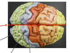
1st bottom
|
Central sulcus
|
|
2nd bottom
|
Primary motor cortex
|
|
1st left top
|
longitudinal fissure
|
|

2nd left
|
sensory association area
|
|

3rd left bottom
|
primary somatosensory cortex
|
|

4th left middle bottom
|
central sulcus
|
|
1st left
|
visual association
|
|

2nd bottom
|
primary visual cortex
|
|

1st
|
Brocas area
|
|

2nd
|
auditory cortex
|
|
3rd
|
wernickes area
|
|

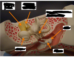
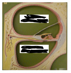
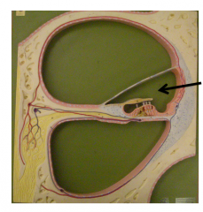
top right
|
Dura matter
|
|
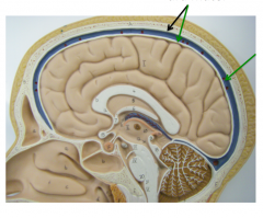
green arrow
|
Dural sinus
|
|

1st top
|
Septum Pellucidum
|
|
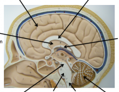
2nd top
|
cerebrum
|
|

3rd top
|
corpus callosum
|
|

4th top
|
fornix
|
|

1st bottom
|
midbrain
|
|

2nd bottom
|
pons
|
|

3rd bottom
|
Medulla oblongata
|
|

4th bottom
|
cerebellum
|
|

1st
|
cerebral aqueduct
|
|
2nd
|
4th ventricle
|
|

left #15
|
optic chiasm
|
|

top left (no number)
|
hypothalamus
|
|
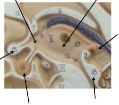
top right (no number)
|
Thalmus
|
|

Right #14
|
Pineal gland
|
|

Bottom left (#16) |
pituitary gland
|
|

bottom right (#18) |
corpora quadrigemina
|
|

top
|
cerebellar cortex (shown in brown) |
|
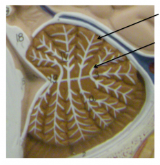
bottom
|
arbor vitae (shown in white) |
|
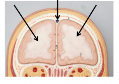
1st
|
right cerebral hemisphere
|
|

2nd
|
Dural sinus
|
|

3rd
|
left cerebral hemisphere
|
|
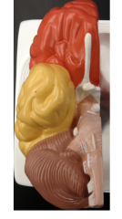
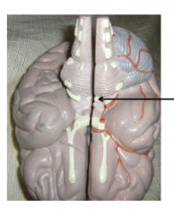
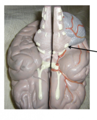
Right side of brain
|
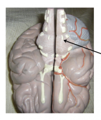
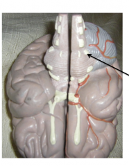
This is midsaggital cut inferior view Cranial nerves are shown in white |
|

1st
|
Frontal lobe
|
|

2nd
|
Temporal lobe
|
|
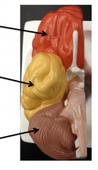
3rd
|
cerebellum
|
|
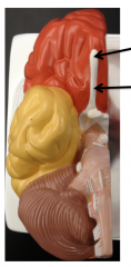
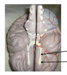
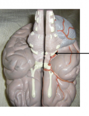
1st
|
olfactory bulb of CNI
|
|

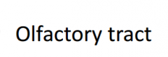
2nd
|
olfactory tract of CNI
|
|

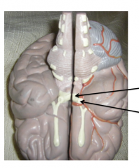
1st
|
optic nerve CNII
|
|

2nd
|
optic chiasm of CNII
|
|

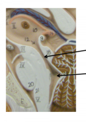
3rd
|
Mammillary body (not a cranial nerve)
|
|
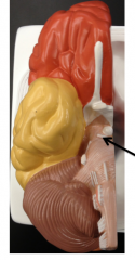
|
oculomotor nerve (CNIII)
|
|
|
trochlear nerve (CNIV)
|
|
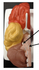
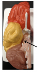
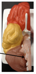
1st
|
trigeminal nerve (CNV)
|
|

2nd
|
abducens nerve (CNVI)
|
|
|
|
|
|
|
|
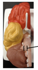
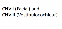
1st
|

|
|

2nd
|

|
|

1st
|
|
|
2nd
|
|
|
1st
|
|
|

2nd
|
|
|
|

|
|
|

|
|
|
|
|
|
|

|
|
|
|
|
1st
|

|
|

2nd
|

|
|
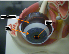
bottom 1st
|
cornea
|
|

2nd bottom
|
superior rectus
|
|

3rd bottom
|
sclera
|
|

1st top left
|
optic nerve
|
|

1st right
|
tendon for superior oblique
|
|
left
|
Lateral rectus
|
|

top
|
superior rectus
|
|

bottom
|
inferior oblique
|
|

right
|
medial rectus
|
|
orange
|
sclera
|
|

bottom left
|
iris
|
|

top right
|
trochlea
|
|

bottom right
|
lacrimal sac / nasolacrimal duct
|
|

top left orange
|
superior oblique
|
|

top right orange
|
trochlea
|
|
middle left
|
medial rectus
|
|

bottom left
|
optic nerve
|
|

bottom right
|
lateral rectus
|
|

bottom middle right
|
superior rectus
|
|

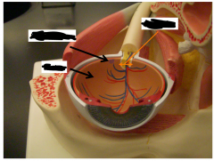
top orange
|
choroid coat
|
|

left bottom
|
iris
|
|
middle
|
pupil
|
|
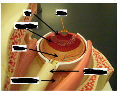
top left
|
macula lutea
|
|

bottom left
|
retina
|
|

top right
|
optic disk
|
|

left 1
|
iris
|
|

left 2
|
ciliary body
|
|

left 3
|
retina
|
|
left 4
|
inferior rectus
|
|

top orange
|
pupil
|
|

right bottom
|
optic nerve
|
|
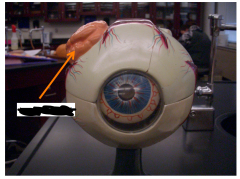
|
lacrimal gland
|
|

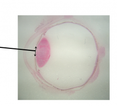
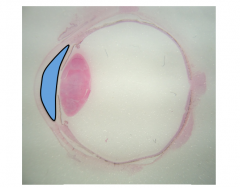
top left |
iris
|
|

bottom left
|
cornea
|
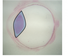
|
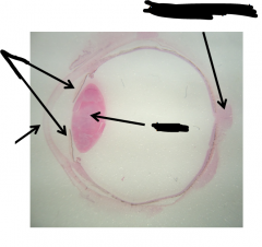
middle of eye
|
lens
|
|

posterior of eye
|
optic nerve
|
|
|
pupil the pupil is the gap in the iris |
|
|
anterior chamber of anterior cavity between iris and cornea filled with aqueous humor |
|
|
posterior chamber of anterior cavity posterior to iris, area immediately surrounding lens filled with aqueous humor |
|
|
posterior cavity filled with vitreous humor |
|

left
|
Eustachian tube
|
|
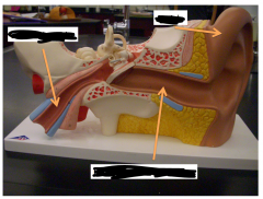
top right
|
auricle
|
|

bottom middle
|
external acoustic meatus
|
|
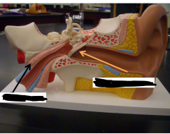
left
|
tensor tympani
|
|

right
|
tympanic membrane
|
|
|
tympanic membrane lateral margin (facing external acoustic meatus) |
|

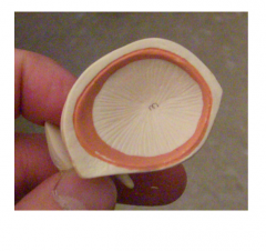
top
|
tympanic membrane
|
|
left bottom
|
incus
|
|

right bottom
|
malleus
|
|

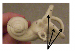
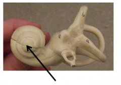
top 1
|
cochlear nerve
|
|

top 2
|
vestibular nerve
|
|

top 3
|
stapes (oval window is underneath)
|
|
top 4
|
incus
|
|

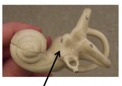
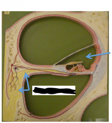
bottom 1
|
cochlea
|
|

bottom 2
|
malleus
|
|
|
stapes oval window is beneath |
|
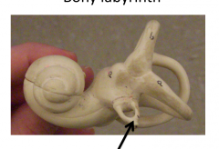
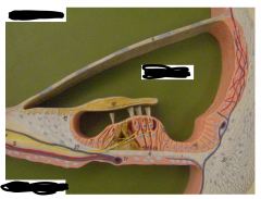
|
semicircular canals
|
|
|
vestibule
|
|
|
cochlea (hearing)
|
|
top |
scala vestibule filled with perilymph |
|

bottom
|
scala tympani filled with perilymph |
|
|
|
|
left |
cochlear nerve
|
|

right
|
filled with endolymph
|
|
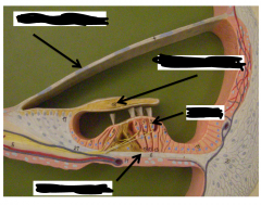
top left
|
scala vestibuli
|
|

bottom left
|
scala tymphani
|
|

right middle
|
scala media
|
|

top left
|
vestibular membrane
|
|
bottom left
|
basilar membrane
|
|

top right
|
tectorial membrane
|
|

bottom right
|
hair cell
|