![]()
![]()
![]()
Use LEFT and RIGHT arrow keys to navigate between flashcards;
Use UP and DOWN arrow keys to flip the card;
H to show hint;
A reads text to speech;
101 Cards in this Set
- Front
- Back
|
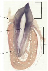
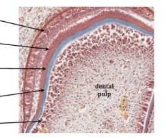
Top to Bottom: 1. [Crown] 2. Gingiva 3. Dental Pulp in the Pulp Cavity 4. [Root] 5. Alveolar Bone |
|
|
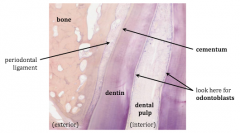
Top to Bottom: 1. Bone 2. Cementum 3. Periodontal Ligament 4. Dentin 5. Odontoblasts 6. Dental Pulp |
|
|
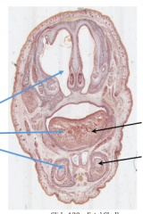
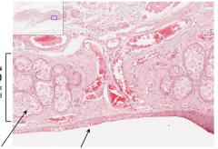
Image is of nasal cavity with cartilaginous nasal septum in the middle; surrounded by intramembranous bone development of the skull Top to Bottom: 1. tongue in the oral cavity 2. Developing teeth with surrounding intramembranous bone development of the jaw |
|
|
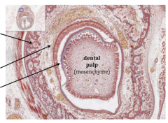
Outside to Inside: 1. Developing bone of jaw via intramembranous ossification 2. Enamel organ (epithelium) 3. Developing Tooth 4. Dental pulp (mesenchyme) |
|
|
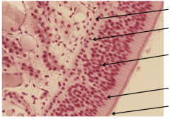
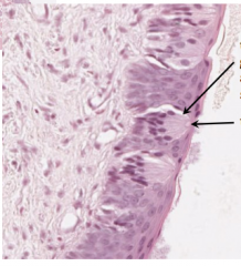
Top to Bottom: 1. ameloblasts (epithelial) 2. enamel 3. dentin 4. predentin 5. odontoblasts (mesenchymal) 6. dental pulp |
|
|
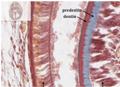
Top to Bottom: 1. Predentin 2. Dentin 3. Ameloblasts (epithelial) 4. Odontoblasts (mesenchymal) |
|
|
Lip. Top to Bottom: 1. oral mucosa (non-keratinized stratified squamous epithelium) 2. Skin (keratinized stratified squamous epithelium w/ hair follicles) |
|
|
Top to Bottom: 1. Salivary glands 2. skeletal muscle |
|
|
soft palate. Top to Bottom: 1. Nasal mucosa with pseudostratified ciliated columnar epithelium 2. skeletal muscle 3. mucous glands 4. oral mucosa with stratified squamous epithelium (non-keratinized) |
|
|
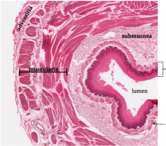
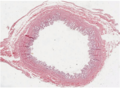
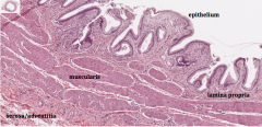
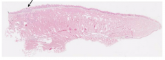
Esophagus. Top to Bottom: 1. adentitia 2. submucosa 3. mucosa 4. muscularis 5. lumen 6. prominent muscularis mucosae |
|
|
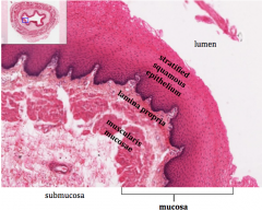
Esophagus. Top to Bottom: 1. Lumen 2. Stratified squamous epithelium 3. lamina propria 4. muscularis mucosae 5. submucosa 6. mucosa |
|
|
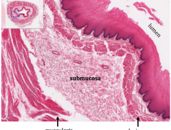
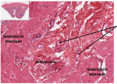
Esophagus. 1. lumen 2. submucosa (between 2 layers of muscle) 3. muscularis (inner circle layer) 4. muscularis mucosae (of mucosa) |
|
|
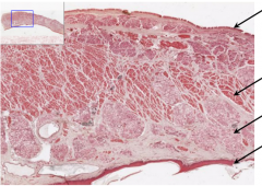
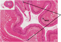
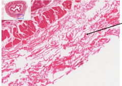
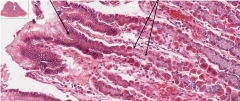
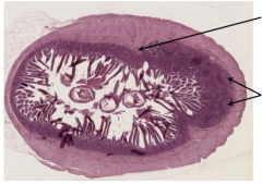
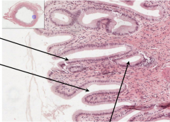
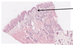
Esophagus. Big arrows: in submucosa, esophageal mucous glands. Small arrow: MALT |
|
|
Esophagus. esophageal mucous glands surrounded by the CT of the sub mucosa |
|
|
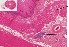
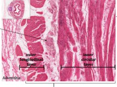
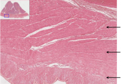
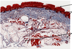
Esophagus. 1. Lumen 2. Auerbach's (myenteric) plexus in the thin later of CT between the 2 muscle layers 3. outer longitudinal layer 4. inner circular layer 5. adventitia 6. muscularis |
|
|
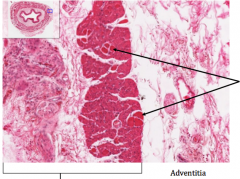
Esophagus. 1. muscularis 2. adventitia 3. skeletal muscle in the muscularis of the upper esophagus as it transitions into only smooth muscle of the lower esophagus |
|
|
Esophagus. most of the esophagus is fixed to adjoining structures so its outer layer is composed of adventita; however, after passing through the diaphragm and entering the abdominal cavity, it become covered by serosa instead, as are all segments of the GI tract within the abdominal cavity |
|
|
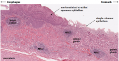
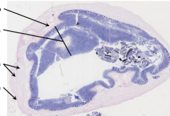
Gastroesophageal Junction. 1. esophagus 2. stomach 3. non-keratinized stratified squamous epithelium 4. lymph nodule 5. simple columnar epithelium 6. MALT 7. cardiac glands 8 & 9. MALT 10. gastric glands 11. muscularis |
|
|
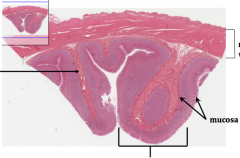
Stomach. 1. muscularis with 3 layers 2. submucosa 3. mucosa 4. rugga: a fold of the mucosa and submucosa; these are not permanent structures but disappear when the stomach is stretched with a bolus of food |
|
|
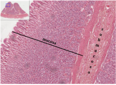
Stomach. 1. mucosa 2. submucosa |
|
|
Stomach. 1. muscularis mucosae 2. ganglion cells of the submucosal plexus (Meissner's plexus) 3. submucosa 4. muscularis mucosae |
|
|
Stomach. Muscularis of stomach has 3 layers. 1. innermost oblique layer 2. inner circular layer 3. outer longitudinal layer |
|
|
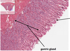
Stomach. 1. Depression = gastric pit. 2. gastric gland, look here for neck mucous cells, parietal cells, and chief cells |
|
|
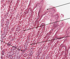
Stomach. 1. surface mucous cells 2. neck mucous cells 3. parietal cells 4. chief cells (peptic cells) |
|
|
Stomach. 1. Surface mucous cells (in gastric pit) 2. mucous neck cells (in gastric gland) |
|
|
Stomach. 1. chief cells 2. parietal cells ("fried egg") |
|
|
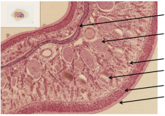
Stomach. 1. gastric pits 2. pyloric (gastric) glands 3. muscularis mucosae is very prominent 4. submucosa 5. muscularis (add. innermost oblique layer)the pylorus is the most distal region of the stomach as it prepares to transition into the small intestine(duodenum); at the junction between the pylorus and the duodenum, the circular layer of smooth muscleof the muscularis thickens to become the pyloric sphincter; as the circular layer thickens, the innermostoblique layer disappears and is not found in the duodenum or remainder of GI tract |
|
|
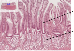
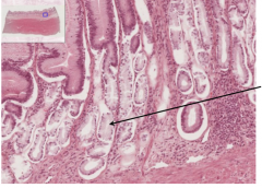
Stomach. 1. gastric pits are deeperin the pyloric regionthan in the body,accounting for nearlyhalf the thickness of themucosa 2. pyloric (gastric) glandsare lined almost entirelyby mucus-secreting cellsand are generallyclassified as branched,coiled glands instead ofthe more tubular glandsof the body and fundus |
|
|
pyloric (gastric) glandswith mucus-secretingcells similar to the neckmucous cells of the bodyand fundus regions; veryfew, if any, parietal cellsare found in the pylorus |
|
|
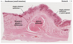
Gastroduodenal Junction. 1. Duodenum (small intestine) 2. Stomach 3. Simple columnar epithelium 4. Simple columnar epithelium w/ pyloric glands 5. Brunner's glands in submucosa 6. Pyloric sphincter |
|
|
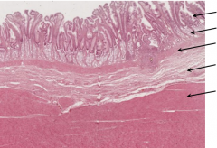
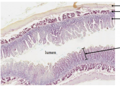
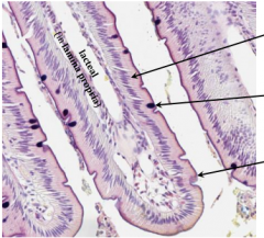
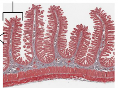
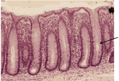
Duodenum. 1. Muscularis 2. Submucosa 3. Mucosa 4. Lumen 5. each tall, foldedprojection of themucosa (epitheliumand lamina propria)is a villus whichcollectively serve toincrease theabsorptive surfacearea of the smallintestine 10x |
|
|
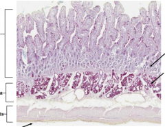
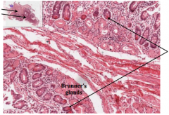
Duodenum. Right side: 1. Mucosa (epithelium + lamina propia (loose CT)) 2. Submucosa 3. Muscularis 4. Serosa Left: 1. intestinal glands/crypts 2. Brunner's glands |
|
|
Duodenum. Left side: lacteal (in lamina propria) Top to bottom of right: 1. enterocytes- cells of the simple columnar epithelium 2. goblet cell 3. the brush border is themicrovilli-covered apicalsurface of the enterocytes;each enterocyte may haveup to 3000 microvilli onits surface; collectively themicrovilli serve toincrease the surface areaof the small intestine 20x |
|
|
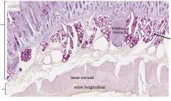
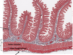
Duodenum. 1. Mucosa 2. Submucosa 3. inner circular 4. outer longitudianl 5. lymphoid nodule 6. Brunner's glands |
|
|
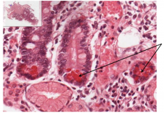
Pancreas & Duodenum. 1. Brunner's glands 2. intestinal crypts with Paneth cells |
|
|
Pancreas & Duodenum. intestinal cryptsw/ Paneth cellswhich are distinguishedby their prominent,intensely-eosinophilicgranules; they serve aspart of the innateimmune defense andsecrete antimicrobialpeptides |
|
|
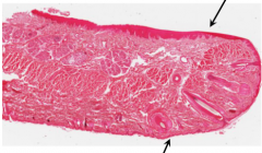
Jejunum. 1. plica circularis 2. villi |
|
|
Jejunum. 1. mucosa 2. submucosa 3. muscularis 4. inner circular 5. outer longitudinal 6. serosa |
|
|
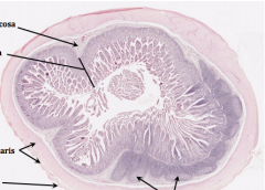
Illeum. 1. submucosa 2. mucosa 3. muscularis 4. serosa 5. Peyer's patches |
|
|
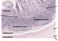
Ileum. 1. Mucosa 2. Peyer's Patches 3. Submucosa 4. Muscularis 5. Lymphoid nodule 6. lymphoid nodule 7. muscularis mucosae 8. inner circular 9. outer longitudinal 10. serosa |
|
|
lleum. 1. muscularis mucosae 2. Peyer's patches |
|
|
Colon. 1. Submucosa 2. Mucosa 3. Muscularis 4. Serosa |
|
|
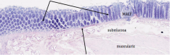
Colon. 1. colonic glands of mucosa -smooth apical surface and lacks villi 2. muscularis mucosae 3. MALT 4. submucosa 5. muscularis |
|
|
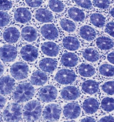
Colon. colonic glandsof the mucosaseen in cross-section;surrounded by laminapropria (loose CT) |
|
|
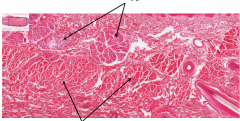
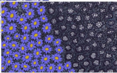
"Colon daisies" |
|
|
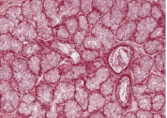
Colon. colonic glands are simple,tubular glands composedlargely of mucous-secretinggoblet cells; the mucusplays an important role inprotecting the mucosa andin regulating the colonicmicrobiome |
|
|
sublingual gland-mucous |
|
|
submandibular gland- serous and mucous |
|
|
parotid gland- serous |
|
|
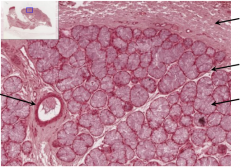
Sublingual Gland. excretory duct. capsule. septum.mucous cells. |
|
|
Submandibular gland. intralobular striated ducts. mucous cells. serous cells (half moon) |
|
|
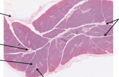
Parotid Gland. capsule. excretory duct. lobule divided by CT septa. striated ducts within lobule. septa |
|
|
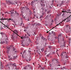
Parotid Gland. striated duct. serous acinusconsists of serous cells(5-10) arranged arounda small, barely-visible,central lumen. intercalated duct |
|
|
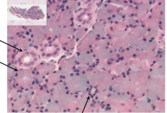
Pancreas. dark cells =secretory acini (excretory). light clusters of cells =pancreatic islets (endocrine)(islets of Langerhans) and willbe discussed in a later lab |
|
|
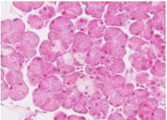
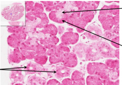
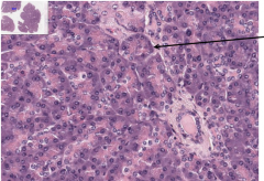
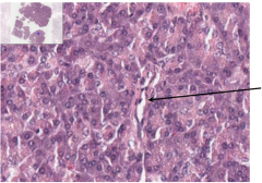
Pancreas. secretory serous acinus |
|
|
intralobular duct |
|
|
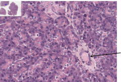
Pancreas. intercalated duct |
|
|
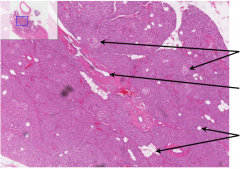
Pancreas. Pancreatic islets. interlobular duct. adipose in the pancreas increases with age |
|
|
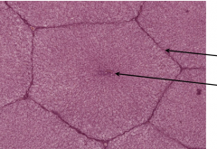
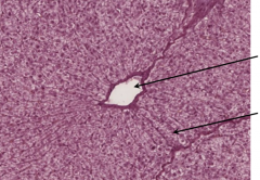
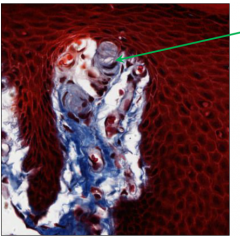
Liver. Septum. Central Vein. |
|
|
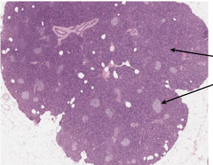
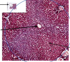
Liver and Gallbladder. 1. look here for gallbladder 2. central vein 3. hepatic lobule 4. hepatic sinusoids between the hepatic plates |
|
|
Liver. 1. Central vein 2. hepatic plates |
|
|
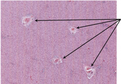
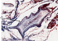
Liver. portal tracts w/ portal triads |
|
|
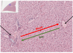
Liver. 1. portal tract w/ portal triad 2. central vein |
|
|
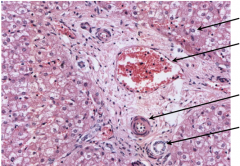
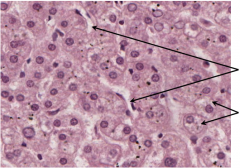
Liver. 1. Hepatocytes 2. branch of hepatic portal vein 3. branch of hepatic artery 4. bile ductule w/ simple cuboidal epithelium |
|
|
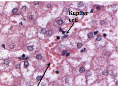
Liver. 1. Kupffer Cell 2. hepatic sinusoid |
|
|
Liver. Hepatic sinusoid lined with endothelium (simple squamous epithelium) |
|
|
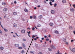
Liver. 1. Kupffer cell 2. hepatic sinusoid 3. endothelial cell |
|
|
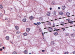
Liver. 1. hepatic sinusoids 2. notice the extensivesmooth endoplasmicreticulum (SER) present inthe hepatocytes; enzymes(e.g., cytochrome P450’s)in the SER are involved inthe liver’s metabolic anddetoxification reactions |
|
|
gallbladder |
|
|
Gallbladder. 1. Serosa/adventitia 2. muscularis 3. epithelium 4. lamina propria |
|
|
Gallbladder. 1. tall simple columnarepithelial cells, withNO goblet cells 2. mucosal fold/rugaare transient folds ofthe mucosa; theydisappear when thegallbladder is full,unlike villi of thesmall intestine 3. glands may seem to be present but thesediverticula/crypts are only deep invaginationsof the mucosa and are not actual glands |
|
|
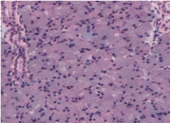
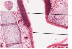
Nasal Cavity. Basement Membrane. Basal cell nuclei. Olfactory neuron nuclei. supporting cell nuclei. cilia |
|
|
Nasal Cavity. nasal concha bone. bundles of unmyelinatedaxons forming the olfactorynerve (CN I). Bowman’s (olfactory) glands. blood vessel, olfactory epithelium. cilia |
|
|
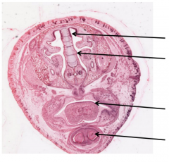
Fetal Pig Head. olfactory epithelium. respiratory epithelium, tongue with developing papillae on superior surface. developing teeth |
|
|
Head. olfactory epithelium. respiratory epithelium |
|
|
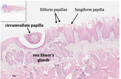
Tongue. circumvallate papilla. von Ebner's glands. filliform papillae. fungiform papilla |
|
|
Tongue. look here on theposterior, superioraspect of the tongue fora circumvallate papilla |
|
|
look along the sides of thecircumvallate papilla to findtaste buds |
|
|
Tongue. taste bud withgustatory cellsand basal andsupporting cells. taste pore |
|
|
Thick Skin. keratinized stratifiedsquamous epithelium. look in the dermal papillaebetween the epithelium andthe underlying connectivetissue to find Meissner's(tactile) corpuscles. look here to seePacinian corpuscles |
|
|
Meissner’s (tactile)corpusclesare receptors for light touchfound in dermal papillae, but arenot seen in every papilla and canbe difficult to distinguish; theyare oval shaped with stackedSchwann cells surrounding acentral nerve fiber; also may bevisible with elongated nuclei offibroblasts wrappingtransversely around |
|
|
Pacinian (lamellated)corpusclesare large receptors forcoarse touch, vibration,and pressure found in thedermis and hypodermis;they consist of 15-20lamellae of Schwann cellsand collagen surrounding abranched unmyelinatednerve fiber |
|
|
Fingertip. 1. fingernail (hard plates of keratin) 2. lateral nail fold withparonychium 3. distal phalanx (bone of finger) 4. nerve 5. Meisnner’s corpuscle(in dermal papilla) 6. Pacinian corpuscle 7. sweat glands and ducts 8. skin with keratinized stratifiedsquamous epithelium |
|
|
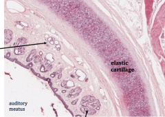
Auditory Meatus. ceruminous glandsare specializedapocrine glands thatproduce cerumen(earwax). hair follice. elastic cartilage |
|
|
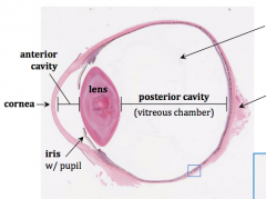
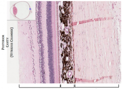
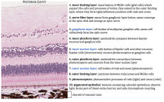
the posterior cavity contains thevitreous humour or body: a largegelatinous mass of transparent CTcovered by the hyaloid membrane;deposits in the vitreous, generally due toage-related degeneration, are seen as“floaters” as they interfere with lightreaching the retina. Optic Nerve -at the optic disc, where the ganglion cellaxons leave the eye through the opticnerve, there are no photoreceptors, sothere is a small blind spot |
|
|
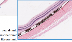
neural-vascular-fibrous |
|
|
eye |
|
|
cuboidal epithelium. capsule. nucleated fibers. non-nucleated fibers (contain crystallin proteins) |
|
|
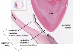
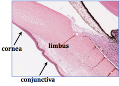
the sclera and cornea are joined at the limbus whichencircles the cornea, serves as the site of stem cells for thecornea, and becomes more stratified at the conjunctivawhich is a stratified columnar mucous membrane withnumerous goblet cells that covers the exposed portion ofthe sclera (not the cornea) and the inside of the eyelid |
|
|
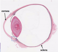
the fibrous tunic is the outermost layerof the eye; it consists of two majorregions:1. sclera - posterior 5/6th; the “white”of the eye; consists of dense CT thatprotects the eye and serves as sitefor extraocular muscle attachment2. cornea - anterior 1/6th; transparentand avascular; composed of fivedistinct layers; density of painreceptors is >100x greater than inskin and >10x in dental pulp |
|
|
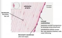
cornea. corneal epithelium. Bowman's membrane. Descemet's membrane. corneal endothelium |
|
|
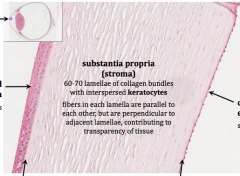
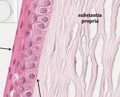
corneal epithelium. Bowman's membrane. substantia propria |
|
|
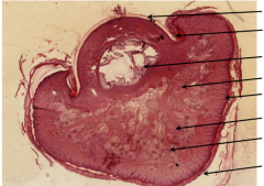
Eye |
|
|
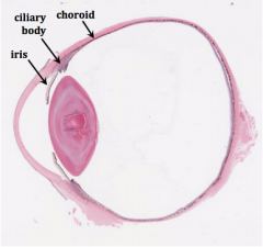
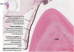
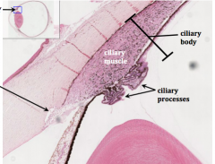
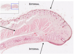
Vascular tunic (uvea) is the middle layer of theeye; it consists of three parts:1. iris: most anterior portion of vascular tunic;contains many melanocytes that provide thecolor of the eye and prevent the passage oflight, leaving only the central pupil (opening)for light to pass through; contains:a. dilator pupillae m.b. sphincter pupillae m.2. ciliary body: expansion of the vascular tunic,encircling the lens; consists of:a. ciliary muscleb. ciliary processesc. ciliary zonule3. choroid: located in posterior 2/3rd of eye;contains:a. loose CTb. lots of vasculaturec. melanocytes |
|
|
eye |
|
|
ciliary body. scleral venous sinus (canal of Schlemm) and trabecular meshwork |
|
|
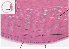
retina-neural tunic. choroid-vascular tunic. sclera-fibrous tunic. the choroid containsloose CT, vasculature, andmelanocytesthe vasculature providesoxygen and nutrients tothe adjacent tissuesthe melanocytes providethe black coloration of thechoroid and serve toabsorb scattered light andreduce reflection |
|
|
Eye Layers |
|
|
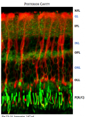
Müller cells are large glial cellsunique to the retina; they extendthrough the full thickness of theretina with their nuclei in theinner nuclear layer (INL)they are critical for retinalfunction by providing trophicand metabolic support for theneurons and their processes,regulating homeostasis andsynaptic activity, and organizingretinal componentsMüller cells are shown in red;axons and dendrites are green |
|
|
skeletal muscle oforbicularis oris m.,and levator palpebrae m.in upper eyelid. look here for skin and follicles of eyelashes. look here for conjunctiva |
|
|
tarsus. tarsal glands. conjunctiva |