Reading...
![]()
Play button
![]()
Play button
![]()
Use LEFT and RIGHT arrow keys to navigate between flashcards;
Use UP and DOWN arrow keys to flip the card;
H to show hint;
A reads text to speech;
10 Cards in this Set
- Front
- Back
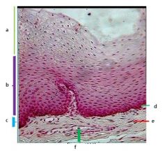
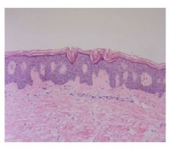
Label
|
a. keratinised layer
b. spinous layer c. basal layer and parabasal layers d. basement membrane e. connective tissue f. blood vessel |
|
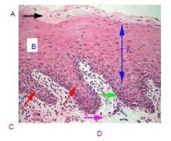
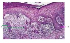
Label
|
a. keratinised layer
c. rete ridges d. connective tissue e. basement membrane f. spinous layer |
|
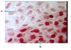
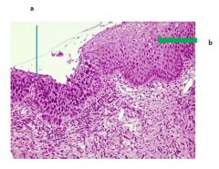
which layer is this?
the cells at the junction a are held by what feature? what's b? |
spinous layer
a. desmosomes b. keratinocyte |
|
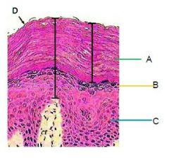
What pathology is this?
Label |
Hyperkeratosis
a. hyper-ortho-Karatinised layer b. granular layer c. spinous layer d. desquamation |
|
This is a picture of?
|
Hyperkeratosis
|
|
What major feature can be seen in this pathoogy?
Label |
Pseudoepitheliomatous Hyperplasia
a. Epithelial islands b. big elongated rete ridges |
|
Label
|
a. Severe dysplasia
b. Moderate dysplasia |
|
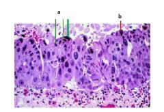
|
a. Pleopmorphism
b. Hyperchromatism |
|
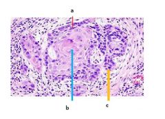
What pathology is this?
Label |
Well differentiated squamous cell carcinoma
a.tumoural cell b. keratin pearl c. tumoural cell |
|
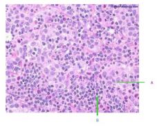
What's the pathology?
Label |
undifferentiated carcinoma
a. tmoural cell b. Inflammatory cels |