Reading...
![]()
Play button
![]()
Play button
![]()
Use LEFT and RIGHT arrow keys to navigate between flashcards;
Use UP and DOWN arrow keys to flip the card;
H to show hint;
A reads text to speech;
58 Cards in this Set
- Front
- Back
|
What is cartilage?
|
A tough but flexible tissue in the body that contains collagen and elastic fibers. The cells, called chondrocytes, are located in depressions, called lacunae, in the matrix.
|
|
|
What are chondrocytes?
|
Cells located in depressions (lacunae) in the matrix in cartilage
|
|
|
What are lacunae?
|
Depressions that hold chondrocytes in the matrix of cartilage
|
|
|
What are the types of cartilage?
|
1. Hyaline cartilage
2. Elastic cartilage 3. Fibrocartilage |
|
Type; ID; Location; Function; Fibers Present; Features to Know
|
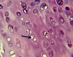
Hyaline Cartilage; ID: Overall smooth, glassy appearance with distinctive lacunae, most occupied by chondrocytes (arrow). Lacunae are often paired. Location: nose, larynx, trachea, between ribs and sternum, and ends of long bones; Function: structural support and cushioning bones; Fibers present: collagen fibers (not visible); Features to know: chondrocyte in lacuna (arrow)
|
|
Type; ID; Location; Function; Fibers Present; Features to Know
|
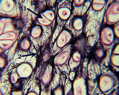
Elastic Cartilage; ID: distinctive large, often paired lacunae, with extensive dark elastic fibers; Location: earlobe and epiglottis; Function: flexibility; Fibers present: elastic fibers (collagen fibers are also present but not visible); Features to know: chondrocyte in lacuna (1) and elastic fibers (2)
|
|
Type; ID; Location; Function; Fibers Present; Features to Know
|
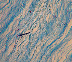
Fibrocartilage; ID: nearly parallel (often blue) collagen fibers and the distinctive chondrocytes in lacunae (arrow); Location: intervertebral disks, pubic symphysis, and meniscus of knee joint; Function: resists compressive forces; Fibers present: collagen; Features to know: chondrocyte in lacuna (arrow)
|
|
|
What is bone tissue?
|
A living tissue consisting of three types of cells. The mature bone cells are called osteocytes. The osteocytes are located within shallow depressions, called lacunae, in the hard, bony matrix.
|
|
|
What is the basic structural unit of compact bone?
|
The Haversian system, or osteon.
|
|
|
What does the Haversian system, or osteon, consist of?
|
A central opening, called the Haversian (or central) canal. Surrounding this canal are concentric rings of bony matrix. These concentric rings are called lamellae. Between the lamellae are the osteocytes within lacunae. Very small canals run perpendicular to the lamellae. These tiny canals are called canaliculi.
|
|
|
What is the Haversian (or central) canal?
|
A central opening in the Haversian system, or osteon, of compact bone.
|
|
|
What surrounds the Haversian (or central) canal?
|
Concentric rings of bony matrix called lamellae
|
|
|
In the Haversian system, or osteon, what are between the lamellae?
|
The osteocytes within lacunae
|
|
|
What run perpendicular to the lamellae in the Haversian system?
|
Very small canals called canaliculi
|
|
Type; ID; Location; Function; Features to Know
|
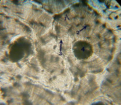
Bone tissue; ID: large, dark circles with concentric circles surrounding it, much like tree rings; Location: bones; Function: support, protection, mineral storage, and attachment sites for muscles; Features to know: lamellae (1), osteocytes in lacunae (2), canaliculi (3), and Haversian (central) canal (4);
|
|
|
What does blood tissue consist of?
|
A liquid matrix, called plasma, and formed elements that include erythrocytes (red blood cells), leukocytes (white blood cells), and platelets (thrombocytes)
|
|
|
Describe erythrocytes:
|
Erythrocytes are small and pink. They lack a nucleus. The center is depressed and usually appears lighter than the rest of the cell.
|
|
|
Describe leukocytes:
|
Leukocytes are usually stained purple. They are fewer and larger than the erythrocytes. Most leukocytes have either a bi-lobed or multi-lobed nucleus.
|
|
|
Describe platelets:
|
Platelets are small cell fragments that are stained blue or purple. They are smaller than the erythrocytes and are scattered between the cells in the tissue.
|
|

Type; ID; Location; Function; Features to know
|
Blood; ID: numerous pink, round red blood cells with larger, purple white blood cells scattered throughout; Location: within the blood vessels; Function: transport of nutrients, gases, wastes, etc.; Features to know: plasma (white background), erythrocytes (round and pink), and leukocytes (1)
|
|
|
What system includes the skin, hair, and nails?
|
The Integumentary System
|
|
|
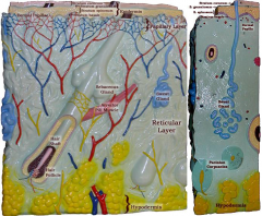
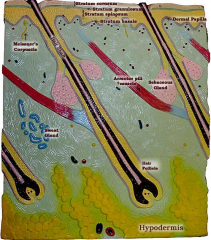
What are the main layers of the integumentary system?
|
1. Epidermis
2. Dermis 3. Hypodermis |
|
|
What is the epidermis?
|
Stratified squamous epithelial tissue consisting of 4-5 distinct layers
|
|
|
What are the layers of the skin, other than the palms and soles?
|
1. Stratum corneum
2. Stratum granulosum 3. Stratum spinosum 4. Stratum basale |
|
|
What is the outermost epidermis layer consisting of several layers of flattened dead cells?
|
Stratum corneum
|
|
|
What is the second, thin epidermis layer consisting of cells within keratin granules?
|
Stratum granulosum
|
|
|
What is the thickest layer of the epidermis?
|
Stratum spinosum
|
|
|
What is the bottom-most dark, thin epidermis layer consisting of pigmented cells?
|
Stratum basale
|
|
|
What is the dermis?
|
Dense irregular connective tissue
|
|
|
What are the main layers of the dermis?
|
1. The papillary layer
2. The reticular layer |
|
|
What is the papillary layer?
|
The thin uppermost dermis layer, folded into peg-like projections called dermal papillae
|
|
|
What are dermal papillae?
|
Peg-like projections of the folded papillary layer of the dermis
|
|
|
What is the reticular layer?
|
The thick layer which accounts for the majority of the dermis, consisting of several appendages, including 1. hair follicles, 2. sebaceous glands, 3. arrector pili muscles, 4. sweat glands, 5. Meissener's corpuscles, and 6. Pacinian corpuscles.
|
|
|
What are most sebaceous glands formed from, and where are they located?
|
Most are formed from the hair follicle and are located beside the hair follicle.
|
|
|
What produces hair?
|
The hair follicle
|
|
|
What forms "goose bumps" and where is it attached?
|
The arrector pili muscle is attached to the hair follicle. When it contracts, it pulls the hair follicle up and forms a "goose bump."
|
|
|
Where are the sweat glands located?
|
Throughout the dermis, with a duct that leads to the outside of the skin
|
|
|
What are the small, encapsulated sensory nerve endings located near the top of the dermis?
|
Meissener's corpuscles
|
|
|
What are Pacinian corpuscles?
|
Larger than Meissener's corpuscles, they are plate-like sensory nerve endings located near the bottom of the dermis
|
|
|
What is the hypodermis?
|
The layer of adipose tissue below the skin. It is not considered part of the skin.
|
|
|
Epidermis; Stratum corneum; Stratum granulosum; Stratum spinosum; Stratum basale; Dermis; Papillary layer; Dermal papillae; Reticular tissue; Meissner's corpuscle; Hair follicle; Sebaceous gland; Arrector pili muscle; Sweat gland; Pacinian corpuscle; Hypodermis
|
|
|
Epidermis; Stratum corneum; Stratum granulosum; Stratum spinosum; Stratum basale; Dermis; Papillary layer; Dermal papillae; Reticular tissue; Meissner's corpuscle; Hair follicle; Sebaceous gland; Arrector pili muscle; Sweat gland; Pacinian corpuscle; Hypodermis
|
|

|
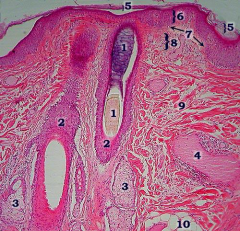
In a cross-section of a bald scalp, you can
see sweat glands (1), sebaceous gland (3), and hair follicle (4). All of these are found in the reticular layer (2). |
|
|
The visible layers of the epidermis include
the outer stratum corneum layer (5), the thick stratum spinosum layer (6), and the thin, dark stratum basale (7). The dermis consists of the upper papillary layer (8) and the main reticular layer (9). You can see the prominent hair follicle (2) and hair (1). You should also be able to see the sebaceous glands (3). Also visible may be the arrector pili muscle (4) and the hypodermis (10). |
|
|
What does the skeletal system consist of?
|
Bones and ligaments
|
|
|
How can bones be classified?
|
On the basis of shape
|
|
|
What are common bone shapes?
|
1. Long bones
2. Short bones 3. Flat bones 4. Irregular bones |
|
|
What are long bones?
|
Bones that are longer than they are wide. Long bones include all bones of the limbs, except the patella (kneecap), wrist bones, and ankle bones.
|
|
|
What are short bones?
|
Bones that are smaller than long bones and many are roughly cube-shaped. Short bones include the wrist bone and ankle bones. Includes sesamoid bone.
|
|
|
What is sesamoid bone?
|
A type of short bone that is shaped like a sesame seed and form in a tendon. The patella is an example of a sesamoid bone.
|
|
|
What are flat bones?
|
Flattened bones. Flat bones include the sternum (breastbone), scapulae (shoulder blades), ribs, and most skull bones.
|
|
|
What are irregular bones?
|
Bones that are irregularly shaped and do not fit into any of the other categories. Irregular bones include the vertebrae and hip bones.
|
|
|
What is the shaft of a long bone called?
|
The diaphysis
|
|
|
What is each knob-like end of a long bone called?
|
An epiphysis
|
|
|
What is the canal that runs through the shaft of a long bone called?
|
The medullary cavity
|
|
|
What is the medullary cavity surrounded by?
|
A thin layer of spongy bone, surrounded by a thicker layer of hard compact bone
|
|
|
What type of bone do the epiphyses contain?
|
Spongy bone
|
|

|
Long bone
|