Reading...
![]()
Play button
![]()
Play button
![]()
Use LEFT and RIGHT arrow keys to navigate between flashcards;
Use UP and DOWN arrow keys to flip the card;
H to show hint;
A reads text to speech;
38 Cards in this Set
- Front
- Back
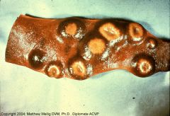
Pig spleen
|
Multifocal granulomatous splenitis (tuberculosis)
Caseous centers surrounded by macros, lymphos, and connective tissue |
|
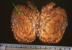
cow lymph node
|
Locally extensive granulomatous lymphadenitis (tubercular)
Node is destroyed. All caseous debris encapsulated by fibrous tissue. |
|
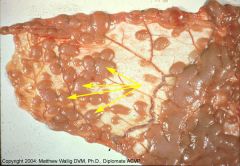
cow omentum
|
Small tubercles caused by systemic tuberculosis.
Probably composed solely of macros |
|
chicken viscera
|
Multple tubercles all over viscera
|
|
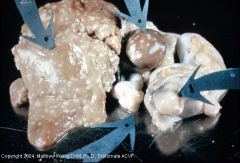
deer lung
|
Granulomatous pneumonia
Has central mineralization and multinuc'd giant cells --> typical of mycobact. |
|
Chicken
|
Hjarre disease (coligranuloma)
necrotic center surrounded by fibrous band with macros and lymphos |
|
Cow intestine
|
Johne's Disease:
thickening of int. ruggae by lots of epith. macros in the mucosa and submucosa |
|
sleep ileum
|
Macro aggregation in ileum of sheep with johne's
|
|
Cow with Actinomycosis
|
Sulfur granule surrounded by macros
|
|
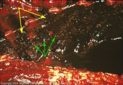
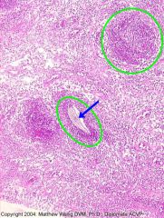
Dog thorax
|
Pyogranulomatous pleuritis caused by nocardia (Nocardiosis)
Sanguinopurulent exudate (yellow) Sulfur granules (green) Also, masses of confluent granulomas could be seen. |
|
Cow mammary gland
|
Chronic pyogranulomatous mastitis (Nocardiosis)
The red strands are the bacteria (modified acid-fast stain) |
|
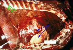
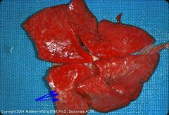
Dog Lungs
|
Blastomycosis with miliary (green) and large (blue) granulomas all over lungs
|
|
dog lungs
|
Blastomycosis again
The granulomas look somewhat like abscesses but are less discrete |
|

dog lungs
|
Blastomycosis again
The granulomas look somewhat like abscesses but are less discrete |
|
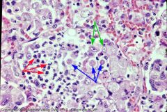
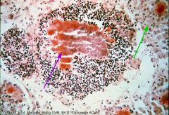
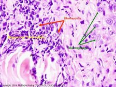
Dog Lungs
Exudate What cells do you see? |
Blastomycosis (histology)
Cellular exudate: Microbe cells (blue) Some inside macros (green) Neutrophils (red) |
|
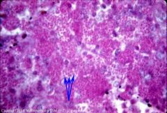
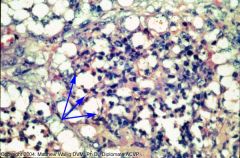
Dog Lung
|
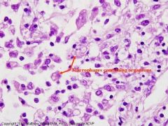
Histoplasmosis
cells inside macros |
|
Silver stain of dog spleen
|
Histoplasmosis
Microbes found inside macros (Silver stains the capsules) |
|
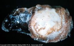
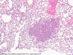
Cow
What is this? |
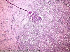
No, it's not a raw oyster
It's a pyogranulomatous lesion within a lymph node caused by coccidiodomycosis. |
|

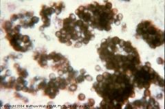
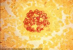
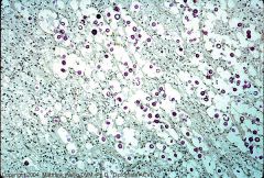
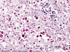
What are these?
|
Endospores of Coccidiodes immitis (without the outer sporocyst wall)
These would be found within a granulomatous or pyogranulomatous lesion. |
|
What are these?
|
Endospores of Coccidiodes immitis (without the outer sporocyst wall)
These would be found within a granulomatous or pyogranulomatous lesion. |
|

What are these?
|
Endospores of Coccidiodes immitis (without the outer sporocyst wall)
These would be found within a granulomatous or pyogranulomatous lesion. |
|
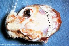
What the hell is going on here?
|
This is a granulomatous lesion eroding the skull caused by Cryptococcus. It originates as a nasal sinus infection.
|
|
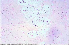
Horse brain
|
Cryptococcus again
The large clear spaces are the capsules of the yeast cells. They inhibit immune response and phagocytosis, so the cellular response is surprisingly weak in this picture. |
|
Cat brain
|
Meningeal cryptococcosis.
Again, the clear space is from this fungus' thick capsule. |
|
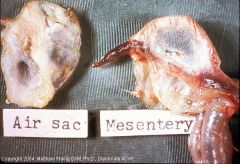
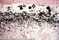
Chicken Air Sac and Mesentery
|
The filamentous growth is from Aspergillus. The underlying tissue will be induced to form granulomatous or pyogranulomatous lesions.
|
|
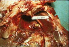
Chicken Lungs
|

Multifocal Aspergillosis
|
|
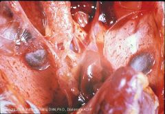
Horse guttural pouch
What are the brown spots |
They are granulomatous nodules caused by Aspergillosis
|
|
Chicken Air Sac
Silver Stain |
Aspergillus mycelia growth with macrophages at the base of the growth
|
|
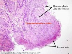
Chronic locally extensive ulcerative and pyogranulomatous dermatitis.
Cutaneous Blastomycosis What will you see? |
These cells are seen in the exudate
|
|
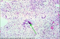
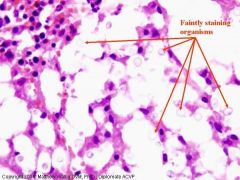
Chronic (multi)focal granulomatous pneumonia.
Pulmonary Histoplasmosis What will you see in the exudate? |
This here's what y'all'll see.
|
|
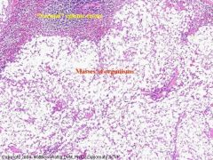
Chronic diffuse granulomatous splenitis
Splenic Cryptococcosis What do the microbes look like? |
Thick capsule
|
|
Chronic locally extensive pyogranulomatous dermatitis
Cutaneous Coccidiomycosis |
FYI, the sporocyst (containing the endospores) illicits a granulomatous response, while the endospores, when released, illicit a suppurative response.
|
|
Cow mammary gland
|
Pyogranulomatous mastitis
The filaments are the nocardia bact. cells |
|
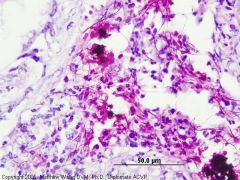
Dog Lung
Mucicarmine stain |
Pulmonary cryptococcosis
The mucin in the capsule of C. neoformans stains red so you can visualize it. |
|
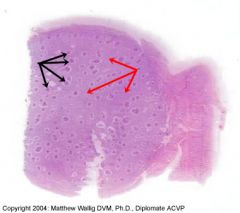
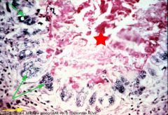
Actinobacillosis
Massive granuloma from cow abomasum with foci of suppuration (black arrows) What is within these suppurative foci? |
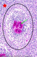
Sulfur granules from the bacteria
|
|
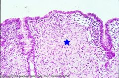
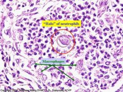
Dog gingival nodule
|
Sections of tissue heavily infiltrated by macrophages (granulomas) with suppurative lesions at the center of them (neutrophils surrounding pieces of plant fiber)
|
|
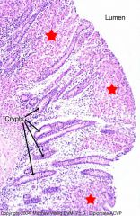
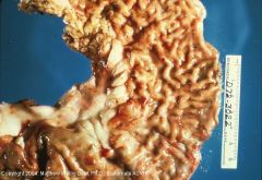
Cow colon
Macrophage infiltration of the lamina propria and submucosa Why? |
Mycobacterium Paratuberculosis (Johne's disease)
Ziehl Nielsen stain reveals them inside the macrophages |
|
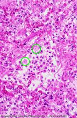
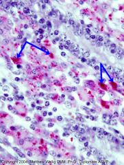
Dog lung
Severe diffuse inflammation with alveolar consolidation. Fibrinopurulent granulomatous exudate. See something in macrophages (green) What? |
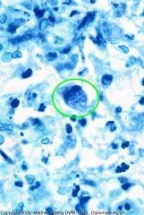
Toxoplasmosis
See the tachyzoites with Giemsa stain |