Reading...
![]()
Play button
![]()
Play button
![]()
Use LEFT and RIGHT arrow keys to navigate between flashcards;
Use UP and DOWN arrow keys to flip the card;
H to show hint;
A reads text to speech;
30 Cards in this Set
- Front
- Back
- 3rd side (hint)
|
Views:
|
LM
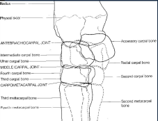
DLMPO DMPLO DP Flexed Lateral Skyline Views: |
Distal row of carpal bones (most common)
Proximal row of carpal bones distal radius |
|
|
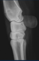
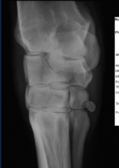
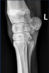
Lateral - Medial
|
two fat pads on dorsal surface
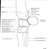
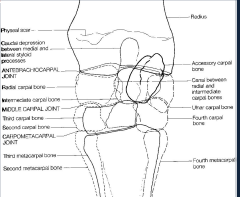
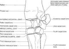
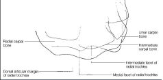
radial carpal bone - convex intermediate carpal bone - concave third carpal bone - convex tendon sheath - extensor carpi radialis three synovial joints |
|
|
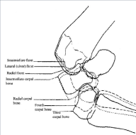
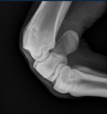
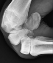
flexed lateral
|
|
|
|
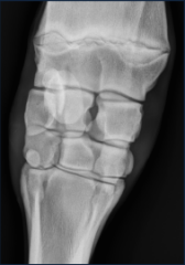
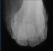
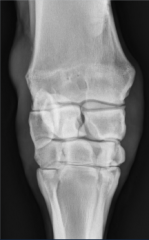
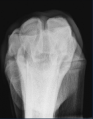
DP - Dorsopalmar
|
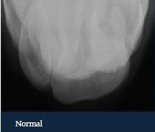
radiolucent area is distal radius is normal
|
|
|
DLPMO
|
|
|
|
DMPLO
|
|
|
|
fifth carpal bone
|
|
|
|
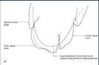
distal row of carpal bones
|
radial facet is medial and the larger of the two facets
|
|
|
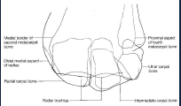
proximal row of carpal bones
|
|
|
|
distal radius
|
|
|
|
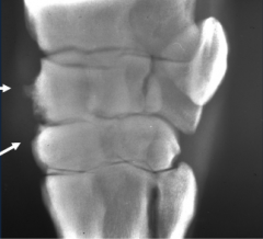
degenerative joint disease
-radiographic findings: . . . . . . -Carpometacarpal joint . . . - |
degenerative joint disease
-radiographic findings: . Osteophytes . Rounding of articular margins .sclerosis or lucent zones of the subchondral trabecular bone .joint capsule distension .periarticular soft tissue swelling .joint space narrowing -Carpometacarpal joint .narrowing, subchondral bone sclerosis and lucencies .sometimes only 2nd and 4th carpal bone .mild but chronic lameness -Correlate findings to age of horse, conformation, previous performance and future work |
|
|
|
osteophytes
|
|
|
|
|
|
|
|
intercarpal ligament desmitis
|
|
|
|
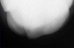
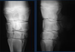
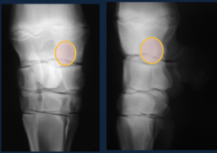
Third carpal bone (C3) Sclerosis
- . - - . . . |
Third carpal bone (C3) Sclerosis
- mild sclerosis is normal modelling of racehorses in training (adaptive) .radial facet -marked sclerosis of radial and/or intermediate facet is abnormal (pathologic) -Radiographic findings . . . |
-Radiographic findings
.loss of trabecular bone and cortiomedullary distinction .lucent zones may be precursors of fractures (necrosis) .middle carpal joint capsule distension |
|
|
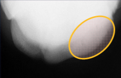
sclerosis radial facet
lucent regions |
|
|
|
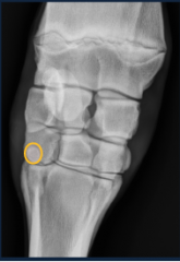
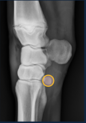
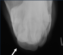
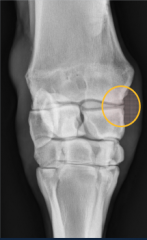
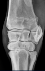
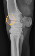
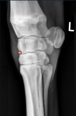
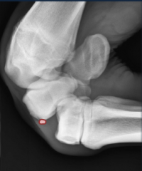
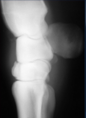
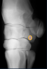
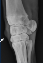
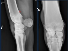
Chip fractures
- - - - . . . . . |
Chip fractures
- occur commonly in racehorses - involve one articular surface -most readily observed on flexed lateral view -common sites: . . . . . |
-common sites:
.distodorsal radius .distodorsal radial carpal bone (medial aspect) .proximodorsal radial facet of third carpal bone .distodorsal intermediate carpal bone .less commonly proximal radial carpal bone and intermediate carpal bone |
|
|
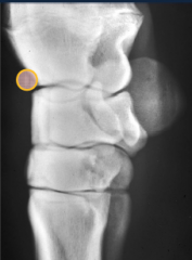
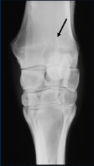
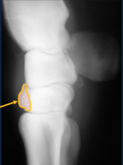
chip fracture
distolateral aspect of the radius |
|
|
|
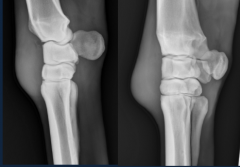
chip fracture distomedial radial carpal bone
|
|
|
|
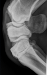
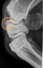
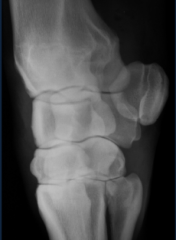
chip fracture
|
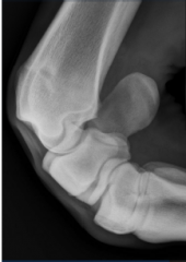
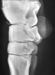
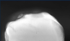
normal
|
|
|
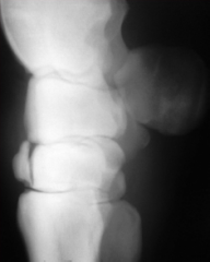
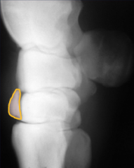
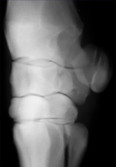
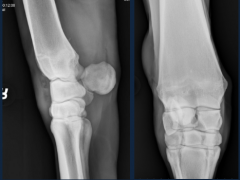
Slab fractures
- - - - - |
Slab fractures
-involve proximal and distal articular surface -can be dorsal (frontal) or sagittal plane -C3 is most common, can be seen on other carpal bones -usually seen with lateral view, but may need skylines -usually develop as terminal event in cascade of nonadaptive remodeling |
|
|
|
slab fracture
-DLPMO |
|
|
|
-LM view
slab fracture |
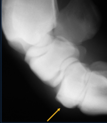
with flexion, fracture reduces
|
|
|
osseous or subchondral cyst-like lesions
- - - . . . |
osseous or subchondral cyst-like lesions
-carpal bones, proximal aspect of the second of fourth metacarpal bones and distal aspect of the radius -developmental abnormality, trauma or infection - can be associated with lameness . . . |
- can be associated with lameness
.articular lesions .distomedial aspect of the radius .ulnar carpal bone lesions with tearing of the lateral palmar intercarpal ligament |
|
|
-DLPMO
osseous or subchondral cyst like lesion |
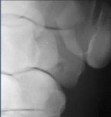
normal
|
|
|
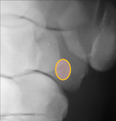
osseous or subchondral cyst like lesion
|
|
|
|
cyst in distal radius
|
|
|
|
other signficant radiographic abnormalities
- . . - - - . . - |
other signficant radiographic abnormalities
-soft tissue swelling . . -radial fractures -physeal fractures -osteochondroma of distal radius . . -infection |
other signficant radiographic abnormalities
-soft tissue swelling .periarticular .articular -radial fractures -physeal fractures -osteochondroma of distal radius .distention of carpal sheath, lameness, resentment to pressure .cartilage covered exostoses -infection |
|
|
soft tissue swelling
-ultrasound or injection of iodinated contrast medium for further diagnostic evaluation -location of soft tissue swelling -evaluate bone and joints |
|
|
|
soft tissue swelling
|
|