![]()
![]()
![]()
Use LEFT and RIGHT arrow keys to navigate between flashcards;
Use UP and DOWN arrow keys to flip the card;
H to show hint;
A reads text to speech;
24 Cards in this Set
- Front
- Back
|
How do you define whether a neurone is PNS or CNS? |
If a neuron is entirely containedwithin the brain and/or spinalcord, it is a CNS neuron. If any part of it (dendrites, axonor cell body) projects outside ofthese structures, it is a PNSneuron. |
|
|
What are the divisions of the central nervous system? |
Brain and spinal cord |
|
|
What are the divisions of the peripheral nervous system? |
Somatic --> sensory and motor Autonomic ---> sympathetic and parasympathetic |
|
|
What are the different planes of the body? |
A sagittal plane is perpendicular to the ground and divides the body into left and right. The midsagittal or median plane is in the midline i.e. it would pass through the midline structures (e.g. navel or spine), and all other sagittal planes (also referred to as parasagittal planes) are parallel to it. Median can also refer to the midsagittal plane of other structures, such as a digit. A coronal or frontal plane is perpendicular to the ground and divides the body into dorsal (posterior or back) and ventral (anterior or front) portions. A transverse plane, also known as an axial plane or cross-section, divides the body into cranial (head) and caudal (tail) portions. It is parallel to the ground, which (in humans) separates the superior from the inferior, or put another way, the head from the fee |
|
|
What are the anatomical compartments the brain can be divided into? |
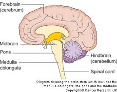
The central nervous system can also bedivided into anatomical compartments |
|
|
How many spinal nerves are there? |
There are normally 31 spinalnerves (8 cervical, 12 thoracic,5 lumbar, 5 sacral and 1coccygeal). |
|
|
Where do the spinal cords leave? |
All leave through a correspondingIntervertebral foramen BUT thespinal cord ends at vertebral levelL1/L2. Therefore……the cauda equina(horse’s tail). |
|
|
Why are Spinal nerves “mixed” nerves? |

|
|
|
How do nerves enter the spinal cord? |
• The spinal cordis segmented • Nerves enter andleave as rootlets |
|
|
What is the outer part of the cerebrum? |
The outer part of the cerebrum isthe cerebral cortex. It is throwninto several ridges (Gyri, sing.gyrus) and grooves (Sulci, sing.sulcus). |
|
|
What are the deeper grooves known as? |
Deeper grooves are known asfissures. |
|
|
What do the gyri and sulci do? |
• The gyri and sulci of the cerebralcortex increase the surface areaof the brain to approx. 2,500 cm2- this is about the same as4 pieces of A4 paper and allowsmuch more neural material to becontained within the skull. |
|
|
Are functions located in just one lobe? |
Some functions are particularlyAssociated with individual lobes, BUT: • No function is only located to just one lobe • No lobe is associated with just one function |
|
|
Why is the spinal cord hollow? |
• The central canalruns the length of thespinal cord. • It is closed caudallyand rostrally iscontinuous with theventricular system ofthe brain |
|
|
What is the organisation of the ventricular system? |
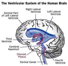
The ventricular system: organisation |
|
|
What is INSIDE the ventricles? |
Ans: Cerebrospinal fluid (CSF)This is made by the choroid plexus(specialised ependymal cells) |
|
|
What are the functions of the CSF? |
Functions of the CSF: • Buoyancy • Protection • Removal of waste products |
|
|
CSF is made continuously – approx. 500 ml/day. Where does it all go? |
CSF is reabsorbed back into thevenous bloodstream and if it isn’t,the result can be……Hydrocephalus(Gr: hydor, water + kephale, head) |
|
|
What are the The meningeal layers of the CNS? |
The CNS has 3 outer layer – the meninges (sing. meninx) 1. The pia mater (innermost layer) - fine, delicate2. The arachnoid mater (middle layer) – silky, web-like 3. The dura mater (outer layer) – tough, hardy |
|
|
What is the subarachnoid space? |
The CSF leaves the ventricles of the brain and exits intoThe space between layers 1 and 2 : the subarachnoid space |
|
|
What is the Pia mater? |
• The innermost meningeal layer • Protects the CNS (together with the other meninges) • A fine, vascular membrane that also allows entry ofblood vessels into the CNS • Provides an impermeable layer for containing CSF |
|
|
What is the arachnoid mater? |
• The middle meningeal layer. • Protects the CNS (together with the other meninges) • A spiders-web like membrane that forms the upperlimits of the sub-arachnoid space. • Is closely associated with the dura mater. |
|
|
What is the dura mater? |
• The outer meningeal layer. • Protects the CNS (together with the other meninges) • In the skull, composed of two layers which separatein places to form sinuses carrying venous blood. • Surrounding the brain, the dura mater is closelyattached to the periosteum of the cranium; surroundingthe spinal cord, it hangs loosely, only attached atthe foramen magnum. |
|
|
How does the CSF re-enter the venous bloodstream? |
• CSF is reabsorbed back intothe venous bloodstream • It goes from the subarachnoidspace into thesuperior sagittal sinus • Its transfer is mediated bythe arachnoid granulations |