![]()
![]()
![]()
Use LEFT and RIGHT arrow keys to navigate between flashcards;
Use UP and DOWN arrow keys to flip the card;
H to show hint;
A reads text to speech;
54 Cards in this Set
- Front
- Back
|
Major excitatory neurotransmitters |
Glutamate (main) |
|
|
Major inhibitory neurotransmitters |
GABA (Brain and spinal cord) Glycine (primarily spinal cord) |
|
|
Glutamate pathways |
Cortico-cortical pathways Corticothalamic pathways Extrapyramidal pathways (related to Parkinsons) Trisynaptic pathway in hippocampus Projections between cortex, substantia nigra, subthalamic nucleus, globus pallidus |
|
|
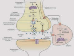
Glutamate neurotransmission |
1. Presynaptic neuron makes glutamate from glutamine via deamination with glutaminase. 2. Glutamate is stored in vesicle. AP opens VGCCs, leading to fusion of vesicles through SNAREs. 3. Release of glutamate in synaptic cleft 4. Glutamate binds to postsynaptic receptors 5. Termination via reuptake of glutamate primarily into neighboring glial cells. 6. In glial cells, glutamine synthetase converts glutamate to glutamine. 7. Glutamine is extruded from glial cells and moves to presynaptic glutaminergic neurons where process starts again. Some glutamate transporters in presynaptic cell as well. |
|
|
Ionotropic glutamate receptors |
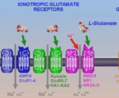
Nonselective cation channels (Na+, K+, Ca2+) NMDA - highly permeable to Ca2+. AMPA Kainate |
|
|
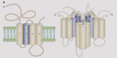
Ionotropic glutamate receptor structure |
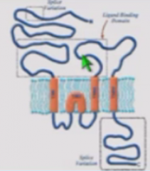
5 subunits. Binding site for glutamate is in extracellular domain which consists of N terminus and loop between third and fourth transmembrane domain. Second transmembrane domain loops back to intracellular - doesn't fully cross membrane. C terminal is thus intracellular as opposed to extracellular. C terminal is highly amenable to post-translational modifications. |
|
|
Metabotropic glutamate receptors |
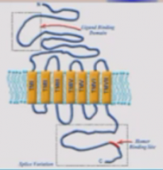
GPCRs, seven transmembrane domains Glutamate binds to N terminal domain Intracellular C terminal domain is highly amenable to post-translational modifications |
|
|
Class 1 of metabotropic glutamate receptors |

mGluR 1 or 5 Signal through Gq - PLC, DAG (PKC), and IP3 (Ca2+) PKC activates other protein kinases and Ca2+ activates CaMK - leads to downstream effects |
|
|
Beta-arrestin |

Binds to metabotropic glutamate receptors and signals for them to be internalized to desensitize cell to glutamate. |
|
|
Class 2 and 3 of metabotropic receptors |

mGluR 2 or 3 mGluR 4, 7, 8 Signal through Gi proteins. mGluR 2 or 3 leads to inhibition of AC, reduction in PKA, and activation of K+ channel causing efflux of K+ and hyperpolarization (inhibitory response) |
|
|
Glutamate receptor activity presynaptically |
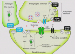
Binding of glutamate to mGluR 2 or 3 Pre-synaptically decreases activity of voltage-gated Ca2+ channels to dampen activity of glutaminegic presynaptic terminal. |
|
|
AMPA receptor subtypes |
Cation channels formed by different subunits that come together to form glutamate-gated channel. Can be calcium-impermeable - has GluA2R (arginine) Or calcium-permeable - has GluA2Q or neither Q or R subunits. These subunits can be modified by RNA editing |
|
|
Agonists of AMPA receptor |
Glutamate (promiscuous) AMPA |
|
|
Competitive antagonists of AMPA receptor |
NBQX, CNQX - relevant for research No clinically relevant antagonists |
|
|
Kainate receptor agonists |
Kainate, glutamate |
|
|
Kainate receptor competitive antagonists |
CNQX (inhibits both AMPA and Kainate) LY - more selective. Interest in producing analogs for treatment of stroke |
|
|
NMDA activation |
Dependent on co-binding of glycine and glutamate Glycine alone will not activate NMDA receptors. Glutamate alone can but small activity. |
|
|
NMDA receptor agonists |
NMDA, glutamate, glycine |
|
|
Main negative regulator of NMDA receptor |
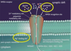
Mg2+ acts as open channel blocker - binds to Mg2+ binding site within channel and keeps channel inactive. |
|
|
Competitive antagonists of NMDA receptor |
APV - research use |
|
|
Noncompetitive antagonists of NMDA receptors |
Mg2+ - binds to site within channel and inactivates Kynurenic acid Pb2+ Ifenprodil Ketamine - anaesthetic Phencyclidine (PCP) - addiction Memantine - Alzheimer's |
|
|
Kynurenic acid |
Antagonist of NMDA receptors Metabolite of kynurenine pathway, comes from tryptophan. Kynurenine crosses brain blood barrier and is converted to Kynurenic acid by astrocytes. Competitive with respect to glycine but noncompetitive with respect to glutamate. |
|
|
NMDA activation in normal conditions |
NMDA receptors are kept in check with lock from Mg2+. When glutamate released from presynaptic terminal, cannot overload postsynaptic cell with Ca2+ because would lead to cell death. Thus, NMDA receptors are only activated by simultaneous glutamate and depolarization (via activation of AMPA receptors (which releases Mg2+ block) |
|
|
AMPA |
AMPA are sensitive to activation by glutamate, depolarize cell and removes Mg2+ block of NMDA Glutamate and glycine fully activates NMDA receptor. |
|
|
Long-term potentiation, NMDA, AMPA (Early effects) |
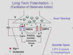
Protein synthesis-independent As glutamate keeps being releases and NMDA continually activated, flow of Ca2+ through NMDA activates calcium-calmodulin kinase which induces NO synthase and phosphorylates AMPA receptors to more stably activate them. Leads to synthesis of NO in postsynaptic cell.
Thus, leads to more sustained, strengthened postsynaptic response. |
|
|
Long-term potentiation (later effects) |
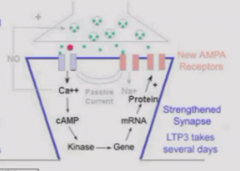
Protein-synthesis dependent Ca2+ influx through NMDA activates Calcium-calmodulin kinase which stimulates the synthesis and insertion of new AMPA receptions onto the postsynaptic membrane. Further increases synaptic transmission. |
|
|
Long-term potentiation purpose |
Underlies memory formation in brain |
|
|
Neurodegeneration in Alzheimer's |
Early stages - not overt or diagnostic Later stages - Overt changes. Brain size decreases by 10-20% due to loss of neuron. |
|
|
Extrasynaptic vs synaptic NMDA receptors |
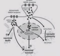
Synaptic NMDA receptors lead to a calcium influx - involved in synaptic plasticity and memory. Extrasynaptic NMDA receptors lead to calcium influx that signals cell death. Shuts down CREB which promotes neuronal survival. Possible because synaptic and extrasynaptic receptors are concentrated in different areas of cell. |
|
|
Memantine |

Alzheimer's drug Blocks activity of extrasynaptic NMDA receptors to prevent neuronal death. Not useful for early stages because also blocks synaptic receptors which are involved in cognitive function. |
|
|
GABA |
Major inhibitory neurotransmitter in brain Mostly in local circuitry as interneurons which modulate activity of excitatory neurons. |
|
|
GABAergic neurotransmission |
1. GABA is synthesized from Glutamate using glutamic acid decarboxylase (GAD). Stored in vesicles 2. Action potential -> VGCCs -> fusion of vesicles with presynaptic membrane 3. GABA binds to postsynaptic ionotropic or metabotropic receptor and exert actions. |
|
|
Termination of GABAergic action |

GABA diffuses from synaptic cleft and is taken up by glial cells or presynaptic axon. In glia, GABA is converted to glutamate which is converted to glutamine via glutamine synthetase. Glutamine travels to presynaptic neuron. In presynaptic neuron, glutamine is converted to glutamate by glutaminase, then to GABA by GAD. |
|
|
Excess of glutamate |
Excess glutamate accumulates in extracellular space and causes overaction of AMPA and NMDA extrasynaptic receptors, causing cell death. NMDA receptor is coactivated by basal le |
|
|
Ionotropic GABA receptors classifications |
GABAa and GABAc (Rho type) GABAa are primary Rho type GABAc receptor are only in retina |
|
|
GABAa receptor structure |
Five subunits, each with four transmembrane domains. Second domain serves as pore. Gating is positively charged so GABAa receptors are permeable to anions like chloride. Alpha subunit is ligand binding. Gamma subunit has barbiturate/benzodiazepine binding site. |
|
|
GABAa receptor positive allosteric modulators |
Barbiturates and benzodiazepines Augment activity of GABA receptor when GABA is present Binds to gamma subunit. Used to prevent convulsions by increasing GABAa receptor activity. |
|
|
Non-competitive antagonists of GABAa receptor |
Picrotoxin Dieldrin (organochlorine pesticide) |
|
|
Competitive antagonists of GABAa receptor |
Bicuculline |
|
|
Result of bicuculline, picrotoxin, dieldrin |
Excitation - proconvulsants |
|
|
Agonists of GABAa receptor |
GABA Muscimol |
|
|
Barbiturate and benzodiapene effect on GABAa receptor |
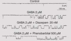
Barbiturates (phenobarbitals) increase duration of GABA induced channel activation Benzodiapines increase frequency of GABA-induced channel openings. Both increase GABAa receptor function |
|
|
Fundamental forms of inhibition in brain |
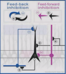
1. Feed-forward inhibition - inhibitory neuron is being excited by outside stimulus. 2. Feedback mechanism - cell that is stimulating the interneuron is inhibited by the interneuron. |
|
|
Chloride transporters in brain |
KCC2 - exports chloride
NKCC1 - imports chloride |
|
|
GABAa receptor action in immature and mature brain |
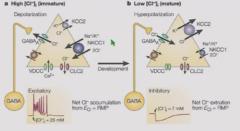
Immature brain - NKCC1 is more active, creating higher chloride inside cell. Chloride flows out through active GABAa channel which has a depolarizing (excitatory) effect. Developedbrain - KCC2 is more active, raising chloride outside cell. Chloride flows in through active GABAa channels which has a hyperpolarizing (inhibitory) effect. |
|
|
GABAb receptor signaling |
Seven transmembrane domain Signals through Gi proteins (blocks AC, decreased cAMP, activated voltage-gated K+ and inactivation of voltage-gated Ca2+ -> inhibition of cell) |
|
|
GABAb receptor agonists |
GABA
Baclogen |
|
|
GABAb receptor antagonists |
Phaclofen |
|
|
Glycine |
Major inhibitoryneurotransmitters at spinal cord |
|
|
Renshawl cells |
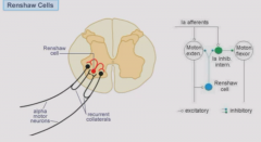
Interneurons that synthesize and release glycine. Excited by motor neurons to release glycine which inhibits the activity of motor neurons. |
|
|
Glycine synthesis and storage |
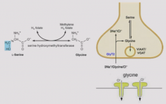
Synthesized from serine by serine hydroxymethyltransferase. Stores in vesicles and binds to ionotropic receptors (Cl- channels) ONLY - no metabotropic receptors. |
|
|
Glycine receptor agonists |
Glycine, serine |
|
|
Glycine receptor antagonists |
Strychnine (rat poison) but not kynurenic acid |
|
|
Hyperekplexia |
Mutation in glycine receptor alpha 1 subunit Hyperreflexia Prolonged response to auditory stimulus |