Reading...
![]()
Play button
![]()
Play button
![]()
Use LEFT and RIGHT arrow keys to navigate between flashcards;
Use UP and DOWN arrow keys to flip the card;
H to show hint;
A reads text to speech;
54 Cards in this Set
- Front
- Back
|
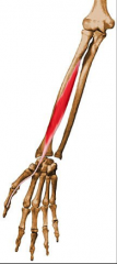
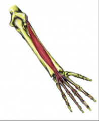
FLEXOR CARPI RADIALIS
|
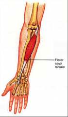
O: medial humeral epicondyle
I: Base of 2nd and 3rd metacarpal A: Flexes wrist and radial abduction Nerve: Median (C6-C7) |
|
|
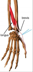
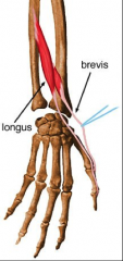
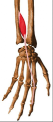
PALMARIS LONGUS
|
O: Medial humeral epicondyle
I: Palmar aponeurosis of hand A: Flexes wrist Nerve: Median nerve (C7-T1) |
|
|
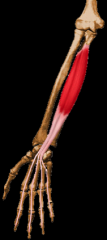
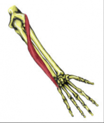
FLEXOR CARPI ULNARIS
|
O: Medial epicondyle of humerus and posterior border of upper 3/5 of ulna
I: Pisiform bone, hamate, 5th metacarpal A: Strongest Flexor; ulnar abductor Nerve: Ulnar nerve (T8) |
|
|
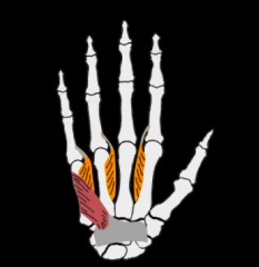
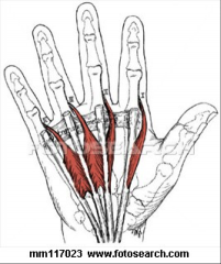
FLEXOR DIGITORUM SUPERFICIALIS
|
O: Humeroulnar head: medial humeral epicondyle and medial border of coronoid process
Radial head: upper part of anterior border of radius I: 4 tendons – on sides of ear middle phalanx of digits 2-5 A: Flexes middle phalanges (PIP); wrist flexion Nerve: Median (C7-T1) |
|
|
FLEXOR POLLICIS LONGUS
|
O: Middle ½ of anterior surface of radius and interosseus membrane
I: Base of DISTAL phalanx of thumb A: Flexes IP of thumb Nerve: Median nerve (anterior interosseus branch) |
|
|
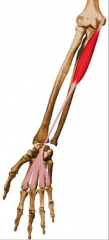
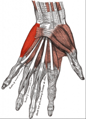
FLEXOR DIGTORUM PROFUNDUS
|
O: Upper 3/4 of anterior ulna and interosseus membrane
I: Bases Distal phalanges of 2-5 A: Flexion of DIP joints of digits 2-5; wrist flexion Nerve: Median nerve to lateral ½ Ulnar nerve to medial ½ |
|
|
EXTENSOR CARPI RADIALIS LONGUS
|
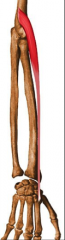
O: Lateral supracondylar ridge and lateral epiridge and lateral epicondyle of humerus
I: Base of 2nd metacarpal A: Extends and radially deviates Nerve: Radial nerve (C6-C7) |
|
|
EXTENSOR CARPI RADIALIS BREVIS
|
O: Lateral humeral epicondyle
I: Base of 3rd metacarpal A: Extends and radially deviates Nerve: Radial (posterior interosseus branch) C6-C7 |
|
|
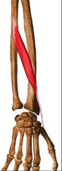
EXTENSOR DIGITORUM Communis
|
O: Lateral epicondyle of humerus
I: Base of distal AND middle phalanges of 2-5 A: Wrist extension, extension of MP joints of digits 2-5 Nerve: Radial nerve (posterior interosseus) |
|
|
EXTENSOR DIGITI MINIMI
|
O: Lateral epicondyle
I: Extensor aponeurosis of little finger A: Extends and abducts little finger Nerve: Radial (posterior interosseus) |
|
|
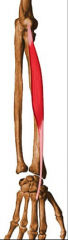
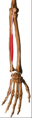
EXTENSOR CARPI ULNARIS
|
O: Lateral epicondyle of humerus
I: Base of 5th metacarpal A: Extends and ulnarly deviates wrist Nerve: Radial (posterior interossseus |
|
|
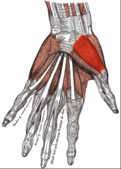
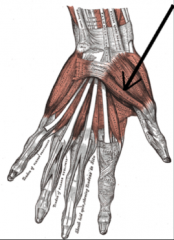
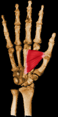
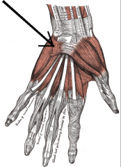
ABDUCTOR POLLICIS LONGUS
|
O: Posterior surface of radius and ulna and the interosseus membrane
I: Radial surface of base of 1st metacarpal and trapezium A: Extension of thumb, abduction of CMC joint of thumb, radial deviation Nerve: Radial (posterior interosseus) |
|
|
EXTENSOR POLLICIS BREVIS
|
O: posterior surface of radius and interosseus membrane
I: Base of proximal phalanx of thumb A: Extension of MP joint of thumb, radial deviation Nerve: Radial (posterior interosseus) |
|
|
EXTENSOR POLLICIS LONGUS
|
O: Posterior surface of ulna and interosseus membrane just distal to abductor pollicis longus
I: Base ofDistal phalanx of thumb A: Extension of IP joint of thumb, radial dev. Nerve: Radial (posterior interosseus) |
|
|
EXTENSOR INDICIS
|
O: Posterior surface of ulna distal to extensor pollicis longus
I: Extensor expansion of index finger A: Extends ALL joints of index finger and independently abducts the index finger Nerve: Radial nerve (posterior interosseus) |
|
|
OPPONENS POLLICIS
|
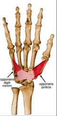
O: Flexor retinaculum and trapezium
I: Length of 1st metacarpal A: Opposition of thumb Nerve: Median nerve (recurrent branch) |
|
|
ABDUCTOR POLLICIS BREVIS
|
O: Tubercle of scaphoid and trapezium, flexor retinaculum, and tendon of abductor pollicis longs
I: Base of 1st proximal phalanx A: Abduction of thumb, opposition Nerve: Median nerve (recurrent branch) |
|
|
FLEXOR POLLICIS BREVIS
|
O: Flexor retinaculum across capitate, trapezoid, and trapezium and their palmar ligaments
I: Shaft of 1st metacarpal; base of 1st proximal phalanx A: MP flexion; thumb oppostion Nerve: Ulnar nerve |
|
|
ADDUCTOR POLLICIS
|
O: Transverse head- from the shaft of the 3rd metacarpal
Oblique head – from the bases of 2nd and 3rd metacarpals, capitate, and palmar ligaments of carpals I: Base of the proximal phalanx of thumb on ulnar side A: Adducts thumb Nerve: Ulnar |
|
|
PALMARIS BREVIS
|
O: Flexor retinaculum and palmar aponeurosis
I: Base of 5th proximal phalanx A: Steadies skin of palm to help with grip Nerve: Ulnar |
|
|
ABDUCTOR DIGITI MINIMI
|
O: Pisiform bone; tendon of FCU and pisohamate ligament
I: base of 5th proximal phalanx; ulnar side A: Abduction of little finger Nerve: Ulnar nerve- deep branch |
|
|
FLEXOR DIGITI MINIMI BREVIS
|
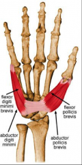
O: Flexor retinaculum and hook of the hamate
I: Base of 5th proximal phalanx A: Flexion of the little finger Nerve: Ulnar nerve- deep branch |
|
|
OPPONENS DIGITI MINIMI
|
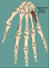
O: Flexor retinaculum and hook of the hamate
I: shaft of 5th metacarpal; ulnar aspect and palmar surface A: Opposition of 5th finger Nerve: Ulnar nerve – deep branch |
|
|
DORSAL INTEROSSEI
|
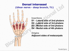
O: Between metacarpal bones
I: 2 are on the 3rd digit, 1 on the 2nd, 1 on the 4th A: (DAB) Abduction of the 2nd, 3rd, and 4th digits and assist in flexion of MP joint and extension of PIP and DIP joints Nerve: Ulnar nerve |
|
|
PALMAR INTEROSSEI
|
O: Base of metacarpals; each muscle has 2 heads from adjacent sides of metacarpals
I: NONE on 3rd digit, Dorsal extensor expansion at the base of the proximal phalanges; 1st and 2nd on radial side- 3rd and 4th on the ulnar side A: (PAD) Adduction of 2nd, 4th and 5th digit and assist with flexion of MP and extension of PIP and DIP joints Nerve: Ulnar |
|
|
LUMBRICALES
|
O: Flexor digitum profundus tendons
I: Extensor digitorum expansion; each muscle attaches distally to the radial side of extensor digitorum A: Extend IP joints (PIP AND DIP) and flex MP joints Nerve: 1 and 2- median nerve 3 and 4 – ulnar nerve |
|
|
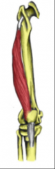
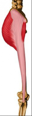
RECTUS FEMORIS
|
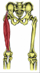
O: AIIS
I: Patellar tendon – tibial tuberosity A: Flex thigh, extend leg Nerve: Femoral |
|
|
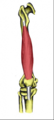
VASTUS MEDIALIS
|
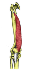
O: linea aspera and trochanteric line
I: Patellar tendon A: Extend leg Nerve: Femoral |
|
|
VASTUS LATERALIS
|
O: Intertrochanteric line
I: Patellar tendon A: Extend leg Nerve: Femoral |
|
|
VASTAS INTERMEDIAS
|
O: Anterior lateral femur
I: Patellar tendon A: Extend leg Nerve: Femoral |
|
|
SARTORIUS
|
O: ASIS
I: Medial surface of tibia inferior to tibial tuberosity A: Flex thigh, flex leg, lateral rotation Nerve: Femoral |
|
|
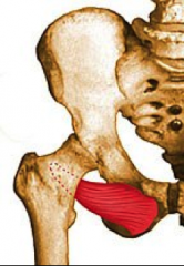
PECTINEUS
|
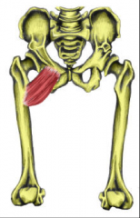
O: Superior ramus of pubis
I: Pectinial line; below less trochanter A: Adduct and medially rotate thingh Nerve: Femoral |
|
|
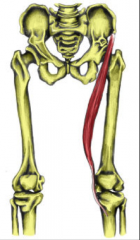
GRACILIS
|
O: Body and inferior ramus of pubis
I: Medial surface of tibia between insertions of Sartorius and semitendinosus A: Adduct and medially rotate thigh Nerve: Obturator |
|
|
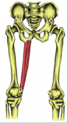
ADDUCTOR BREVIS
|
O: Body and inferior ramus of pubis (same as gracilis)
I: Inferior part of pectineal line and superior part of linea aspera A: Adduction and medial rotation of thigh Nerve: Obturator |
|
|
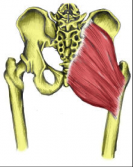
ADDUCTOR LONGUS
|
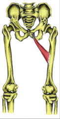
O: Body and Inferior ramus of pubis (same as gracilis)
I: Whole length of linea aspera in line with pectineus A: Adduct and medially rotate thigh Nerve: Obturator |
|
|
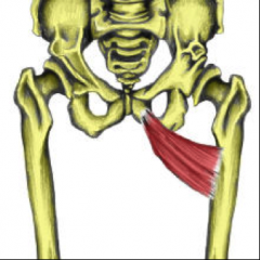
ADDUCTOR MAGNUS
|
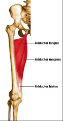
O: Ischiopubic ramus and ishial tuberosity
I: Linea aspera and adductor tubercle A: Adduction and medial rotation of thigh Nerve: Obturator (except for the part on the ishial tuberosity=tibial) |
|
|
OBTURATOR EXTERNUS
|
O: External surface of obturator membrane and surrounding margins of pubis and ischium
I: Passes BEHIND femoral neck to trochanteric fossa A: Lateral rotator of thigh; holds head of femur in acetabulum Nerve: Obturator |
|
|
GLUTEUS MAXIMUS
|
O: Posterior gluteal line, dorsal surface of the sacrum and coccyx, and sacrotuberous ligament
I: Gluteal tuberosity of femur and iliotibial tract A: Extends thigh Nerve: Inferior gluteal nerve |
|
|
GLUTEUS MEDIUS
|

O: Wing of ilium and deep fascia of the muscle
I: Greater trochanter A: Abduction and medial rotation of the thigh Nerve: Superior gluteal nerve |
|
|
GLUTEUS MINIMUS
|
O: Wing of Ilium
I: Greater trochanter A: Abduction and medial rotation of thigh Nerve: Superior gluteal nerve |
|
|
TENSOR FASCIA LATA
|
O: Anterolateral Iliac crest and ASIS
I: Iliotibial tract A: Flexion and Abduction of thigh Nerve: Superior gluteal nerve |
|
|
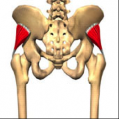
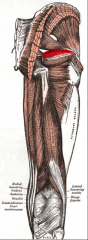
PIRIFORMIS
|
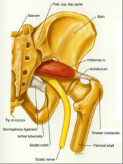
O: anterior surface Sacrum and Sacrotuberous ligament
I: Medial side of greater trochanter A: Lateral rotation of thigh Nerve: Nerve to piriformis |
|
|
OBTURATOR INTERNUS
|
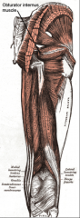
O: : inside the obturator foramen and emerges through the lesser sciatic foramen
I: Greater trochanter A: Lateral rotation of thigh Nerve: Nerve to obturator internus |
|
|
SUPERIOR GEMELLUS
|
O: Ischial spine
I: Inserts to GT by way of superior obturator internus tendon A: Lateral rotation of the thigh Nerve: nerve to obturator internus |
|
|
INFERIOR GEMELLUS
|
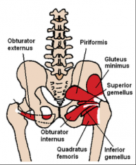
O: Ishcial tuberosity
I: Inserts to GT by way of superior obturator internus tendon A: Lateral rotation of thigh Nerve: Nerve to quadratus femoris |
|
|
QUADRATUS FEMORIS
|

O: Ischial tuberosity
I: Intertrochanteric crest A: Lateral rotation of thigh Nerve: nerve to quadratus femoris |
|
|
SEMITENDINOSIS
|
O: Ishial tuberosity
I: Medial surface of tibia just posterior to insertion of gracilis and Sartorius A: Extend thigh, flex leg Nerve: Tibial division of sciatic |
|
|
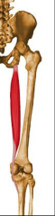
BICEPS FEMORIS
|
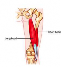
O: Long head- Ischial tuberosity
Short head- Lateral lip of linea aspera I: Lateral tibial condyle and head of fibula A: Extend thigh and flex leg Nerve: Long head – Tibial division of sciatic Short head – Peroneal division of sciatic |
|
|
SEMIMEMBRANOSIS
|
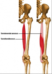
O: Ishial tuberosity
I: Medial and posterior surface of medial condyle of tibia A: Extend thigh, flex leg Nerve: Tibial division of sciatic |
|
|
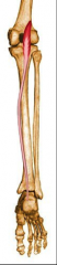
TIBIALIS ANTERIOR
|
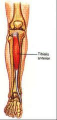
O: Upper 2/3 of lateral shaft of tibia and interosseus membrane
I: Medial side of medial (1st) cuneiform and base of 1st metatarsal A: Dorsiflexion and inversion of foot Nerve: Deep peroneal nerve |
|
|
PLANTARIS
|
O: Lower lateral supracodylar line and popliteal surface of femur
I: Calcaneus tendon A: NONE! Nerve: Tibial |
|
|
POPLITEUS
|
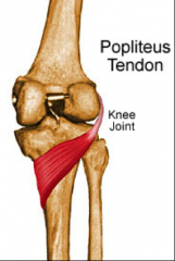
O: Just inferior to lateral epicondyle of femur
I: Posterior tibia just above popliteal line A: Flexion of leg and medial rotation of tibia; unlocks the knee Nerve: Tibial |
|
|
PSOAS MAJOR
|
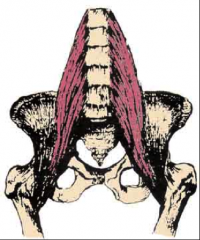
O: Bodies and transverse processes of lumbar vertebrae
I: Lesser trochanter of femur A: Flexion of thigh and slight adduction Nerve: L2-L4 |
|
|
ILIACUS
|
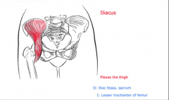
O: Iliac fossa
I: Lesser trochanter A: Flexion and slight adduction Nerve: Femoral |