Reading...
![]()
Play button
![]()
Play button
![]()
Use LEFT and RIGHT arrow keys to navigate between flashcards;
Use UP and DOWN arrow keys to flip the card;
H to show hint;
A reads text to speech;
116 Cards in this Set
- Front
- Back
|
What is ventral and dorsal mean in the brain?
|
according to the embryological development. dorsal-superior
ventral-inferior |
|
|
How do we name anterior and posterior in the brain?
|
rostral - front
caudal- back |
|
|
Is there CT in the CNS?
|
No, it is just neurons and glial cells
|
|
|
What does anterograde mean?
|
when something goes AWAY from the cell body (like an AP or transport of neurotransmitters)
|
|
|
What does somatic mean?
|
the body wall, including muscle and skin
|
|
|
What are the visceral motor fibers also called?
|
autonomic NS
|
|
|
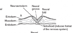
What germ layer becomes the whole nervous system?
|
The ectoderm
|
|
|
Envision what the neural plate and notochord look like before folding into the neural tube.
|
|
|
|
What is neuralation?
|
the thickening of the central ectoderm and it's invagination to become the neural tube
|
|
|
What induces neuralation? What germ layer is this from?
|
The notochord from the mesoderm
|
|
|
imagine Step 1 neuralation.
|

|
|
|
imagine Step 2 neuralation.
|

|
|
|
What just happened?
|
neural grooves came out and there are two neural crest dots on top
|
|
|
Imagine step 4
|

|
|
|
Where are the neural crest cells? What will they become?
|
outside the neural tube. They will become cranial and peripheral nerves.
|
|
|
When the neural tube forms, is it on the dorsal or ventral part of the body?
|
dorsal, the whole baby plate hasn't folded yet
|
|
|
Recap- what forms the eventual CNS and PNS?
|
CNS- neural tube
PNS- neural crests |
|
|
What is the most common birth defect with the NS?
|
Neural tube closing defects
|
|
|
What part of the neural tube clses first and when does it do so in fetal development?
|

|
|
|
Draw what the neural crest looks like after neuralation
|

|
|
|
What is the top vs bottom of that tube called?
|
rostral and caudal
|
|
|
Which closes first and how long does it take for the other one to close?
|
Rostral (brain) closes first and then the Caudal 4 days later.
|
|
|
What happens when the rostral end fails to close?
|
anencephaly (lacks a brain, but spinal cord is ok)
|
|
|
What happens when the caudal end fails to close?
|
very severe spina bifida
|
|
|
essentially what do you have when you have a defect of closing the neural tube?
|
a body wall defect
|
|
|
What will leak out when you have a body wall defect? To where?
|
Alpha fetoprotein by the fetal liver will leak out into the amnionic fluid
|
|
|
So how would you test for neural tube defects?
|
check for elevated alpha feto-protein in amniocentesis
|
|
|
do these elevated lvels of afp necessarily mean a neural tube defect?
|
No it can mean a bodywall defect in any part of the fetus
|
|
|
What does polyhydraminos mean?
|
too much amnionic fluid
|
|
|
How is amnionic fluid regulated?
|
fetus will swallow it to remove and urinate it out
|
|
|
What neural tube defect can result in polyhydramios? Why?
|
failure of rostral end to close because then they don't have a brain to regulate swallowing.
|
|
|
Would there be polyhydramios in caudal closure failure?
|
No
|
|
|
What are the two plates that form dorsal/ventral polarity in the neural tube? What kind of nerves do they end up forming?
|

|
|
|
What is in the neural tube?
|
Nothing! it is a hollow tube so far.
|
|
|
how do you remember the 4 things in the diencephalon?
|
They all end in -alamus
|
|
|
What is the role of the thalamus?
|
To filter and relay all the info going to the telencephalon
|
|
|
What does the hypothalamus do?
|
regulates the endocrine and autonomic systems
|
|
|
What is another name for the epithalamus? Why?
|
Pineal gland (that's all it contains)
|
|
|
What is another name for the subthalamus? How can I remember this?
|
basal ganglia (it is at the base, it is sub to all the other thalami)
|
|
|
What are the lobes of the brain named for?
|
the bones of the skull that cover them
|
|
|
Where is the central sulcus?
|
exactly between the frontal and parietal
|
|
|
What else is derived from the ectoderm?
|
skin, hair, and all glands.
|
|
|
Will MS affect the eyes? Why?
|
Yes, because they affect oligodendrocytes that supply myelin to the eyes.
|
|
|
What is the difference in embryological origin of the PP and AP glands? What does this mean for how they can secrete things?
|
AP comes from ectoderm of mouth, which means it can secrete to the blood in veiscles
PP comes from neuroectoderm, which means it releases things with axons. |
|
|
What is the name for the outpouching of oral cavity cells that make the AP?
|
rathke's pouch
|
|
|
What are the two types of ganglia in the body?
|
sensory or autonomic
|
|
|
What "gland" is actually from the neural crest cells? How do you know this?
|
The adrenal medulla., which is an autonomic ganglion They secrete epi and norepi.
|
|
|
What surprising cells (that have a lot of connections) are from neural crest cells that migrated to the skin?
|
melanocytes
|
|
|
What glial cells are from the neural crest?
|
Schwann cells
|
|
|
What meningeal layers are from the neural crest?
|
The pia and arachnoid
|
|
|
What organ surprisingly gets a lot of contribution from neural crest cells?
|
the heart (endocardial cushion)
|
|
|
Now we go into the different forms of spina bifida! (there are 4)
|
yay!
|
|
|
What is the most severe, but least common type of spina bifida?
|
myeloschosis?
|
|
|
dissect the word myeloschisis?
|
schisis- split
myelo- spinal cord |
|
|
What is common between the rest of the 3 types of spina bifida?
|
They all have a complete spinal cord, but just failed to secrete enough inductive signals to develop a spinous process.
|
|
|
What does occulta mean?
|
mysterious
|
|
|
What is the only sign of spina bifida occulta? (picture!)
|
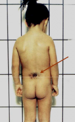
tuft of hair grows out where it failed to grow a spinous process
|
|
|
Why is spina bifida occulta called what it is? Mnemonic?
|
People are usually asymtommatic and don't know they have it. It also looks kinda freaky that they have that tuft of hair and can be occult.
|
|
|
What does the word -cele mean?
|
cyst
|
|
|
What is the common thing between spina bifida meningocele and meningomyelocele?
|
they both have cyst protrusions out where the spinous process should be.
|
|
|
What is the difference between these two cyst spina bifidas?
|
the meningomyelocele one has the spinal cord outside of where it should be
|
|
|
Show a picture of this.
|

|
|
|
What meningeal layers are outpouched? Logical mnmonic?
|
dura and arachnoid because the cyst is filled with CSF pushing both those layers out
|
|
|
Which of the common spina bifidas is most likely to cause problems in the lower leg?
|
spina bifida meningeomyelocele
|
|
|
Would you have elevated alpha feto protein in these 3 common spina bifidas? Why?
|
You may if the cyst leaks fluid out (it does tend to thin the ectoderm layer)
|
|
|
What illicit drugs can cross the BBB?
|
heroin, ethanol,
|
|
|
Are there only 2 choroid plexuses in each ventricle like in Najeeb's drawings?
|
no, there are many of them along the walls where he drew the 2.
|
|
|
What is the main cause of communicating hydrocephalus?
|
problem in drainage
|
|
|
Who gets prblems in CSF drainage? Why?
|
Older people who halve their granulations calcify
People who get infections or hemorrhages that plug up their granulations |
|
|
What are the 3 main sx of someone with normal pressure hydrocephalus?
|
wackly, wobbly and wet
|
|
|
Why do they get these 3 W's?
|
degradation of cortical control over these things from all the CSF not being drained/
|
|
|
Specifically what kind of "wet" will they get?
|
infantile bladder that is a loss of being able to choose when they will go. (but reflexes still intact)
|
|
|
Specifically what kind of "wobbly" will they get?
|
apraxic gait that look like they are shuffling and can't take their feet off the groun
|
|
|
What other disease results in apraxic gait?
|
parkinson's
|
|
|
What kind of pt will classically have hydrocephalus ex vacuo?
|
people with alzheimer's or other cortical loss.
|
|
|
What kind of hydrocephalus is ex vacuo?
|
Neither communicating nor noncommunicating because it starts with the brain and is neither a bloackage or drainage problem
|
|
|
What type of organelle is especially prominent in neuronal cell bodies? WHat is their special name here?
|
the rER called nissel bodies
|
|
|
Why are the rER so prevalent in the neuronal cell bodies?
|
because they synthesize neurotransmitters
|
|
|
are there any nissl substances in the axon?
|
no
|
|
|
How are the NT's moves to the terminal of the neuron? What is the train track and what powers it?
|
It is moved by anterograde transport down microtubules which is powered by kinesin.
|
|
|
What other cell uses a similar mode of tranportation?
|
melanocytes transporting melanin
|
|
|
Do neurons d retrograde axonal transport? For what?
|
to transport waste back to the cell body for processing
|
|
|
What provides the energy for retrograde axonal transport?
|
dyenin
|
|
|
Which direction of axonal transport travels faster? Why is this so?
|
anterograde because it is more important to resupply the neurotransmitters
|
|
|
Which types of axonal transport is more important clinically? Why?
|
retrograde because the neuron can actually pick up viruses from it's terminals and retrograde transport them back to the cell body to infect the cell.
|
|
|
What are two viruses that are taken up through skeletal motor fibers?
|
polio and rabies
|
|
|
How do you remember polio being transported this way?
|
polio results in the degradation of skeletal nerves
|
|
|
How do you remember rabies being transported this way?
|
wild, rabid animals will bite you in your muscle
|
|
|
What toxin can be transported retrograde from skeletal nerves? Mnemonic?
|
tetanus toxin. You stab yourself in the muscle with a rusty nail.
|
|
|
What virus will travel retroograde from sensory nerves?
|
herpes
|
|
|
Where will herpes hide out?
|
In the sensory ganglia
|
|
|
How would you transmit rabies to someone else? Where does the virus travel?
|
It travel anterograde to salivary glands so you can infect someone with your bite.
|
|
|
How would herpes manifest itself? Where does the virus travel?
|
it would end up as erupted skin vesicles by travelling anterigrade from it's sensory ganglia to all the nerves that go to it
|
|
|
What skin distribution does this explain for herpes?
|
it explains the sensory dermotomal distribution.
|
|
|
What does synapse mean?
|
To fasten together
|
|
|
Does the function of the synapse depend on the NT or the receptor?
|
the receptor!
|
|
|
What excitatory NT is present in more then 50% of the CNS?
|
glutamate
|
|
|
What is the main NT is present in the CNS?
|
GABA
|
|
|
Is ACh present in the PNS or CNS?
|
Both!
|
|
|
What does it do in the PNS? (3 functions)
|
1. NT of all ganglia
2. NT at all neuromuscular junctions using nicotinic receptors 3. NT for all PS post ganglionic cells using muscarinic receptors |
|
|
What does ACh do in the CNS? (2)
|
Neuromodulation
1. memory consolidation 2. sleep/wake cycles |
|
|
What kind of receptors for ACh are present in both these functions in the CNS?
|
Both nicotinic and muscarinic subtypes.
|
|
|
In what disease are the cholinergic neurons aiding in memory consolidation lost?
|
alzheimer's
|
|
|
What can you say about the function of ACh in the nervous system?
|
There is a huge distribution of functions depending on the synapse and receptor you are at.
|
|
|
What receptor is the majority of cholinergic pharmacology aimed at?
|
muscarinic receptors.
|
|
|
Why don't we want to do anything to the nicotinic receptors?
|
they are widely distributed and can cause huge musculoskeletal consequences
|
|
|
What disease happens when there is a lack of dopamine in the midbrain?
|
parkinson's
|
|
|
What is the pleasure center of our brain called? What NT is released here?
|
nucleus occumbens, which responds to dopamine
|
|
|
What may people do to activate the nucleus occumbens?
|
take illicit drugs
|
|
|
What is the most mysterious NT?
|
serotonin
|
|
|
Why is serotonin so mysterious?
|
We are not entirely sure what it does because so many different cell types interact with it.
|
|
|
what does histamine do in the brain?
|
it is an excitatory NT that helps you maintain an awake state
|
|
|
How can you remember what histamine does in the brain with an OTC drug side effect?
|
antihistamines taken for allergies may make you very drowsy.
|
|
|
What kind of NT is glycine?
|
inhibitory
|
|
|
how is glycine different is distribution from the other inhbitory NT, GABA?
|
it is not as widespread, but it does cover the spinal cord!
|
|
|
How many types of neuropeptides are there and what do they usually come in?
|
many! over 50! They are usually packages along with the neuron's main NT's.
|