![]()
![]()
![]()
Use LEFT and RIGHT arrow keys to navigate between flashcards;
Use UP and DOWN arrow keys to flip the card;
H to show hint;
A reads text to speech;
102 Cards in this Set
- Front
- Back
Explain ** |
Frontal coronal plane |
|
|
Describe frontal coronal plane ** |

|
|
|
Types planes ** |
Coronal (frontal): divides anterior and posterior Median / Sagittal/ parasagittal / paramedian: divides left and right Transverse (horizontal): divides superior and inferior |
|
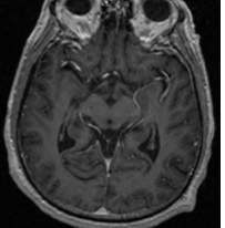
Type scan shown ** |
MRI |
|
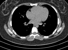
Type scan shown ** |
CT |
|
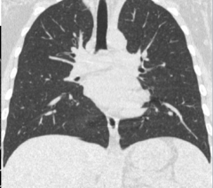
Type scan shown ** |
CT lung window |
|
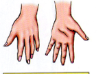
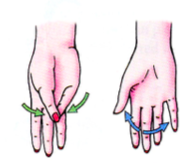
Explain ** |
Left: dorsum Right: palm |
|
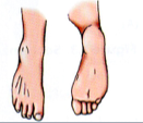
Explain ** |
Left: dorsum Right: sole |
|
|
Hand and foot relationships* |
Hand: Dorsum Palmer Foot: Dorsum Sole |
|
|
Rostral refers to what ?** |
Brain along w/ caudal |
|
|
Flexion and extension relationships ** |

|
|
Explain** |
Top: Extension Bottom: Flexion |
|
Explain ** |
Top: Flexion Bottom: Extension |
|
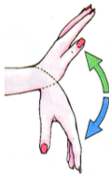
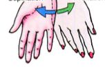
Explain ** |
Left: opposition Right: extension |
|
Explain ** |
Left: Supination Right: pronation |
|
|
Supination, Pronation ** |

|
|
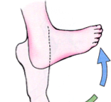
Explain ** |
Top: Dorsiflexion Bottom: Plantarflexion |
|
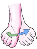
Explain ** |
Blue: Inversion Right: Eversion |
|
Explain ** |
circumduction |
|
Explain ** |
lateral bending |
|
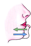
explain ** |
Top: Retrusion Bottom: Protrusion |
|
|
Explain variation in individuals ** |
> Significant anatomical variations exists between individuals > Insufficient knowledge of anatomical variation is a major cause of iatrogenic injury and malpractice |
|
|
Fascia ** > properties > importance |
Properties > 'a bandage that holds things together' or > 'a CT that surrounds, supports every organ of our body' > composed of C/T > envelopes muscles, bones, organs, joints, continuous over the entire body Importance > provides attachments for muscles, serves as an elastic sheath for muscle, forms retinacula and fibrous sheaths for tendons, provide passage for vessels and nerves, allows structures to glide for each other and provides continuity > Important for OMM > Used in acupuncture |
|
|
provide passage for vessels and nerves, allows structures to glide for each other and provides continuity ** |
fascia |
|
|
Important for OMM** |
fascia |
|
|
Types fascia ** |
Superficial fascia > subcutaneous loose fatty C/T, that covers entire body > most distinct in abdomen, perineum, limb > Connects skin to subjacent body wall, facilitating movement of skin, allowing the passage of vessels and nerves to the integument Deep fascia > found deep superficial fascia, covering entire body, thickness variety and betst observed in limbs > important in the 'musculovenous' pump > forms inter muscular septa, defines fascial or muscular compartments (inner component) > forms neurovacqlar sheaths and attachments for muscles |
|
|
Where is superficial fascia most distinct ** |
Abdomen, perineum, limbs |
|
|
Connects skin to subjacent body wall, facilitating movement of skin, allowing the passage of vessels and nerves to the integument ** |
Superficial fascia |
|
|
** 1. Important distinguishing characteristic of deep investing fascia? 2. Important distinguishing characteristic of superficial fascia? a. variable thickness b. covers entire body c. distinct in abdomen, perineum and limbs d. forms inter muscular septa e. facilitates movement of skin |
1. forms inter muscular septa 2. distinct in abdomen, perineum and limbs + facilitates movement of skin > a, b, applies to both |
|
|
Properties of fascial planes ** |
> between fascial compartments or layers > avascular regions (vessels, nerves located within the fascia) > allows a surgeon to separate adjacent structures, in different planes w/ blunt dissection > govern the spread of infections from within a fascial compartment => predictable |
|
|
govern the spread of infections from within a fascial compartment => predictable ** |
fascial planes |
|
|
agonists ** |
prime movers, contract to move desired movement |
|
|
antagonists ** |
oppose the action of the prime mover, makes movement smooth |
|
|
synergists ** |
when muscle passes over 2 joints , muscles preventing action of the intervening joint |
|
|
fixators ** |
steady proximal parts of a limb while action occurs at more distally |
|
|
steady proximal parts of a limb while action occurs at more distally ** |
fixators |
|
|
Flexor digitorum example of what?** a. agonist b. antagonist c. fixator d. synergists e. opposer |
synergist when muscle passes over 2 joints , muscles preventing action of the intervening joint |
|
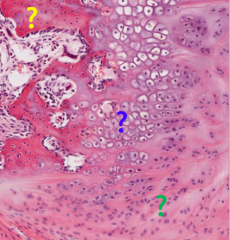
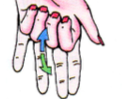
label 3 colors ** |
yellow: compact bone blue: hyaline cartilage green: fibrocartilage |
|
|
parts femur ** (diagram shown) |

|
|
|
periosteal arteries ** |
numerous small vessels that nourish the compact tbone |
|
|
nutrient arteries ** |
enters bone via nutrient foramen near middle body, supplying spongy bone and marrows, canals |
|
|
Epiphyseal and metaphyseal ** |
form articular vessels that supply joints |
|
|
nerves travel where in the bone ** |
to periosteum |
|
|
1. To periosteum 2. Leave near articular end bone 3. Numerous small vessels that nourish the compact bone 4. Supply spongy bone and marrow 5. Supply joint a. periosteal arteries b. nerves c. veins d. nutrient arteries e. fascia f. epiphyseal vessels g. metaphyseal vessels |
1. nerves 2. veins 3. periosteal arteries 4. nutrient arteries 5. metaphyseal and epiphyseal arteries (supply joint) |
|
|
suture ** |
bone in skull
|
|
|
gomphosis ** |
teeth, mandible |
|
|
primary cartilaginous articulation ** |
aka synchondroses temporary unions, little - no movement, 2 bones connected by hyaline cartilage, epiphyseal plates |
|
|
secondary cartilaginous articulation ** |
aka symphyses slightly moveable joints united by fibrocartilage , intervertebral symphysis, pubic symphysis |
|
|
slightly moveable joints united by fibrocartilage , intervertebral symphysis Type of ** |
secondary cartilaginous articulation (aka symphyses) |
|
|
pubic symphysis type of ** |
secondary cartilaginous articulation (symphyses) slightly moveable joints united by fibrocartilage , intervertebral symphysis |
|
|
syndesmoses ** |
2 bones held together w/ a ligament or band of fibrous tissue: vertebral column (longitudinal ligament), interosseous membrane |
|
|
** 1. What are the fibrous articulations? 2. What are the cartilaginous articulations? 3. What are the synovial articulations? 4. High degree mobility a. suture b. syndesmosis c. gomphosis d. synchondroses e. symphyses f. plane g. hinge h. pivot i. hinge j. condyloid k. ball and socket |
1. suture, syndesmoses, gomphosis 2. synchondroses (primary cartilaginous), symphyses (secondary cartilaginous) 3. plane, hinge, pivot, hinge, condyloid, ball and socket) 4. ball and socket |
|
|
Describe picture hand after mastectomy ** ** E1 Q |
Lymphedema |
|
|
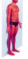
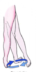
Person on heels. Describe anatomical position ** E1 Q |
Plantar flexion |
|
|
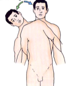
How to cut in a way to show ears and nose together ** ** E1 Q |
Transverse section (NOT FRONTAL/CORONAL SECTION) |
|
|
What lacks lymph vessels ** ** E1 Q |
Epidermis |
|
|
What has end arteries ** ** E1 Q |
brain |
|
|
What is in the growing bone ** ** E1 Q |
synchondroses |
|
|
Describe ligament between bones ** ** E1 Q |
syndesmoses |
|
|
What is the level of spinal cord/nerve innervating muscle ** ** E1 Q |
Myotome |
|
|
Explain anatomical position ** ** E1 Q |
Arms extended |
|
|
What type of muscle in large veins *** ** E1 Q |
Longitudinal muscle |
|
|
Fascia ** |
> bandage that holds things togeteher = a CT that surrounds, supports every organ of our body > composed of C/T > envelops muscles, organs, organs, joints, continuous over entire body > provides attachments for muscles > serves as elastic sheath for muscle, forms retinacula and fibrous sheaths for tendons, provides passage for vessels and nerves, allows structures to glide over each other and provides continuity |
|
|
a CT that surrounds, supports every organ of our body ** |
fascia |
|
|
envelops muscles, organs, organs, joints, continuous over entire body** |
fascia |
|
|
serves as elastic sheath for muscle, forms retinacula and fibrous sheaths for tendons, provides passage for vessels and nerves ** |
fascia |
|
|
important for OMM ** |
fascia |
|
|
important for acupuncture ** |
fascia |
|
|
components fascia ** |
superficial fascia > 2 components = fatty superficial layer (connects skin to adjacent body wall, facilities movement of skin, allows passage vessels and nerves to the integument) = deep membranous layer ( ) deep investing fascia > found deep superficial fascia covering entire body > thickness varies and best observed in limbs where this layer forms a stocking like covering over the muscles > role 'musculovenous' pump > forms inter muscular septa, defines fascial or muscular compartments (inner component) > forms neuromuscular sheaths and attachments for muscles (inner component) |
|
|
** 1. best observed in limbs 2. 3. 4. important role in the 'musculovenous' pump 5. avascular regions 6. |
1. deep investing fascia (where layer forms stocking like covering over muscles) 2. 3. 4. deep investing fascia 5. fascial planes (vessels nerve located within fascia) 6. |
|
|
** 1. form neurovascular sheaths 2. between fascial compartments or layers 3. 4. forms a stocking like covering over muscles 5. |
1. deep investing fascia 2. fascial planes (vessels nerve located within fascia) 3. 4. deep investing fascia 5. |
|
|
fascial planes ** |
> between fascial compartments or layers > avascular regions (vessels nerve located within fascia) > allows surgeon to separate adjacent structures, in different planes with blunt dissection > govern the spread of infections from within a fascial compartment ==> predictable |
|
|
** 1. unilateral 2. ipsilateral 3. contralateral 4. dorsal surface 5. palmar surfaces |
1. only 1 2. same side 3. different side 4. back hand 5. palm |
|
|
** 1. gallbladder what? 2. kidneys what? |
1. unilateral (only 1) 2. bilatearl (both sides) |
|
|
** meaning flexion, extension |
flexion > reduces the 180 angle when standing and arm down (all except knee) |
|
|
eversion, inversion ** |
away/toward midline |
|
|
Foot movements ** |
dorsiflexion: up to ceiling plantar flexion: down to floor |
|
|
what goes to skin, lymphatics and blood vessels?** |
fatty superficial layer of superficial fascia |
|
|
allows skin to move ** |
fatty superficial layer of superficial fascia |
|
|
** what contains deep investing fascia |
all parts body |
|
|
pathology deep investing fascia significance ** |
infection deep investing fascia can cause permanent nerve damage |
|
|
significance fascial planes ** |
govern the spread of infections from within a fascial compartment ==> predictable |
|
|
govern the spread of infections from within a fascial compartment ==> predictable ** |
fascial planes |
|
|
endochondral ossificatino ** |
> start w/ primary center of ossification in shaft > then at proximal and distal ends shaft (diaphyses), we have secondary centers of ossification = after secondary centers of ossification, we have some cartilage left out, epiphyseal line (hyaline cartilage between secondary centers of ossification), which close at different points in life |
|
|
gender differences in bone ** |
male > thicker, rugged, bigger protrusions, for more muscle attachments = But there can be variations where females have manlike bones and males have female bones |
|
|
Classification bones ** |
long bones > femur, humerus short bones > ankle, wrist bones flat bones > skull bones (via intramembranous ossification) irregular bones > facial bones sesamoid bones > bones within tendons > types: patella |
|
|
*** What type bound? 1. patella 2. fascial bones 3. skull bones |
1. sesamoid bone 2. irregular bones 3. flat bones |
|
|
Support of bone ** |
A > Periosteal A. = Nourish compact bone > Nutrient A. = enter bone via nutrient foramen near middle of body, supplying spongy bone and marrow > Epiphyseal, metaphyseal A. V > accompany nutrient arteries |
|
|
Periosteal arteries ** |
nourish compact bone |
|
|
Nutrient arteries ** |
enter bone via nutrient foramen near middle body, supplies spongy bone and marrow |
|
|
Veins of bones ** |
accompany nutrient arteries leave carrying from marrow |
|
|
articulations ** |
description > where bones come together, allowing some movement types > simple: 2 bones involved > complex: 3 bones > cartilaginous joints = primary (growth plates, epiphyseal plates, temporary joints, limited movement) = secondary (slightly moveable joints united by fibrocartilage) (types: intevertebral symphysis, pubic symphysis) > fibrous joints = suture (between flat bones skull, complex ends bones, inner digitation that are put together, flat bones of skull can be manipulated via OMT) = syndesmoses (bones held together w/ ligament of band of fibrous tissue) = gomphosis (fibrous attachment of tooth into socket) > synovial |
|
|
adventitia contain what ** |
longitudinal muscle fibers |
|
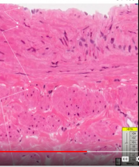
label ** |

|
|
|
structure veins ** |
thinner tunica media larger lumen |
|
|
lymphangitis ** |
inflammation lymph vessel |
|
|
lymphadenitis ** |
inflammation lymph node
|
|
|
lymphedema ** |
fluid in interstitial space not being drainaged, stays in lymph |
|
|
Parts neuron ** |
dendrite, soma, axon, termianl branch |
|
|
Types neurons ** |
multipolar: motor pseudounipolar: sensory |
|
|
Compare image elastic vs muscular arteries ** |

|
|
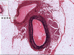
Describe ** |
Muscular arteries > tunica intima = internal elastic lamina (wavy in tunica intima) > tunica media = many layers circular arranged smooth muscle cells |