![]()
![]()
![]()
Use LEFT and RIGHT arrow keys to navigate between flashcards;
Use UP and DOWN arrow keys to flip the card;
H to show hint;
A reads text to speech;
19 Cards in this Set
- Front
- Back
- 3rd side (hint)
|
Plantar aponeurosis fascia Origin Pathway Insertion Action
|
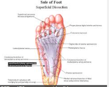
Plantar aponeurosis/fascia Origin •Medial process of tuberosity of the calcaneus
Pathway •Fans outwards from its origin as it moves distally
Insertion • Inserts into plantar aspect of digits 1 → 5
Action • Supports the longitudinal arch of the foot • Protects the plantar muscles and deep structures in the sole of the foot • Plays an important role in maintaining stability during propulsion • Common site of injury (heel pain)
|
|
|
|
First layer |
First layer Most superficial layer of the plantar muscles Lies deep to the plantar fascia Contains three muscles Abductor hallucis,
flexor digitorum brevis,
abductor digiti minimi(medial → lateral) |
|
|
|
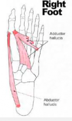
Abductor hallucis Origin Pathway Insertion
Action innervation |
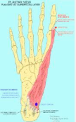
Abductor hallucis
Origin Medial process of the tuberosity of the calcaneus
Pathway
Runs along the plantar/medial aspect of the foot. Soft tissue bulge on the medial aspect of the foot
Insertion Medial/plantar aspect of base of 1st proximal phalanx Action
Abduction of the hallux. Flexion of the 1st metatarsal phalangeal joint innervation Medial plantar nerve (S1, S2, S3)
|
|
|
|
Flexor digitorum brevis Origin Pathway Insertion Action innervation |
Flexor digitorum brevis Origin
Medial process of the tuberosity of the calcaneus and plantar fascia Pathway
Runs between the plantar fascia and tendons of flexor digitorum longs Flat muscle belly splits into four tendons (digits 2 → 5)
Insertion At the base of each proximal phalanx, the tendons split into two, inserting into the plantar aspect of the middle phalanx Action Flexion of the proximal interphalangeal joints of digits 2 → 5 Innervation Medial plantar nerve (S1, S2, S3)
|
|
|
|
Abductor digiti minimi Origin Pathway Insertion Action innervation |
Abductor digiti minimi Origin Medial and lateral processes of tuberosity of the calcaneus Pathway Runs along the plantar surface of the foot over a groove on the base of the 5th metatarsal
Insertion Lateral aspect of the base of the 5th proximal phalanx Action Abduction of the 5th digit (at the 5th metatarsal phalangeal joint
innervation Lateral plantar nerve (S1, S2, S3) |
|
|
|
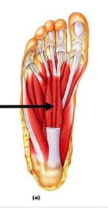
Second layer |
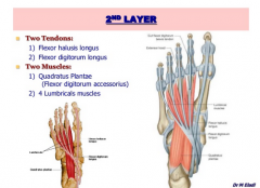
Second layer • Contains two muscles • Quadrates plantae • Two heads • Lumbricals (4) • 1st lumbrical → 4th lumbrical |
|
|
|
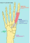
Quadrates plantae Origin Pathway Insertion Action innervation
|
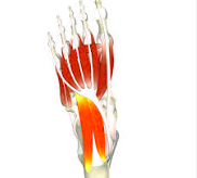
Quadrates plantae Origin • Medial surface of calcaneus, inferior to STT • Inferior surface of calcaneus, anterior to lateral tubercle
Pathway • Flat, quadrangular muscle belly projecting distally
Insertion • Lateral aspect of flexor digitorum longus (FDL) • Where the tendon of FDL divides Action • Assists FDL in the flexion of digits 2 → 5 • Consider the location of FDL as it enters the foot from the medial aspect of the ankle • Quadratus plantae alters the ‘line of pull’ by attaching to the lateralaspect of the tendon
Innervation • Lateral plantar nerve (S1, S2, S3)
Branches of the posterior tibial artery
|
|
|
|
Lumbricals Origin Pathway Insertion Action Innervation
|
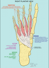
Lumbricals
Origin 1st lumbrical: medial aspect of FDL tendon (2nd digit) 2nd-4th lumbricals: medial aspect of adjacent FDL tendons
Pathway Run distally (medial to adjacent FDL tendons)
Insertion Medial aspect of the extensor hoods of digits 2 → 5 Inserts more dorsally than planetary
Action Flexion of metatarsalphalangeal joints (2 → 5) Extension of the interphalangeal joints (consider the slightly dorsal insertion)
Innervation 1st lumbrical: medial plantar nerve (S2, S3) 2nd-4th lumbricals: lateral plantar nerve (S2, S3)
|
|
|
|
Third layer |
Third layer • Contains three muscles
• Flexor hallucis brevis (FHB) • Medial and lateral heads • Sesamoid bones
• Adductor hallucis • Transverse and oblique heads
• Flexor digiti minimi |
|
|
|
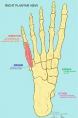
Flexor hallucis brevis Origin Pathway Insertion Action innervation
|
Flexor hallucis brevis
Origin Lateral head: plantar aspect of the cuboid, lateral cuneiform Medial head: tendon of tibialis posterior (as it enters the foot)
Pathway Medial and lateral heads join to form a muscle belly,containing a medial and lateral parts Insertion Medial and lateral, plantar surfaces of the base of the proximal phalanx Each tendon contains a sesamoid bone (medial and lateral)
Action Flexion of the 1st metatarsal phalangeal joint • Forms a groove between the medial and lateral heads, allowing flexor hallucis longs to pass through
Innervation • Medial plantar nerve (S1, S2) |
Two heads Each tendon contain |
|
|
Adductor hallucis Origin Pathway Insertion Action innervation
|
Adductor hallucis Origin Transverse head: plantar ligaments of digits (3 → 5) and adjacent transverse ligaments Oblique head: plantar aspect, bases of metatarsals (2 → 4)
Pathway Transverse head runs medially, oblique head runs anteriorly/medially Both join near the base of the proximal 1st phalanx Insertion Lateral aspect of base of proximal 1st phalanx, FHB Enclose lateral sesamoid Action Adduction of the 1st metatarsal phalangeal joint
Innervation • Lateral plantar nerve (S2, S3)
|
|
|
|
Flexor digiti minimi Origin Pathway Insertion Action innervation
|
Flexor digiti minimi Origin Base of the 5th metatarsal • Adjacent sheath of fibulas longus
Pathway Runs distally, parallel to 5th metatarsal
Insertion Lateral aspect of the base of the 5th proximal phalanx
Action • Flexion of the 5th metatarsal phalangeal joint
Innervation • Lateral plantar nerve (S2, S3)
|
|
|
|
Fourth layer |
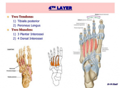
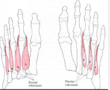
Fourth layer • Two muscles • Plantar interossei (3) • Dorsal interossei (4) |
|
|
|
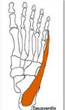
Plantar interossei Origin Pathway Insertion Action innervation
|
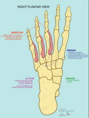
Plantar interossei Origin Plantar/medial aspect of the bases of metatarsals (3 → 5)
Pathway Run medial to the corresponding metatarsal shaft Insertion Plantar/medial aspect of the bases of the proximal phalanges (3 → 5)
Action • Adduction of digits 3 → 5 (metatarsal phalangeal joints)
Innervation • Lateral plantar nerve (S2, S3)
|
|
|
|
Dorsal interossei Origin Pathway Insertion Action innervation
|
Dorsal interossei
Origin • Bipinnate muscles, adjacent surfaces of the metatarsals Pathway Run between the adjacent metatarsal shafts
Insertion • 1st interossei: medial aspect 2nd proximal phalanx • 2nd-4th interossei: lateral aspect 2nd-4th proximal phalanx
Lateral aspect 1st metatarsal shaft and medial aspect 2nd metatarsalshaft → medial aspect 2nd proximal phalanx Lateral aspect 2nd metatarsal shaft and medial aspect 3rd metatarsal shaft → lateral aspect 2nd proximal phalanx Lateral aspect 3nd metatarsal shaft and medial aspect 4th metatarsal shaft → lateral aspect 3rd proximal phalanx Lateral aspect 4th metatarsal shaft and medial aspect 5th metatarsalshaft → lateral aspect 4th proximal phalanx
Action • Abduction of digits 2 → 4 (metatarsalphalangeal joints)
innervation • Lateral plantar nerve (S2, S3)*1st and 2nd dorsal interossei are innervated by the deep fibular nerve
|
|
|
|
Interossei |
Interossei • Resist extension of metatarsalphalangeal joints • Resist flexion of interphalangeal joints • Consider the extensor hoods |
|
|
|
Extensor hoods |
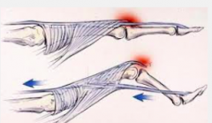
Extensor hoods • Triangular shaped covers for the extensor tendons of the foot • Apex, fans out over the middle/proximal phalanx, wraps around the metatarsal phalangeal joint attaching to the deep transverse metatarsal ligament • Site of attachment, leverage, prevention of excessive motion |
|
|
|
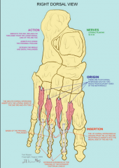
Dorsal layer |
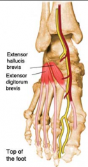
Dorsal layer • Extensor hallucis brevis (EHB) • Extensor digitorum brevis (EDB) • Only intrinsic muscles located in the dorm of the foot • Both share a common origin |
|
|
|
Extensor hallucis/digitorum brevis Origin Pathway Insertion Action innervation |
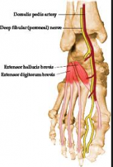
Extensor hallucis/digitorum brevis Origin •Superior lateral surface of the calcaneus •Lies lateral to sinus tarsi
Pathway •Muscle belly runs anteriorly/medially over the foot •Runs deep to extensor digitorum longs
Insertion • EDB Inserts into the lateral aspect of the tendons of extensor digitorum longs (as they insert into digits 2, 3 and 4) EHB to the base of the proximal phalanx of the great toe.
Action • Extension of the 1st-4th metatarsalphalangeal joints
Innervation • Deep fibular nerve (S1, S2)
|
|