![]()
![]()
![]()
Use LEFT and RIGHT arrow keys to navigate between flashcards;
Use UP and DOWN arrow keys to flip the card;
H to show hint;
A reads text to speech;
53 Cards in this Set
- Front
- Back
- 3rd side (hint)
|
The contraction of the ventricles is referred to as _______. |
systole
|
|
|
|
The period of ventricular relaxation is called the _____.
|
diastole.
|
|
|
|
The first heart sound is a result of closure of the ___ valves.
|
AV
|
|
|
|
The second heart sound is caused by closure of the ____ valves.
|
SL
|
|
|
|
The heart chambers that have just been filled when you hear the first heart sound are the the _____.
|
ventricles.
|
|
|
|
The pacemaker of the intrinsic conduction system is the ______.
|
SA node
|
|
|
|
The point in the intrinsic conduction system where the impulse is temporarily delayed. The atria contracts.
|
AV node
|
|
|
|
This node initiates depolarization.
|
SA node
|
|
|
|
This links the SA to the AV node.
|
internodal pathway
|
|
|
|
This is the link between atria and ventricles.
|
AV bundle or bundle of his.
|
|
|
|
This conveys impulse down the septum.
|
bundle branchees.
|
|
|
|
This conveys impulse throughout the ventricular walls.
|
Purkinje fibers
|
|
|
|
AV valve with 2 flaps.
|
mitral
|
|
|
|
AV valve with 3 flaps.
|
tricuspid
|
|
|
|
_____ and ____ prevent backflow into the ventricles when the heart is relaxed.
|
Pulmonary valve and aortic valve |
|
|
|
____ and ____ prevent backflow into the atria when the ventricles are contracting. |
mitral |
|
|
|
n |
n |
|
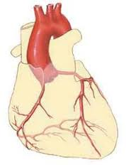
What vessels are these? |
n |
n |
|
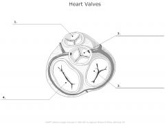
What valves are these? |
n |
n |
|
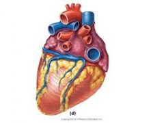
What vessels are these? |
n |
n |
|
|
SA Node |
In right atrium inferior to the entrance to the superior vena cava; provides the stimulus for contraction. Often referred to as pacemaker because it sets the rate of depolarization for the heart as a whole |
n |
|
|
AV node |
In the lower atrial septum at the junction of the atria and ventricles; After impulse arrives at this node atrial contraction happens; at this node the impulse is delayed (0.1 sec) to allow atria to complete contraction |
n |
|
|
AVBundle/bundle of his |
located in interventricular septum; conduction velocity increases to highest speed |
n |
|
|
Right and Left branches |
located in interventricular septum |
n |
|
|
Purkinje fibers |
Long strands of barrel-shaped cells called purkinje myocytes which ramify within muscle bundles of the ventricular walls; these fibers are much denser and more elaborate in the left ventricle because of the larger size of this chamber |
n |
|
|
ECG |
the graphic recording of electrical changes(depolarization followed by repolarization) occurring during the cardiac cycle, records voltage and time nothing else; |
n |
|
|
Deflection waves |
Record voltage and time of electrical events of large amounts of muscle tissue- might not allow us to see nodal issues Abnormalties of deflection waves and changes in time intervals of ECG are useful in detecting myocardial infarcts or problems with the conduction system of the heart |
n |
|
|
Tachycardia |
>100 bpm; prolonged may progress to fibrillation |
n |
|
|
Bradycardia |
Below 60 bpm
in athletes is positive finding because it indicates an increased efficiency of cardiac functioning. Because stroke volume (the amount of blood ejected by a ventricle with each contraction) increases with physical conditioning the heart can contract more slowly and still meet circulatory commands. |
n |
|
|
Fibrillation |
a condition of rapid uncoordinated heart contractions which makes the heart useless as a pump |
n |
|
|
Heart block-- Partial AV |
Seen on ECG: p-r interval longer than0.18 seconds may suggest this and damage to AV node |
n |
|
|
Total heart block |
no impulses are transmitted through the AV node; and the atria and ventricles beat independently of one another- the atria at the SA node rate and the ventricles at their intrinsic rate which is considerably slower. |
n |
|
|
Right and left bundle branch block |
on ECG it is a prolonged QRS complex (norm 0.08 sec); one ventricle is contracting later than the other |
n |
|
|
Standard Limb leads: indirect leads |
electrodes connected to each arm and to the left leg are considered to connect to the triangle vertices
these record the voltages generated in the extracellular fluids surrounding the heart by the ion flows occurring simultaneously in many cells between any two of the connections
Recording using lead I RA-LA is most sensitive to activity occurring horizontally across the heart
Lead II (RA-LL) and lead III (LA-LL) record activity along the vertical axis from the base of the heart to its apex but from different orientations |
|
|
|
electrodes |
electric conductor in which electric current enters or leaves |
|
|
|
Einthoven's law |
Einthovens triangle- the heart lies in the center of a triangle with sides of equal lengths
Recording connections are made at the vertices (corners) of that triangle
The sum of the voltages of leads I and III equals that in lead II-- Einthoven's law |
n |
|
|
Ground (RL) |
aka earth |
n |
|
|
define intrinsic |
it does not depend on impulses from the nervous system to initiate its contraction and will continue to contract rhythmically even if all nerve connections are severed |
|
|
|
functional syncytium |
n |
n |
|
|
2 types of controlling systems that exert effects on the heart activity |
1. Nerves of the ANS: accelerate or decelerate the heartbeat rate depending on which division is activated 2.Intrinsic conduction system/nodal system consisting of specialized noncontractile myocardial tissue; ensures that heart muscle depolarizes in an orderly and sequential manner(from atria to ventricles) and that the heart beats as a coordinated unit |
n |
|
|
Sequence of events in intrinsic conduction |
SA-AV-AV BUNDLE/BUNDLE OF HIS- RIGHT AND LEFT BUNDLE BRANCHES-PURKINJE FIBERS= VENTRICULAR CONTRACTION |
|
|
|
Depolarization |
the return of heart muscle cells to resting state following stimulation |
|
|
|
Damage to the AV node-bundle results in ... |
partial/total insulation of the ventricles from the influence of the SA node |
|
|
|
Autorythmic |
Found throughout the heart; rates of spontaneous depolarization differ |
|
|
|
p-R interval |
represents the time between the beginning of atrial depolarization and ventricular depolarization, including the period during which the depolarization wave passes to the AV node, atrial systole, and the passage of the excitation wave to the balance of the conducting system; generally 0.16-0.18 seconds-- A longer interval may suggest a partial AV heart block caused by damage to the AV node
start of p deflection to start of Q deflection
P-r segment: end of p wave to start of Q deflection |
|
|
|
QRS complex |
ventricular depolarization and atrial repolarization
duration normally 0.08 sec; if prolonged may indicate R or L bundle branch block in which one ventricle is contracting later than the other |
n |
|
|
S-T segment |
time of ventricular contraction
End of S deflection and start of T wave. |
n |
|
|
Q-T Interval |
Start of Q deflection to end of T wave
This is the period from the beginning of ventricular depolarization through the repolarization which includes the time of ventricular contraction (S-T segment)
HR 70 bpm- interval normally 0.31-0.41 sec; as rate increases this interval becomes shorter and vise versa |
n |
|
|
T wave |
ventricular repolarization |
n |
|
|
End T to next R |
n |
n |
|
|
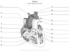
Fossa Ovalis/ foramen ovale |
n |
|
|
|
QRS interval |
the repolarization of the atria is generally obsecured by the large QRS complex |
|
|
|
P wave |
atrial depolarization |
|