Reading...
![]()
Play button
![]()
Play button
![]()
Use LEFT and RIGHT arrow keys to navigate between flashcards;
Use UP and DOWN arrow keys to flip the card;
H to show hint;
A reads text to speech;
21 Cards in this Set
- Front
- Back
- 3rd side (hint)
|
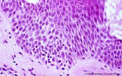
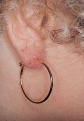
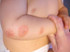
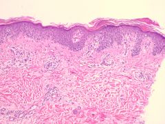
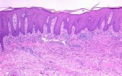
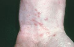
Allergic contact dermatitis
Note the extensive spongiosis, lymphocyte exocytosis, papillary dermal edema |
|
|
|
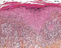
Allergic contact dermatitis
|
|
|
|
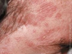
Seborrheic Dermatitis
Associated with? |
family history
Obesity, Parkinson's, HIV, malabsorption, Alcoholism Pitysporum ovale infection |
|
|
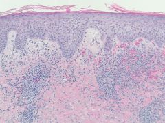
Seborrheic dermatitis
Describe: |
Spongiotic
with hyperkeratosis, psoriasiform hyperplasia, exocytosis, dermal perivascular lymphocytic infiltrate |
|
|
Nummular eczema
Histology shows: |
spongiotic dermatitis with parakeratosis, acanthosis, exocytosis, perivascular lymphocytic infiltrate
|
|
|
Lichen simplex chronicus
|
|
|
|
Lichen simplex chronicus
Note: irregular psoriasiform acanthosis, para/ortho keratosis, minimal spongiosis, dermal fibrosis |
|
|
|
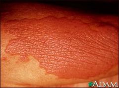
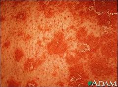
Pityriasis rosea
|
|
|
|
Pityriasis rosea
Note: subacute spongiotic change with mounds (instead of diffuse) parakeratosis. |
|
|
|
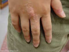
Lichen striatus
|
|
|
|
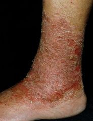
Stasis dermatitis
|
|
|
|
Stasis dermatitis
Note the spongiotic dermatitis picture with dermal changes including capillary proliferation, hemorrhage, hemosiderin, and fibrosis-mainly in upper dermis |
|
|
|
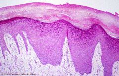
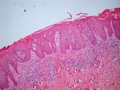
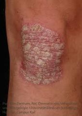
Psoriasis vulgaris
|
|
|
|
Psoriasis vulgaris
Note the epidermal parakeratosis, absence of granular layer, regular psoriasiform epidermal acanthosis with suprapapillary plate thinning, increased dermal vessels, sparse inflammation, comb-like appearance. |
|
|
|
Pityriasis rubra pilaris
|
|
|
|
Pityriasis rubra pilaris
Note the alternating checkerboard para/orthokeratosis, acrotrichial parakeratosis also present |
|
|
|
What are the two important features of lichenoid interface dermatitis?
|
Destruction of basal keratinocytes (cytoplasm becomes dyskeratotic-bright pink-and then nucleus extrudes)
Band-like lymphocytic infiltrate in papillary dermis |
|
|
|
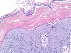
Lichen planus
|
|
|
|
Lichen planus
Note: acanthosis, lichenoid interface dermatitis, hyperkeratosis, sawtooth BM zone, Colloid bodies |
|
|
|
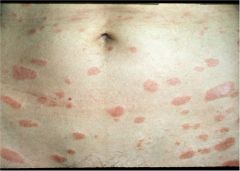
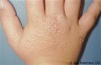
Lichen nitidus
flesh colored pin-point papules on trunk, abdomen, forearms, genitals |
|
|
|
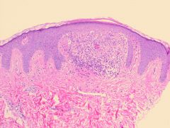
Lichen nitidus
Note clawlike configuration of epidermis surrounding a papillary dermal infiltrate. |
|