![]()
![]()
![]()
Use LEFT and RIGHT arrow keys to navigate between flashcards;
Use UP and DOWN arrow keys to flip the card;
H to show hint;
A reads text to speech;
240 Cards in this Set
- Front
- Back
- 3rd side (hint)
|
Functions of bone |
Support the body shape
System of levers for muscle action Protection of internal organs Site of blood formation Mineral storage pool |
5 |
|
|
Mechanical properties of bone |
Cable-like flexibility and resistance to tension (collagen) Pillar-like flexibility and resistance to compression (hydroxyapatite) |
2 |
|
|
Two main types of bone tissue |
Woven (immature) - more random, weaker, more flexible Lamellar (mature) - in normal skeleton |
|
|
|
Two main layers of lamellar bone |
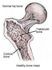
Cortical bone Cancellous bone |
|
|
|
Session 3 |
Session 3 |
Session 3 |
|
|
List the proximal row of carpal bones from lateral to medial |
Scaphoid Lunate Triquetrum Pisiform |
4 |
|
|
List the distal row of carpal bones from lateral to medial |
Trapezium Trapezoid Capitate Hamate |
4 |
|
|
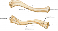
Describe the linear roughening on the posterior surface of the Humerus |
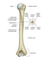
For the attachment of the lateral head of the triceps brachii Begins just inferior to the surgical neck - passes diagonally across the bone - deltoid tuberosity |
Function Anatomy |
|
|
Describe the anatomy of the radial groove |

Marks the margin between the middle posterior surface + adjacent part of the anterolateral surface Parallel to the posterior margin of the deltoid tuberosity |
2 |
|
|
What lies in the radial groove? |
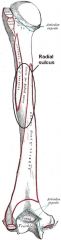
Radial nerve Profunda brachii artery |
2 |
|
|
Session 2 |
Session 2 |
Session 2 |
|
|
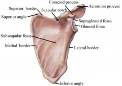
Describe the scapula |
Overlies ribs 2 - 7 Angles: Inferior, Superior, Lateral Border: Superior, Medial, Lateral Processes: Acromion, Coracoid Surfaces: Anterior, Posterior |
Ribs it overlies Angles Boreders Processes Surfaces |
|
|
Articulations of the scapula |
Gleno-humeral joint Acromio-clavicular joint Scapulo-thoracic joint |
3 |
|
|
Tubercles of the scapula |
Infraglenoid tubercle - site of attachment of the long head of the tricep Supraglenoid tubercle - site of attachment of the long head of the bicep |
2 |
|
|
Scapula: Superior border Acromion process Glenoid fossa Inferior angle Lateral border Supraglenoid fossa Coracoid process Subscapular fossa Scapular notch Superior angle Medial border |
|
|
|
|
Acromial end Shaft body Sternal end Acromial facet Subclavian groove Impression for costoclavicular ligament Sternal facet |
|
|
|
|
Joints of the clavicle |
Sterno-clavicular joint Acromio-clavicular joint |
|
|
|
How is the shaft of the clavicle divided |
Medial 2/3 Lateral 1/3 |
|
|
|
Bodies on the inferior surface of the lateral 1/3rd of the clavicle |
Conoid tubercle - attachment for coracoclavicular ligament Trapezoid line - attachment for trapezoid ligament |
2 |
|
|
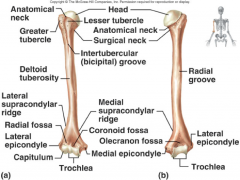
Articulations of the head of the humerus |
Glenoid cavity of scapula |
|
|
|
Articulations of the capitulum and trochlea |

Radius and Ulna respectively |
|
|
|
Four nerves the humerus is in contact with |
Axillary nerve Radial nerve Ulnar nerve Median nerve |
|
|
|
Axillary nerve |

Winds around the surgical neck of the humerus Runs with posterior circumflex humeral artery Can be damaged in shoulder dislocation Test for damage by asking patient to abduct injured shoulder against resistance |
Where does it run What does it run with How can it be damaged How do you test for damage |
|
|
Radial nerve |

Runs in the radial groove Runs with profunda brachii artery Damaged in humeral shaft fractures |
Where does it run What does it run with How can it be damaged |
|
|
Ulnar nerve |

Runs posterior to the medial epicondyle and is very superficial Damaged in fractures and dislocations around the elbow joint |
Where does it run How can it be damged |
|
|
Median nerve |

Runs anterior to the distal humerus Damaged in fractures and dislocations around the elbow joint |
Where does it run How can it be damaged |
|
|
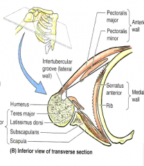
Attachments of the bicipital (intertubercular) groove |
Pectoralis major - lateral lip of the groove Teres Major - medial lip of the groove Latissimus dorsi - floor of the groove |
3 |
|
|
Most common site of fracture on the humerus |
Surgical neck |
|
|
|
Four joints of the pectoral girdle |
Summation of: Sterno-clavicular joint (SCJ) Acromio-clavicular joint (ACJ) Scapulo-thoracic joint Gleno-humeral joint If one is damaged the whole unit is impaired |
|
|
|
Components of a synovial joint |
Cartilage - capsule (synovial membrane + fibrous membrane) - synovial fluid |
3 |
|
|
Ligaments of the acromio-clavicular joint |
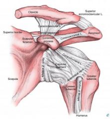
Acromio-clavicular ligament Coraco-clavicular ligament (conoid + trapezoid) Coraco-acromial ligament |
3 |
|
|
How is the acromio-clavicular joint commonly injured |
Falls onto outstretched hands |
|
|
|
What injuries predispose to minor and major dislocations |

Minor dislocation: acromio-clavicular ligament alone is torn Major dislocations: coraco-clavicular ligament alone is torn |
|
|
|
Which structures help provide stability for the gleno-humeral joint |
Tendons of the rotator cuff muscles Long head of biceps Ligaments Capsule Coraco-acromial ligament Glenoid labrum |
6 |
|
|
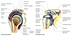
Two extensions of the shoulder joint capsule |

Subacromial bursa - capsule extends above the humeral head to form a bursa between it and the overlying acromion process (site of shoulder impingement) Extenstion around the long head of biceps and lies in the bicipital groove |
|
|
|
Ligaments that blend with the shoulder joint capsule |
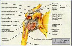
Gleno-humeral ligament - strengthens the anterior portion Coraco-humeral ligament - strengthens superiorly Transverse humeral ligament |
|
|
|
Function of the transverse humeral ligament |
Holds the tendon of the long head of biceps in the inter-tubercular (bicipital) groove |
|
|
|
Muscles involved in flexion of the shoulder joint |
Clavicular head of pectoralis major Anterior fibres of deltoid Coraco-brachialis Biceps brachii |
4 |
|
|
Muscles involved in extension of the shoulder joint |
Latissimus dorsi Posterior fibres of the deltoid |
2
|
|
|
Muscles involved in abduction of the shoulder joint |
Supraspinatus (first 15 degrees) Central fibres of deltoid (after 15 degrees) |
2 |
|
|
Muscles involved in adduction of the shoulder joint |
Pectoralis major Latissimus dorsi |
2 |
|
|
Muscles involved in internal rotation of the shoulder joint |
Subscapularis |
1 |
|
|
Muscles involved in external rotation of the shoulder joint |
Infraspinatus |
1 |
|
|
Other muscles involved in resisting dislocation of the shoulder joint |
Rotator cuff - holds and depresses humeral head in glenoid Deltoid Coraco-brachialis Short head of biceps Long head of biceps |
5 |
|
|
Scapulo-thoracic joint |
Theoretical concept that represents the articulation between the scapula and chest wall |
Definition |
|
|
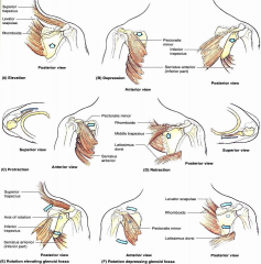
Muscles involved in elevation of the scapula |
Superior trapezius Levator scapulae Rhomboids |
3 |
|
|
Muscles involved in depression of the scaupla |
Inferior trapezius Pecoralis minor Serratus anterior |
3 |
|
|
Muscles involved in protraction of the scapula |
Pectoralis minor Serratus anterior |
2 |
|
|
Muscles involved in retraction of the scapula |
Rhomboids Middle trapezius Latissimus dorsi |
3 |
|
|
Muscles involved in upwards rotation of the scapula |
Superior trapezius Inferior trapezius Serratus anterior |
3 |
|
|
Muscles involved in downwards rotation of the scapula |
Pectoralis minor Latissimus dorsi Rhomboids Levator scapulae |
4 |
|
|
Muscles involved in flexion at the elbow joint |
Biceps Brachiails Brachioradialis (pronator teres) |
3 |
|
|
Muscles involved in extension at the elbow joint |
Triceps (anconeus) |
1 |
|
|
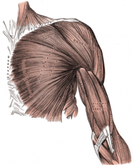
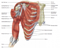
Pectoralis major |
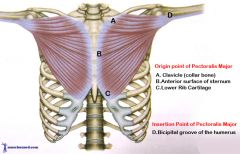
Origin: Clavicle, sternum, upper six costal cartialges Insertion: Lateral lip of intertubercular sulcus of humerus Action: adducts arm, rotates arm medially, flexion of humerus Innervation: Medial and lateral pectoral nerves |
Origin (3) Insertion (1) Action (3) Innervation (2) |
|
|
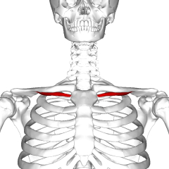
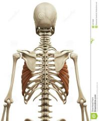
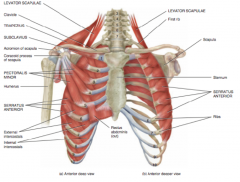
Pectoralis minor |
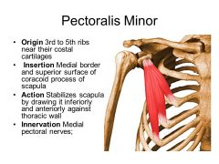
Origin: Ribs 3, 4 and 5 Insertion: Coracoid process of scapula Action: Depression and protraction of scapula Innervation: medial pectoral nerve |
Origin (1) Insertion (1) Action (2) Innervation (1) |
|
|
Subclavius |
Origin: First costal cartilage Insertion: Inferior border of clavicle Action: Depresses and stabilises clavicle Innervation: Nerve to subclavius |
Origin (1) Insertion (1) Action (1) Innervation (1) |
|
|
Serratus anterior |
Origin: Upper eight ribs Insertion: Medial border and inferior angle of scapula Action: Pulls scapula forwards and rotates it Innervation: Long thoracic nerve (C5,C6,C7) |
Origin (1) Insertion (2) Action (1) Innervation (1) |
|
|
Anterior pectoral muscles |
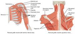
Pectoralis major Pectoralis minor Subclavius Serratus anteriro |
4 |
|
|
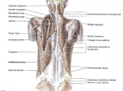
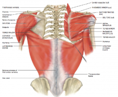
Posterior pectoral muscles |

Trapezius Latissimus dorsi Levator scapulae Rhomboids |
4 |
|
|
Trapezius |
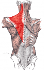
Origins: Occipital bone, ligamentum nuchae, spinous processes of thoracic vertebrae Insertion: Lateral third of clavicle, anterior of acromion, spine of scapula Action: Elevates scapula, pulls scapula medially and pulls medial border of scapula downwards Innervation: Spinal part of accessory nerve and C2 and C3 |
Origin (3) Insertion (3) Action (3) Innervation (3) |
|
|
Latissimus dorsi |
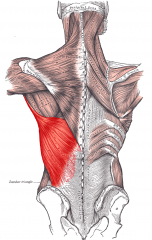
Origin: Iliac crest, lumbar fascia, spinal processes of lower six thoracic vertebrae, lower ribs, scapula Insertion: floor of intertubercular sulcus of humerus Action: Extends, adducts and medially rotates arm (palpated in posterior axillary fold Innervation: Thoracodorsal nerve (C6,C7,C8) |
Origin (4) Insertion (1) Action (3) Innervation (1) |
|
|
Origin of the Levator scapulae |
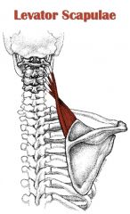
Transverse processes of C1 - C4 |
1 |
|
|
Insertion of levator scapulae |
Medial border of scapula |
1 |
|
|
Action of levator scapulae |
Elevates scapula |
1 |
|
|
Innervation of levator scapulae |
C3 C4 Dorsal scapular nerve |
3 |
|
|
Origin of the rhomboids |

Major: Spines of T2-T5 Minor: Ligamentum nuchae Spines of C7 + T1 |
Major (1) Minor (2) |
|
|
Insertion of the rhomboids |
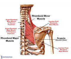
Medial border of scapula |
1 |
|
|
Action of the rhomboids |

Elevates and retracts the medial border of scapula |
2 |
|
|
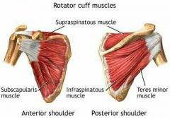
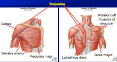
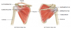
Intrinsic shoulder muscles |
Deltoid Teres major (rotator cuffs): Supraspinatous Infraspinatus Teres minor Subscapularis |
6 |
|
|
Origin of deltoid |
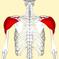
Posterior of clavicle Acromion Inferior spine of scapula |
3 |
|
|
Insertion of deltoid |
Deltoid tuberosity of humerus |
1 |
|
|
Action of deltoid |
Abducts Flexes Medially rotates Extends Laterally rotates the arm |
5 |
|
|
Innervation of deltoid |
Axillary nerve (C5,C6) |
1 |
|
|
Origin of teres major |
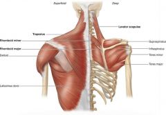
Lateral side of inferior angle of scapula |
1 |
|
|
Insertion of teres major |
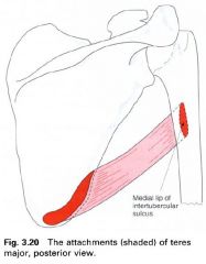
Medial lip of intertubercular sulcus of humerus |
1 |
|
|
Action of teres major |
Medially rotates Adducts the arm
|
2 |
|
|
How is teres major palpated |
Palpated in lower axillary fold |
1 |
|
|
Innervation of teres major |
Lower subscapular nerve (C5,C6) |
1 |
|
|
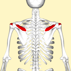
Origin of supraspinatus |

Supraspinous fossa of scapula |
1 |
|
|
Insertion of supraspinatus |
Greater tubercle of humerus Capsule of shoulder joint |
2 |
|
|
Action of supraspinatus |
Abduction of arm for the first 15 degrees |
1
|
|
|
Innervation of supraspinatus |
Suprascapular nerve |
1 |
|
|
Origin of infraspinatus |
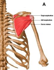
Infraspinous fossa of scaupla |
1 |
|
|
Insertion of infraspinatus |

Greaters tubercle of humerus Capsule of shoulder joint |
2 |
|
|
Action of infraspinatus |

Laterally rotates arm |
1 |
|
|
Innervation of infraspinatus |
Suprascapular nerve |
1 |
|
|
Origin of teres minor |

Lateral border of scapula |
1 |
|
|
Insertion of teres minor |

Greater tubercle of humerus Capsule of shoulder joint |
2 |
|
|
Action of teres minor |
Laterally rotates arm |
1 |
|
|
Innervation of teres minor |
Axillary nerve |
1 |
|
|
Origin of subscapularis |
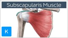
Subscapular fossa |
1 |
|
|
Insertion of subscapularis |

Lesser tubercle of humerus |
1 |
|
|
Action of subscapularis |
Medially rotates arm |
1 |
|
|
Innervation of subscapularis |
Upper and lower subscapular nerves |
2 |
|
|
Muscles of the anterior compartment of the upper arm |
Biceps brachii Brachialis Coraco-brachialis |
3 |
|
|
Origin of biceps brachii |
Long head: Supraglenoid tubercle of scapula Short head: Coracoid process of capula |
2 |
|
|
Insertion of biceps brachii |
Radial tuberosity Deep fascia of forearm via bicipital aponeurosis |
2 |
|
|
Bicipital aponeurosis |
Broad aponeurosis of biceps brachii located in the cubital fossa Seperates most superficial from deep structures and protects brachial artery and median nerve |
3 |
|
|
Action of biceps brachii |
Supinator (return to anatomical position) of flexed forearm Flexor of elbow joint Weak flexor of shoulder joint |
3 |
|
|
Innervation of biceps brachii |
Musculocutaneous nerve |
1 |
|
|
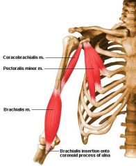
Origin of brachialis |
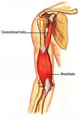
Anterior surface of humerus |
1 |
|
|
Insertion of brachialis |

Ulnar tuberosity Ulnar coronoid process |
2 |
|
|
Action of brachialis |

Flexes elbow joint |
1 |
|
|
Innervation of brachialis |
Musculocutaneous nerve |
1 |
|
|
Origin of coracho-brachialis |

Coracoid process of scapula |
1 |
|
|
Insertion of coraco-brachialis |

Shaft of humerus |
1 |
|
|
Action of coraco-brachialis |
Flexes and adducts shoulder joint |
1 |
|
|
Innervation of coraco-brachialis |
Musculocutaneous nerve |
1 |
|
|
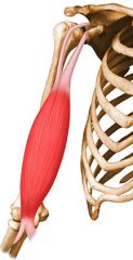
Muscles of the posterior compartment of the uppper arm |
Triceps brachii Anconeus |
2 |
|
|
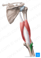
Origin of triceps brachii |
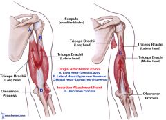
Long head: Infraglenoid tubercle of scapula Medial head: Posterior surface of humerus (lower part) Lateral head: Posterior surface of humerus (Upper part/linear roughening) |
3 |
|
|
Insertion of triceps brachii |

Olecranon process |
1 |
|
|
Action of triceps brachii |

Extends elbow joint |
1 |
|
|
Innervation of triceps brachii |
Radial nerve |
1 |
|
|
Origin of anconeus |

Lateral epicondyle of humerus (proximal end) |
1 |
|
|
Olecranon |
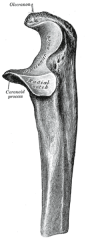
Curve bony eminence of the forearm that projects behind the elbow joint (opposite of cubital fossa) |
1 |
|
|
Insertion of anconeus |
Lateral surface of olecranon |
1 |
|
|
Action of anconeus |

Stabalise elbow during pronation and supination |
1 |
|
|
Innervation of anconeus |
Radial nerve |
1 |
|
|
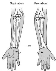
Define pronation and supination |
|
Or demonstrate them
|
|
|
|
|
|
|
|
|
|
|
|
|
|
|
|
|
|
|
|
|
|
|
|
|
|

|
|
|
|
|
Shape of the Axilla + wallls |

Square based pyramid with: Base Apex Anterior wall Posterior wall Medial wall Lateral wall |
7 |
|
|
Contents of the base of axilla |
Skin Subcutaneous tissue Fascia extending from arm to chest |
3 |
|
|
Contents of the apex of axilla |
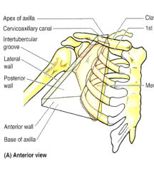
Between: First rib Clavicle Superior border of subscapularis |
3 |
|
|
Contents of the anterior wall of axilla |

Pectoralis major Pectoralis minor |
2 |
|
|
Contents of the posterior wall of axilla |
Scapula Subscapularis (superiorly) Teres minor (inferiorly) Latissimus dorsi (inferiorly) |
4 |
|
|
Contents of medial wall of axilla |

Chest wall (ribs 1-4) Serratus anterior |
2 |
|
|
Contents of lateral wall of axilla |

Humerus |
1 |
|
|
Contents of axilla |
Axillary artery Axillary vein Lymphatics Brachial plexus |
4 |
|
|
When does the subclavian artery become the axillary artery |
When it passes over the first rib |
|
|
|
When does the axillary artery become the brachial artery |
When it passes the inferior border of teres major |
|
|
|
When does the axillary artery divide |
When it passes pectoralis minor (3 parts) |
|
|
|
When and into what does the brachial artery divide |
Divides at the elbow into:
Radial artery Ulnar artery |
3 |
|
|
Which nerve does the brachial artery run alongside in the distal part of the arm |
Median nerve |
|
|
|
Biggest muscular branch of the brachial artery and with which nerve does it run |
Profunda brachii artery Runs with the radial nerve |
2 |
|

|
|
|
|
|
Describe the anastomosis between the subclavian and axillary artery |
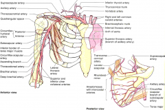
Forms a network around the scapula Between the: Subclavian - deep branch of transverse cervical + suprascapular and Axillary - subscapular + circumflex scapular |
Location Arteries involved (6) |
|
|
Why are the anastomosis between the axillary and subclavian artery useful |
Provide collateral circulation and prevent ischemia When the first part of the subclavian and thrid part of the axillary are obstructed |
2 |
|
|
How is the axillary artery divided into three parts
|
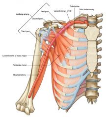
Divided by pectoralis minor |
|
|
|
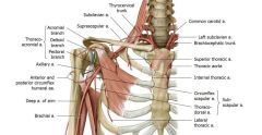
6 branches of the axillary artery |
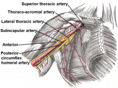
Superior thoracic artery Thoraco-acromial artery Lateral thoracic artery Subscapular artery Anterior humeral circumflex artery Posterior humeral circumflex artery |
Some Times Life Seems A Pain |
|
|
Venae comitantes |
A pair (or more) of veins that closely accompany an artery such that pulsations of the artery aid venous return |
|
|
|
How is the axillary vein formed |

As the basilic vein (superficial) passes through deep fascia to join the venae comitantes of the brachial artery at the lower border of teres major |
Preceding vein Path To join what At what level |
|
|
Where does the axillary vein end and what does it become |
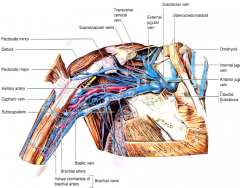
First rib Where it become the subclavian vein |
|
|
|
Tributaries of the axillary vein and their clinical importance |

Large number of tributaries Including cephalic vein And some abdominal veins (clinically important if abdominal veins are occluded by a tumour) |
|
|
|
How does the Cephalic vein join the axillary vein |
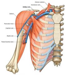
Is superficial, passes on lateral aspect of arm Passes deep into the delto-pectoral triangle to join axillary vein |
Path along arm Where it starts to run deep |
|
|
Describe lymph drainage from the hands |
Superficial lymphatics which run alongside the cephalic and basilic veins |
Where they run (1) Next to what (2) |
|
|
Location of three lymph nodes in the arm and shoulder |
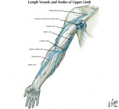
Cubital Axillary Delto-pectoral |
C A D |
|
|
Where does lymph drainage of that arm ultimately go |
Axillary
|
|
|
|
Clinical significance of Axillary lymph nodes |
Breast carcinomas as lymphatic drainage of the breast includes axillary lymph nodes |
Cancer |
|
|
Five groups of axillary lymph nodes |
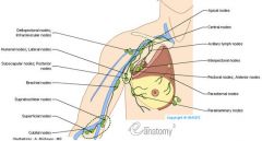
Apical (all drain via this - subclavian lymphatic trunk - right lymphatic duct/thoracic duct) Pectoral Subscapular Humeral Central |
A P S H C |
|
|
2 nerves in close relation to the axillary lymph node region and their clinical significance |
Long thoracic nerve - serratus anterior (section = winging of scapula) Thoraco-dorsa nerve - Latissimus dorsi May be cut when axillary lymph nodes are being sampled in patients with breast carcinomas |
Nerve and muscle supplied Nerve and muscle supplied Clinical significance |
|
|
What is the brachial plexus formed from |

Anterior primary rami of C5 - T1 spinal nerve |
|
|
|
Where and from what are brachial plexus roots formed |

In the neck From spinal nerve roots |
|
|
|
Where and from what are brachial plexus trunks formed |
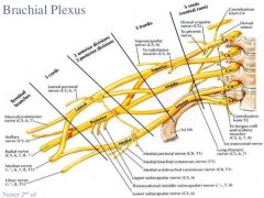
In the inferior portion of the neck From the roots |
|
|
|
Where and from what are brachial plexus divisions formed |

Behind the clavicle From the trunks |
|
|
|
Where and from what are brachial plexus cords formed |

In close proximity to the axillary artery From the divisions in the axilla |
|
|
|
Important nerves of the brachial plexus |
Long thoracic nerve Suprascapular nerve Lateral pectoral nerve Axillary nerve Musculo-cutaneous nerve Ulnar nerve Median nerve Radial Nerve |
9 |
|
|
What does the long thoracic nerve supply |
Serratus naterior |
1 |
|
|
What does the suprascapular nerve supply |
Supraspinatus Infraspinatus |
2 |
|
|
What does the lateral pectoral nerve supply |
Pectoralis major |
1 |
|
|
What does the thoraco-dorsal nerve supply |
Latissimus dorsi |
1 |
|
|
What does the axillary nerve supply |
(C5) Teres minorDeltoid Dermatome over deltoid |
3 |
|
|
Musculocutaneous nerve of the brachial plexus |

(C5-C7) Supplies anterior compartment of the upper arm Continues as the lateral cutaneous nerve in the forearm Lies close to the subscapularis tendon Can be damaged in surgery |
Spinal roots What does it supply What does it continue as in the forearm Which tendon does it lie close to How can it be damaged |
|
|
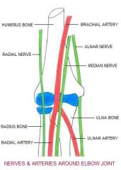
Ulnar nerve of the brachial plexus |

(C8, T1) Anterior compartment of forearm but mainly a nerve of the hand Passes through the anterior compartment of upper arm initially but then posterior compartment more distally Lies alongside brachial artery at the elbow Can be damaged in elbow fractures/dislocations |
Spinal roots What does it supply Path in the upper arm What does it lie alongside at the elbow How can it be damaged |
|
|
Median nerve of the brachial plexus
|
(C6-T1) Anterior compartment of the forearm and hand Passes through the anterior compartment of upper arm Lies alongside the brachial artery at the elbow Can be damaged in elbow fractures and dislocations |
Spinal roots What does it supply Path in the upper arm What does it lie alongside at the elbow How can it be damaged |
|
|
Radial nerve of the brachial plexus |
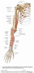
(C5-T1) Posterior compartment of the upper arm and forearm Lies in the radial groove of the humerus Can be damaged there Divides into the Superficial radial nerve (sensory) and Posterior inerosseus nerve (motor) Just above the elbow |
Spinal roots What does it supply Where does it lie in the upper arm Where can it be damaged What does it divide into At what level does it divide |
|
|
Significance of a synovial ball and socket joint |
Allows for movement in many planes |
1 |
|
|
Describe an acromio-clavicular dislocation |
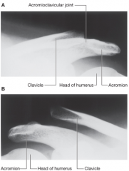
Dislocation of joint after trauma Lateral third of clavicle is joined to the scapula by the Coracoclavicular ligament (Conoid + Trapezoid ligaments) Minor injury: tear the fibrous joint capsule and ligaments of the acromioclavicular joint Major injury: Disrupt the conoid and trapezoid ligaments - elevation and upward subluxation of clavicle |
Ligaments of the coracoclavicular ligament Describe a minor injury Describe a major injury |
|
|
Describe a gleno-humeral joint dislocation |
Most frequent is anterior dislocation Anterior inferior glenoid labrum is torn Once the joint capsule is disrupted - more susceptible to recurrent dislocations Axillary nerve may be injured by direct compression by humeral head in anterior and inferior dislocation Radial nerve may be damaged by "lengthening" of the humerus since it is tightly bound in radial groove - radial nerve paralysis Posterior dislocation is extremely rare - focus on cause - vigorous muscle contraction - epileptic seizure - from electrocution |
Most frequent type of dislocation What is torn in this location More susceptible to what after dislocation How can the axillary nerve be damaged How can the radial nerve be damaged Cause of posterior dislocation |
|
|
Fibrocartilaginous glenoid labrum |
Fibrocartilaginous rim around the glenoid fossa that acts to deepen the socket Joint is still susceptible to dislocation even with fibrocatilaginous glenoid labrum and ligamentous support |
1 |
|
|
Describe rotator cuff impingement with regards to the rotator cuff tendon |
Rotator cuff tendon is trapped in the subacromial space Tendon is repeatedly scraped against shoulder blade - fraying - weakening and susceptibility to tear + tendinopathy Most commonly involves supraspinatus as it runs beneath acromion and acromioclavicular joint |
Definition Complications (5) Muscle most commonly involved |
|
|
Describe rotator cuff impingement with regards to the supraspinatus ligament |
Space beneath which supraspinatus ligament passes if of fixed dimensions Swelling of supraspinatus Excessive fluid in subacromial/subdeltoid cavity Subacromial bony spurs Leads to impingement in abduction This can cause degenerative change - calcium deposition - pain |
Describe the space beneath which the supraspinatus ligament passes Causes of impingement of the supraspinatus ligament When does this impingement occur What can this lead to |
|
|
Define Neer's impingement test |
Patient straigtens arm Doctor raises arm forward keeping palm away from body Pain = positive for rotator cuff impingement |
2 actions 1 result |
|
|
Describe the painful arc test to diagnose tendonitis |
Start with arm by side Lift arm outwards from side in an arc Pain usually felt at a maximum between 70-120 degrees in cuff tendonitis |
2 actions 1 result |
|
|
Anatomical basis of a frozen shoulder |
Adhesive capsulitis Shoulder capsule surrounding glenohumeral joint becomes inflamed and stiff Restricts motion + chronic pain |
|
|
|
Causes of winged shoulder blade |
Serratus anterior paralysis due to damage to long thoracic nerve Trapezius palsy involving the accessory nerve Rhomboid palsy involving the dorsal scapular nerve |
3 |
|
|
Cortical bone |
Outer hard layer of compact lamellar bone (arranged in osteon subunits) Makes 80% of the skeleton Slow turnover rate high resistance to torsion and bending |
Definiton How much of the skeleton does it make Turnover rate Resistance to which types of forces |
|
|
Cancellous bone |
Spongy/trabecular Inner layer of interlacing struts of lamellar bone Less dense Higher turnover rate Consists of spicule with marrow between |
Other names Definition Density Turnover rate What does it consist of |
|
|
Muscles of the anterior compartment of the forearm |
Flexor carpi ulnaris Flexor carpi radialis Pronator teres Palmaris longus Flexor digitorum superficialis Pronator quadratus Flexor digitorum profundus Flexor pollicis longus |
F F P P F P F F |
|
|
Muscles of the posterior compartment of the forearm |
Brachioradialis Extensor carpi radialis longus Extensor carpi radialis brevis Extensor digitorum Extensor digiti minimi Extensor carpi ulnaris Anconeus Supinator Abductor pollicis longus Extensor pollicis longus Extensor pollicis brevis Extensor indicis |
B E E E E E A S A E E E |
|
|
Muscles of the hand |
Lumbricals Opponens pollicis Abductor pollicis Flexor pollicis brevis Opponens digiti minimi Abductor digiti minimi Flexor digiti minimi Palmaris brevis Adductor pollicis Dorsal interosseous Palmar interosseous |
L O A F O A F P |
|
|
Superficial muscles of the gluteal region |
Tensor Fascia Lata Gluteus maximus Gluteus medius Gluteus minimus |
T G G G |
|
|
Deep muscles of the gluteal region |
Piriformis (S1+S2) Gemellus superior (L5+S1) Obturator internus (L5+S1) Gemellus inferior (L5+S1) Quadratus femoris (L5+S1) |
P G O G Q |
|
|
Muscles of the anterior compartment of the thigh |
(Femoral nerve) Iliopsoas (Iliacus + Psoas major) Quadriceps femoris (Rectus femoris + Vastus lateralis + Vastus medialis + Vastus intermedius Sartorius |
I P R V V V S |
|
|
Muscles of the medial compartment of the thigh |
Gracilis Pectineus Adductor longus Adductor brevis Adductor magnus Obturator externus |
G P A A A O |
|
|
Muscles of the posterior compartment of the thigh |
Biceps femoris Semitendinosous Semimembranosous |
B S S |
|
|
Superficial muscles of the posterior compartment of the leg |
Gastrocnemius Plantaris Soleus |
G P S |
|
|
Deep muscles of the posterior compartment of the leg |
Popliteus Flexor hallucis longus Tibialis posterior Flexor digitorum longus |
P F T F |
|
|
Tensor fascia lata |
Iliac crest - Iliotibial tract Stabilise knee in extension Stabilise femur in acetabulum Superior gluteal nerve |
Origin + insertion Functions (2) Innervation |
|
|
Gluteus maximus |
Iliac crest + Coccyx - posterior iliotibial tract + gluteal tuberosity of femur Extends thigh Inferior gluteal nerve |
Origin + insertion Function (1) Innervation |
|
|
Gluteus medius |
Side of iliac crest - lateral greater trochanter Abduction of thigh SGN |
Origin + insertion Function (1) Innervation |
|
|
Gluteus minimus |
Lateral ilium - lateral greater trochanter Abduction SGN |
Origin + insertion Function (1) Innervation |
|
|
Piriformis |
Anterolateral surface of sacrum (between foramen) - lateral greater trochanter Lateral rotation of thigh Nerve to piriformis (S1 + S2) |
Origin + Insertion Function Innervation |
|
|
Gemellus superior |
Ischial spine - tendon of obturator internus - greater trochanter Lateral rotation of thigh Nerve to obturator internus (L5 + S1) |
Origin + Insertion Function Innervation |
|
|
Obturator internus |
Medial side of obturator membrane - greater trochanter Lateral rotation of thigh Nerve to obturator internus (L5 + S1) |
Origin + Insertion Function Innervation |
|
|
Gemellus Inferior |
Upper ischial tuberosity - tendon of the obturator internus - greater trochanter Lateral rotation of thigh Nerve to quadratus femoris (L5 + S1) |
Origin + Insertion Function Innervation |
|
|
Quadratus femoris |
Lateral ischium - intertrochanteric crest + quadrate tubercle + quadrate line Lateral rotation of thigh Nerve to quadratus femoris (L5 + S1) |
Origin + Insertion Function Innervation |
|
|
Path of sciatic nerve in gluteal region |
Through the greater sciatic foramen, below the piriformis |
|
|
|
Path of the superior gluteal nerve in the gluteal region |
Through the greater sciatic foramen, above the piriformis |
|
|
|
Iliopsoas |
Iliacus Psoas |
What is it comprised of |
|
|
Iliacus |
Iliac fossa - lesser trochanter Flexion of thigh |
Origin + Insertion Function |
|
|
Psoas Major |
Transverse processes of T12 - L5 - Lesser trochanter Flex thigh |
Origin + Insertion Function |
|
|
Quadriceps femoris |
Rectus femoris Vastus lateralis Vastus medius Vastus medialis |
What is it comprised of R V V V |
|
|
Rectus Femoris |
AIIS (straight head) + Superior acetabulum fossa (reflected head) - Quadriceps femoris tendon - patella Flexion of thigh Femoral nerve |
Origin + Insertion Function Innervation |
|
|
Vastus lateralis |
Femur - Quadriceps femoris - patella Extension of leg |
Origin + Insertion Function |
|
|
Vastus medius |
Femur - quadriceps femoris tendon - patella Extension |
Origin + Insertion Function |
|
|
Vastus medialis |
Femur - quadriceps femoris tendon - patella Extension |
Origin + Insertion Function |
|
|
Sartorius |
ASIS - Medial proximal tibia Flexion at hip and knee joint |
Origin + Insertion Function |
|
|
Gracilis |
Medial body of pubis - medial proximal tibia Adduction + flexion at knee joint Obturator nerve |
Origin + Insertion Function Innervation |
|
|
Pectineus |
Pectineal line - medial aspect of femur Adduction + flexion of thigh Femoral nerve |
Origin + Insertion Function Innervation |
|
|
Adductor longus |
Pubis - middle third of femur Adduction Obturator nerve |
Origin + Insertion Function Innervation |
|
|
Adductor brevis |
Pubis - upper third of femur Adduction Obturator nerve |
Origin + Insertion Function Innervation |
|
|
Adductor magnus |
Hamstring part: Ischial tuberosity - adductor tubercle on medial condyle (tibial nerve) Extend hip Adductor part: Ischiopubic ramus - lower gluteal line + linea aspera (obturator nerve) Adduction + Medial rotation (forms adductor hiatus for femoral artery and vein) |
Origin + Insertion (2) Functions (2) Innervation (2) |
|
|
Obturator externus |
Lateral obturator membrane - trochanteric fossa Lateral rotation Obturator nerve |
Origin + Insertion Function Innervation |
|
|
Muscles of the lateral compartment of the leg |
Peroneus/Fibularis longus Peroneus/Fibularis brevis |
P P |
|
|
Muscles of the anterior compartment of the leg |
Tibialis anterior Extensor hallucis longus Extensor digitorum longus Peroneus/Fibularis tertius |
T E E P |
|
|
Foot |
|
|
|
|
Fascia and retinacula of the foot |
Plantar aponeurosis Superficial transverse metatarsal ligaments Deep transverse metatarsal ligaments Superior extensor retinaculum Inferior extensor retinaculum Flexor retinaculum Superior fibular retinaculum Inferior fibular retinaculum |
P S D S I F S I |
|
|
Contents of the tarsal tunnel from medial to lateral |
Extensor digitorum longus Tibialis posterior Posterior tibial vein Posterior tibial artery Tibial nerve Flexor hallucis longus |
E T P P T F |
|
|
Tarsal bones |
Talus Calcaneus Navicular Cuneiform (3) Cuboid (2) |
T C N C C |
|
|
Describe the locking and unlocking of the knee |
Femur is medially rotated on the tibia at full extension - locking it in place Femur is laterally rotated by the popliteus muscle to unlock it |
2 |
|
|
What are the femoral condyles separated by? |
intercondylar fossa |
1 |
|
|
Function of the femoral intercondylar fossa |
Attachment for: Anterior cruciate ligament Posterior cruciate ligament |
2 |
|
|
Function of the tibial intercondylar region |
Attachment for: Cartilage of menisci/semilunar cartilage Anterior cruciate ligament Posterior cruciate ligament |
3 |
|
|
Define the tibial plateau |
Superior surface of the tibia
|
1 |
|
|
Function of the transverse ligament of the knee |
Connect the lateral and medial semilunar cartilage |
1 |
|
|
Ligaments of the knee |
Lateral collateral Medial collateral Anterior cruciate Posterior cruciate Patella |
L M A P P |
|
|
2 components of the knee capsule |
Fibrous membrane Synovial membrane |
F S |
|
|
Attachment site of the sartorius, gricilis and semitendinosous |
Pes anserinus |
P |
|
|
Muscles that attach to the pes anserinus |
Sartorius Gricilis Semitendinosous |
S G S (T) |
|
|
What reinforces the fibrous membrane of the knee capsule medially |
Medial collateral ligament |
M |
|
|
What reinforces the fibrous membrane of the knee capsule anteriorly |
Patella ligament Retinacula of the vastus lateralis Retinacula of the vastus mediallis |
P R R |
|
|
What reinforces the fibrous membrane of the knee capsule laterally |
Iliotibial tract (not lateral collateral because it is separated from the capsule by the fibular bursa) |
I |
|
|
What reinforces the fibrous membrane of the knee capsule posteriorly |
Oblique popliteal ligament (extension of the semimembranosous) |
O |
|
|
Important bursae of the knee |
Formed by the synovial joint: Suprapatellar bursa Subpopliteal recess Subcutaneous prepatella bursa Deep infrapatellar bursa Superficial infrapatella bursa |
S S S D S |